
Quick answer
What Is Pulse Volume Recording?
A PVR (pulse volume recording) test records the pulse of blood flow in several parts of the leg using blood pressure cuffs. The wave shapes help your doctor find narrowed leg arteries. It uses no needles or radiation, is often done with an ABI test, and is useful when arteries are stiff from diabetes or kidney disease.
Sources01 / Understanding the procedure
About Pulse Volume Recording
What Is a PVR Test
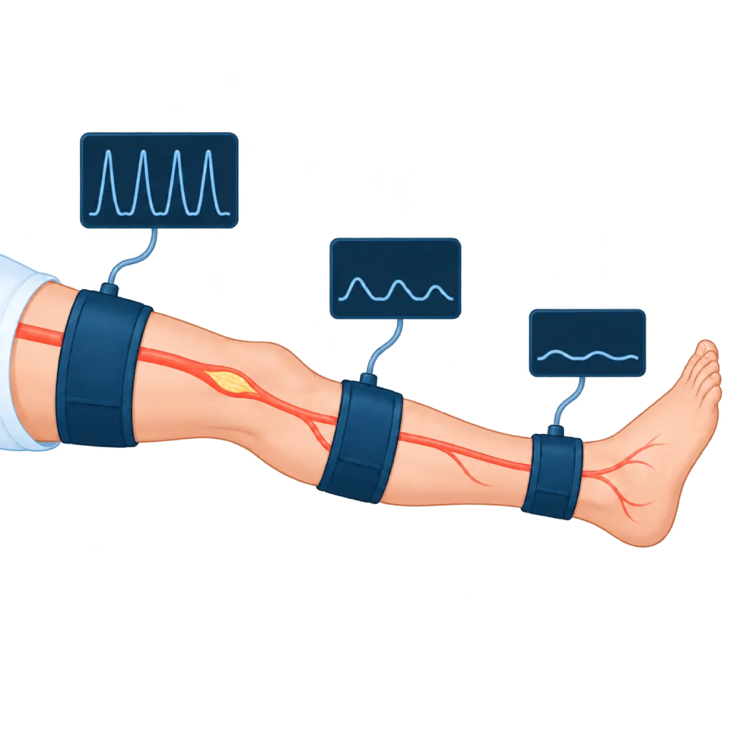
A PVR (pulse volume recording) test uses cuffs like those used to measure blood pressure, placed on several parts of the leg. Each time the heart beats, the volume of blood in the leg increases slightly. This small change is recorded as a wave. The shape of the wave in each part of the leg shows how well blood is flowing to that area.
How It Works at a Glance
The cuffs are inflated with low pressure, and the device records the pulse wave at each level. The doctor compares the wave shapes from the thigh down to the ankle.
Reading the Wave Shapes
- Normal wave: rises sharply, has a pointed peak, and then falls with a small notch.
- Flattened wave: rises more slowly with a blunt, low peak. This indicates narrowing or blockage above that cuff.
- Comparison between levels: if the wave at the thigh is normal but the wave at the calf is flattened, the narrowing is likely between those two points.
PVR, ABI, and TBI
PVR is often done together with the ankle-brachial index (ABI) test, which compares the blood pressure at the ankle and the arm. In people with diabetes or chronic kidney disease, and in older adults, the leg arteries can harden, so the ABI result looks normal or too high. PVR does not depend on pressure to compress the artery, so it can still show impaired flow in these situations, together with the toe-brachial index (TBI) test.
Further Testing
PVR shows whether there is impaired blood flow and roughly where it is, but it does not produce images of the blood vessels. If a procedure is being considered, your doctor may follow up with duplex ultrasound, CT angiography, or catheter angiography.
02 / Understanding the procedure
When Is This Procedure Considered?
A doctor may consider this procedure in the situations below. The decision is always made after a thorough assessment.
- Leg Pain or Aching When WalkingSymptoms that improve when you stop walking can indicate narrowing of the leg arteries.
- Unreliable ABI ResultsFor example, stiff leg arteries in diabetes or chronic kidney disease, which make the ABI look normal or too high.
- Locating the NarrowingComparing the waves at the thigh, calf, and ankle helps estimate where the narrowing is.
- Foot Wounds That Are Slow to HealTo assess whether blood flow to the foot is enough to support wound healing.
- Monitoring After a ProcedureAfter balloon angioplasty, stenting, or leg bypass surgery, to monitor blood flow over time.
Situations to Consider First
- Wounds, Bandages, or a Cast Where the Cuff GoesThe cuff is not placed over a wound or cast, so the test is adjusted or that level is skipped.
- Suspected Blood Clot in a Leg VeinThe cuff is not inflated on a leg suspected of having a new clot. Duplex ultrasound is done beforehand.
- Recent Vascular Surgery or ProcedureThe doctor decides whether it is safe to place a cuff over the area of a recent operation or procedure.
- Severe Leg SwellingCan reduce the accuracy of the recording, so the results are read together with other tests.
- Sudden Severe Pain, Paleness, and Coldness in the FootThis can indicate a sudden blockage. Do not wait for a scheduled test; go to the emergency room right away.
03 / Before the procedure
How Should You Prepare?
A PVR test does not require special preparation or fasting.
Clothing. Wear loose clothing that is easy to roll up or remove from your legs. You may be asked to wear an examination gown and take off your socks.
Smoking. Avoid smoking for two hours or more before the test, because smoking temporarily narrows the blood vessels.
Rest. Arrive early. You will lie down and rest for about 10 minutes before the recording starts.
Medicines. Take your regular medicines as usual, unless your doctor gives other instructions.
Important information. Tell the staff if you have had leg blood vessel surgery or procedures, have a wound on your foot, or are being treated for a blood clot.
Shoes. If a treadmill walking test is planned, wear comfortable walking shoes.
Unsure about the preparation instructions? Ask our team before the procedure.
04 / During the procedure
How Is the Procedure Done?
The test usually takes about 30 minutes, including rest time. If it is followed by a treadmill walking test, it takes longer.
Rest and Positioning
You lie on your back and rest for a few minutes so your blood flow is stable before the recording.
Placing the Cuffs
Cuffs are placed on the thigh, calf, and ankle, sometimes also on the foot or toes, and on the arm for comparison.
Recording the Waves
Each cuff is inflated with low pressure in turn, and the device records the pulse wave for a few seconds. You will only feel light pressure. Stay still and breathe normally during the recording.
Blood Pressure Measurement
At the same visit, blood pressure at the arm and ankle is usually measured to calculate the ABI.
Walking Test If Needed
If the results at rest are normal but symptoms appear when you walk, you may be asked to walk on a treadmill, and the recording is repeated right afterward.
05 / Benefits and risks
Benefits and Risks Should Be Discussed Together.
Every medical procedure has benefits and risks. Your doctor will explain both for your situation before the procedure.
Benefits You Can Expect
- No Needles and No RadiationIt only uses cuffs like a blood pressure cuff, so it is safe to repeat.
- Still Useful in Stiff ArteriesHelps assess blood flow when ABI results are unreliable, for example in diabetes or chronic kidney disease.
- Estimates Where the Narrowing IsHelps your doctor decide which part of the leg needs further testing.
- Long-Term MonitoringSuitable for monitoring blood flow in the legs after treatment or a procedure.
- Quick and ComfortableFinished in about 30 minutes, and you can return to your activities right away.
The benefits of a procedure are not a promise of a cure. Ask how the results will affect your care plan.
Risks You Should Know
- Uncomfortable Cuff PressureOnly brief. Tell the staff if it feels painful.
- Does Not Show Images of the VesselsPVR shows impaired flow, not the shape of the vessels, so duplex ultrasound or CT angiography may be needed.
- Results Affected by the Condition of the LegSevere swelling, excess body weight, or movement during the recording can reduce the accuracy of the results.
- Symptoms During the Treadmill TestIf a walking test is done, leg pain or chest symptoms may appear. The test is stopped right away if needed.
- Minor Bruising on Fragile SkinVery rare, mainly in older adults or people taking blood thinners.
Risks can differ from person to person. A full explanation and consent are given after an individual assessment.
06 / After the procedure
What Is Recovery Like?
There is no recovery period. You can return to your activities right away.
Skin. Marks from the cuff pressure may be visible on the skin for a short time and fade on their own.
Discussing the results. The doctor reads the results together with your symptoms and other tests, then explains whether you need treatment, structured walking exercise, or further tests.
Healthy habits. If narrowing of the leg arteries is found, quitting smoking, regular walking exercise, and controlling blood sugar, blood pressure, and cholesterol help a great deal.
Warning signs. Go to the emergency room right away or contact the medical team if you have:
- Sudden severe pain, paleness, coldness, or numbness in the leg or foot
- Foot pain at rest, especially at night
- A foot wound that is getting worse, turning black, or oozing pus
- Chest pain or shortness of breath
Know the Signs That Need Prompt Care.
Follow the team's guidance on warning signs. If severe or sudden symptoms appear after the procedure, go to the nearest emergency department right away or call Heart Attack Center 1500 258.
07 / Team and support
Doctors and Procedure Support
The doctors below are listed for this procedure. Listed by name, not by ranking.

dr. Debby C. Soemitha, Sp.PD
Internal Medicine
Clinical focusDiabetes and Diabetes Complications · Obesity and Metabolic Disease
View Profile
dr. Emanoel Oepangat, Sp.JP(K)
Interventional Cardiology
Vascular Diagnostic and Therapeutic Center +1 center
Clinical focusCoronary Intervention · Peripheral Vascular Intervention
View Profile
dr. Suko Adiarto, Sp.JP(K), Ph.D.
Interventional Cardiology
Vascular Diagnostic and Therapeutic Center +1 center
Clinical focusCoronary Intervention · Peripheral Vascular Intervention
View ProfileAnkle-Brachial Index (ABI) Testing System
A needle-free test that compares blood pressure at the ankles and arms. The result is an index number that helps doctors detect…
The availability of technology and test support is confirmed with the team. Technology is chosen according to clinical need.
08 / Common questions
Before You Plan the Procedure.
Frequently asked questions about the PVR test. The team will give you instructions specific to your test.
What is the difference between PVR and an ABI test?
An ABI test compares the blood pressure at the ankle with the pressure in the arm and produces a number. PVR records the shape of the pulse wave in several parts of the leg. The two tests complement each other and are often done together. PVR is especially helpful when the leg arteries are stiff, because in that situation the ABI number can be misleading.
Does a PVR test hurt?
No. You will only feel the cuff tighten briefly around your thigh, calf, or ankle, similar to having your blood pressure measured. There are no needles, injections, or radiation. If the cuff pressure feels uncomfortable, for example on a painful leg, tell the staff so it can be adjusted.
Do I need to fast before a PVR test?
No. You can eat, drink, and take your medicines as usual. What matters is not smoking for two hours or more before the test and arriving early to rest. Wear loose clothing so the cuffs can be placed on your legs easily, and bring the results of any previous blood vessel tests.
What does a flattened wave mean?
A flattened wave, with a low and blunt peak, shows that blood flow to that part of the leg is reduced because of narrowing or blockage above it. By comparing the waves at the thigh, calf, and ankle, your doctor can estimate where the problem is. These results are then read together with your symptoms and other tests.
Do people with diabetes need a PVR test?
People with diabetes are at higher risk of narrowing in the leg arteries, and their arteries often harden, making ABI results less reliable. For this reason, your doctor may request a PVR or TBI test, especially if you have leg pain when walking, a foot wound that is slow to heal, or weak foot pulses.
How long does the test take?
The test usually takes about 30 minutes, including rest time before the recording and the ABI measurement. If your doctor requests a treadmill walking test, it takes longer. The team will give you an estimated time when scheduling, so set aside enough time and arrive before your appointment.
Sources and Medical Review
Translated from the Indonesian edition medically reviewed by dr. Emanoel Oepangat, Sp.JP(K).
Sources as cited in the Indonesian edition.
- Mazzolai L, Teixido-Tura G, Lanzi S, et al. 2024 ESC Guidelines for the management of peripheral arterial and aortic diseases. Eur Heart J. 2024;45(36):3538-3700.
- Gornik HL, Aronow HD, Goodney PP, et al. 2024 ACC/AHA/AACVPR/APMA/ABC/SCAI/SVM/SVN/SVS/SIR/VESS Guideline for the Management of Lower Extremity Peripheral Artery Disease. Circulation. 2024;149(24):e1313-e1410.
- Aboyans V, Criqui MH, Abraham P, et al. Measurement and Interpretation of the Ankle-Brachial Index: A Scientific Statement From the American Heart Association. Circulation. 2012;126(24):2890-2909.
- Sumpio BE, Benitez E. Pulse volume recording for peripheral vascular disease diagnosis in diabetes patients. J Vasc Diagn. 2015.