
Quick answer
What Is Peripheral MR Angiography?
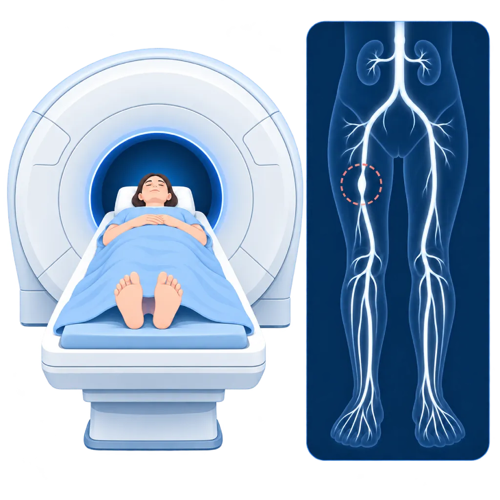
Peripheral MR angiography examines the leg arteries with an MRI scanner, which uses a magnetic field and radio waves, not X-rays. Gadolinium contrast is usually injected through an IV in the arm. The result is an arterial map from the abdomen to the feet that helps the doctor find narrowings and plan treatment, with no catheter inside the artery.
Sources01 / Understanding the procedure
About Peripheral MR Angiography
What Is Peripheral MR Angiography
Peripheral MR angiography, or peripheral MRA, examines the arteries that carry blood to the legs, from the aorta in the abdomen through the pelvic and thigh arteries down to the feet. This test uses an MRI scanner, meaning a strong magnetic field and radio waves, so there is no X-ray exposure. No catheter is inserted into the artery.
How It Works in Brief
You lie on a table that moves into the MRI scanner. Gadolinium contrast is injected through an IV line in your arm, and the scanner takes images in stages as the contrast flows from the abdomen to the feet. A computer then assembles the images into a single map of the arteries.
What Can Be Seen
- Narrowings and blockages: their location, length, and severity, from the abdomen to the feet.
- Arteries below the knee: useful for assessing foot wounds that are slow to heal.
- Collateral vessels: small pathways that form to carry blood around a blockage.
- Widened arteries: for example, an aneurysm of the artery behind the knee.
- Arteries for bypass: helps choose the connection sites if surgery is planned.
With or Without Contrast
The majority of peripheral MRA scans use gadolinium contrast because the images are obtained faster and are clearer. When contrast needs to be avoided, for example in severe kidney impairment or pregnancy, there are non-contrast MRA techniques that use the blood flow itself. Your doctor will choose the technique that suits your condition.
MRA, CT Angiography, or Catheter Angiography
Duplex ultrasound is usually the initial test. When a more complete map of the arteries is needed, the doctor may choose CT angiography or MRA. MRA does not use X-rays and is not affected by heavy calcification in the artery walls, but it takes longer and cannot be done with some implanted devices. Catheter angiography is usually chosen when a procedure to open the narrowing has already been planned.
02 / Understanding the procedure
When Is This Procedure Considered?
A doctor may consider this procedure in the situations below. The decision is always made after a thorough assessment.
- Limiting Leg Pain When WalkingWhen a procedure is being considered and the doctor needs a complete map of the leg arteries.
- Slow-Healing Foot Wounds or Pain at RestTo map the arteries down to the feet and look for a route to restore blood flow.
- Planning Balloon, Stent, or Bypass SurgeryHelps the doctor choose the type of procedure and the right route before it is performed.
- Severe Arterial CalcificationIn diabetes or kidney disease, MRA can assess arteries that are hard to assess with CT because of calcification.
- CT Contrast or X-Rays Need to Be AvoidedFor example, with a history of reactions to iodinated contrast, or when X-ray exposure is to be avoided.
- Widened or Inflamed ArteriesWhen a leg artery aneurysm or blood vessel inflammation such as vasculitis is suspected.
Situations to Consider First
- Implants That Are Not Safe for MRIFor example, some older implants, ear implants, drug pumps, or metal fragments in the eye. The team checks your type of implant beforehand.
- Pacemaker or ICDMany newer devices can go into the MRI under special conditions. The arrhythmia team assesses and adjusts the device before and after the test.
- Severe Kidney Impairment or DialysisGadolinium contrast is considered carefully, or a non-contrast MRA technique is chosen.
- PregnancyGadolinium contrast is avoided. MRI without contrast may be considered if it is really needed.
- Fear of Enclosed SpacesA mild sedative can help. Tell the team early so it can be arranged.
- History of a Severe Reaction to GadoliniumThe doctor considers other tests, or gives anti-allergy medication beforehand.
03 / Before the procedure
How Should You Prepare?
The team will give you instructions and an MRI safety form before the test.
MRI safety form. You will be asked about any devices or metal in your body, for example a pacemaker, ICD, artificial heart valve, stent, blood vessel clip, artificial joint, ear implant, or metal fragments. Bring your implant ID card if you have one.
Pacemaker or ICD. The test is scheduled together with the arrhythmia team so the device can be checked and adjusted before and after the MRI.
Kidney function. If contrast will be used and you have kidney disease or diabetes, or are older, your kidney function may be checked beforehand.
Medications and food. Regular medications are generally taken as usual. Fasting is usually not required unless the team gives other instructions.
Clothing and metal items. Wear clothing without metal or change into a hospital gown. Remove jewelry, watches, bank cards, hearing aids, and other metal items before entering the MRI room. Some medicated patches also need to be removed.
Pregnancy, breastfeeding, and fear of enclosed spaces. Tell the team if you are or may be pregnant, are breastfeeding, or are afraid of enclosed spaces.
Unsure about the preparation instructions? Ask our team before the procedure.
04 / During the procedure
How Is the Procedure Done?
The test generally takes about 30 to 60 minutes, depending on the technique and the arteries being examined.
Preparation in the MRI Room
After the safety form is checked, an IV line is placed in your arm for the contrast. You lie on your back on the table, and pad-shaped signal receivers are placed over your abdomen and legs.
Inside the Scanner
The table moves into the tunnel-shaped scanner. The scanner makes loud knocking sounds, so you are given earplugs or headphones. You can still talk to the staff through an intercom and hold a call button.
Taking the Images
You are asked to stay still, and sometimes to hold your breath briefly while the abdomen is imaged. Contrast is injected through the IV, then the table moves in stages so images are taken from the abdomen to the feet, following the flow of contrast. The contrast injection may feel cold in your arm.
After the Test
The IV is removed and you can change your clothes right away. The images are then processed into a map of the arteries, analyzed, and reported by a doctor.
05 / Benefits and risks
Benefits and Risks Should Be Discussed Together.
Every medical procedure has benefits and risks. Your doctor will explain both for your situation before the procedure.
Benefits You Can Expect
- No X-RaysThere is no radiation exposure, so it can be repeated for monitoring if needed.
- No Catheter in the ArteryContrast is simply injected through an IV in the arm, with no puncture of an artery.
- Arterial Map from the Abdomen to the FeetA single test shows the entire course of the leg arteries.
- Stays Clear Despite Heavy CalcificationCan assess arteries with heavily calcified walls, for example in diabetes or kidney disease.
- No Iodinated ContrastAn option for patients who have reacted to CT or catheterization contrast.
The benefits of a procedure are not a promise of a cure. Ask how the results will affect your care plan.
Risks You Should Know
- Reaction to Gadolinium ContrastUsually mild, such as nausea, dizziness, or a rash. Severe reactions are very rare and are treated right away by the team.
- Risk in Severe Kidney ImpairmentVery rarely, the contrast can trigger thickening of the skin and tissues (NSF). The risk is very low with the contrast agents used today.
- Metal and Implant HazardsThe magnetic field can pull metal objects or interfere with implanted devices, so safety screening is done carefully.
- Anxiety in Enclosed SpacesSome people feel anxious inside the scanner. The team can help, including with a mild sedative.
- Less Clear Images Around StentsMetal stents or body movement can blur the images, so another test is sometimes needed.
Risks can differ from person to person. A full explanation and consent are given after an individual assessment.
06 / After the procedure
What Is Recovery Like?
There is no special recovery period. The majority of patients can go home right away and resume their usual activities.
Drink water. Drink enough water to help your kidneys clear the contrast, unless your doctor limits your fluids.
If you were given a sedative. Arrange for someone to take you home, and do not drive on the day of the test.
Pacemaker or ICD. The device is checked and returned to its original settings before you go home.
Breastfeeding. You can generally continue as usual. Discuss it with your doctor if you are unsure.
Results. Your doctor explains the findings and next steps, for example medication, walking exercise, a catheter-based procedure, or bypass surgery.
Warning signs to watch for. Go to the emergency room right away or contact the medical team if you experience:
- A rash, itching, facial swelling, or shortness of breath after the test
- Pain, swelling, or redness at the IV site
- A leg that suddenly feels cold, pale, or very painful
Know the Signs That Need Prompt Care.
Follow the team's guidance on warning signs. If severe or sudden symptoms appear after the procedure, go to the nearest emergency department right away or call Heart Attack Center 1500 258.
07 / Team and support
Doctors and Procedure Support
The doctors below are listed for this procedure. Listed by name, not by ranking.

dr. Debby C. Soemitha, Sp.PD
Internal Medicine
Clinical focusDiabetes and Diabetes Complications · Obesity and Metabolic Disease
View Profile
dr. Emanoel Oepangat, Sp.JP(K)
Interventional Cardiology
Vascular Diagnostic and Therapeutic Center +1 center
Clinical focusCoronary Intervention · Peripheral Vascular Intervention
View Profile
dr. Suko Adiarto, Sp.JP(K), Ph.D.
Interventional Cardiology
Vascular Diagnostic and Therapeutic Center +1 center
Clinical focusCoronary Intervention · Peripheral Vascular Intervention
View Profile08 / Common questions
Before You Plan the Procedure.
Frequently asked questions about peripheral MR angiography. The choice of test is made together with your doctor.
What is the difference between MRA and peripheral CT angiography?
Both map the leg arteries without a catheter inside the artery. CT angiography uses X-rays and iodinated contrast, and image acquisition lasts only a few minutes. MRA uses a magnetic field and gadolinium contrast, with no X-rays, and stays clear in heavily calcified arteries. However, MRA takes longer and cannot be done with some implants. Your doctor chooses the one that suits you.
Can I have an MRA if I have a pacemaker or a stent?
Stents in both the heart arteries and the leg arteries are generally safe for MRI, although stents in the leg can blur the images around them. Many newer pacemakers and ICDs can also go into the MRI under special conditions. The team will check your type of device, and the arrhythmia team adjusts it before and after the test. Bring your implant ID card.
Is the test painful?
No. All you feel is the needle prick when the IV is placed and a cold sensation when the contrast is injected. The scanner makes loud knocking sounds, so you are given ear protection. The main challenge is lying still for quite a long time. If you are afraid of enclosed spaces, tell the team early so they can help, for example with a mild sedative.
What if my kidney function is reduced?
Gadolinium contrast is used in small amounts, and its risk to the kidneys is very low. In severe kidney impairment or dialysis, the doctor weighs the benefits carefully, chooses a safer type of contrast, or uses a non-contrast MRA technique. Tell the team if you have kidney disease or are on regular dialysis.
Why does the test take quite a long time?
The scanner needs to take images in stages from the abdomen to the feet, following the flowing contrast. Before that, preparatory images are taken to set the right position and timing. With a non-contrast technique, it can take longer. Overall, plan to spend about one to one and a half hours at the hospital, including preparation.
When will the results be available?
The images need to be processed into a map of the arteries and read carefully, so the written report is not ready the moment the test ends. The team will tell you when the results will be available. The doctor who requested the test then explains the results and discusses next steps with you, for example medication, walking exercise, a catheter-based procedure, or bypass surgery.
Sources and Medical Review
Translated from the Indonesian edition medically reviewed by dr. Emanoel Oepangat, Sp.JP(K).
Sources as cited in the Indonesian edition.
- Mazzolai L, Teixido-Tura G, Lanzi S, et al. 2024 ESC Guidelines for the management of peripheral arterial and aortic diseases. Eur Heart J. 2024;45(36):3538-3700.
- Gornik HL, Aronow HD, Goodney PP, et al. 2024 ACC/AHA/AACVPR/APMA/ABC/SCAI/SVM/SVN/SVS/SIR/VESS Guideline for the Management of Lower Extremity Peripheral Artery Disease. Circulation. 2024;149(24):e1313-e1410.
- Menke J, Larsen J. Meta-analysis: Accuracy of Contrast-Enhanced Magnetic Resonance Angiography for Assessing Steno-occlusions in Peripheral Arterial Disease. Ann Intern Med. 2010;153(5):325-334.
- Weinreb JC, Rodby RA, Yee J, et al. Use of Intravenous Gadolinium-based Contrast Media in Patients with Kidney Disease: Consensus Statements from the American College of Radiology and the National Kidney Foundation. Radiology. 2021;298(1):28-35.
- Indik JH, Gimbel JR, Abe H, et al. 2017 HRS expert consensus statement on magnetic resonance imaging and radiation exposure in patients with cardiovascular implantable electronic devices. Heart Rhythm. 2017;14(7):e97-e153.