
Quick answer
What Is Rotational Atherectomy?
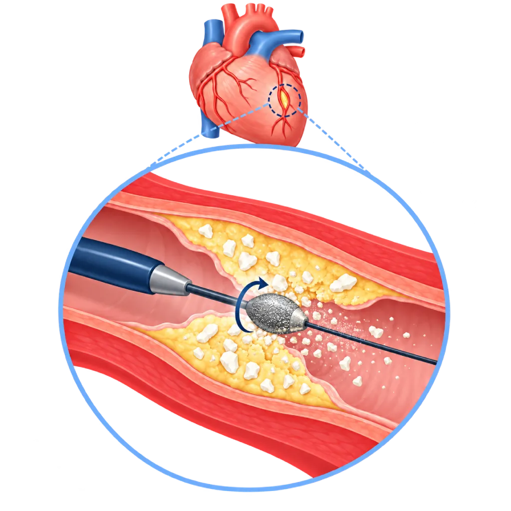
Rotational atherectomy (rotablation) is part of stent placement in heavily calcified coronary arteries. A small diamond-coated burr is passed through a catheter and spins very fast to grind hard calcium into fine particles. This allows the balloon and stent to expand properly. The procedure is done in a cath lab under local anesthesia.
Sources01 / Understanding the procedure
About Rotational Atherectomy
What Is Rotablation
Rotablation, or rotational atherectomy, is an additional technique used during coronary stent placement. It is used when the wall of a coronary artery contains very hard calcium, so a balloon cannot pass through or expand the narrowed section. Without good preparation, the stent may not expand fully, making it more likely to narrow again or become blocked.
How It Works in Brief
Through a catheter inserted at the wrist or groin, the doctor places a special guidewire across the narrowing. An olive-shaped burr (a tiny drill tip) coated with diamond dust, about 1.25 to 2.5 mm in size, is advanced along the wire and spins hundreds of thousands of times per minute. The burr grinds away hard, stiff tissue, while the flexible artery wall tends to be pushed aside. The calcium is ground into particles smaller than red blood cells, which are carried away by the bloodstream and cleared by the body.
When Rotablation Is Chosen
- Heavy calcium: seen on angiography or on imaging from inside the artery with IVUS or OCT.
- Balloon gets stuck: the balloon cannot pass through the narrowing or cannot expand fully.
- Long, stiff narrowing: often found in older adults and in people with diabetes or chronic kidney disease.
- Other options for calcium: special balloons or intravascular lithotripsy are considered depending on the shape and location of the calcium, and are sometimes used together.
After the Artery Is Prepared
Once the calcium has been ground away, the doctor widens the artery with a balloon, places a drug-eluting stent, and then confirms that the stent is fully expanded, often with the help of IVUS or OCT. Rotablation requires special skills, so it is performed by trained interventional cardiologists, with careful planning before the procedure.
02 / Understanding the procedure
When Is This Procedure Considered?
A doctor may consider this procedure in the situations below. The decision is always made after a thorough assessment.
- Heavily Calcified Coronary NarrowingCalcium prevents the balloon and stent from expanding fully.
- Balloon Cannot Cross the NarrowingThe burr opens a path for the balloon and stent.
- Narrowing That Does Not Open with a BalloonStiff calcium keeps the balloon from expanding fully.
- Angina That Persists Despite MedicationWhen a calcified narrowing needs to be opened with a stent.
- Coronary Disease with Diabetes or Kidney ProblemsConditions that are often accompanied by heavy calcium in the coronary arteries.
Situations to Consider First
- Unable to Take Dual Antiplatelet TherapyThis medication is needed after stent placement.
- Blood Clot in the Artery to Be TreatedThe burr can worsen blood flow; the clot is treated in another way.
- Existing Tear in the Artery WallDrilling can make the tear larger.
- Vein Bypass GraftsRotablation is usually not performed in these grafts.
- Severe Contrast Allergy or Severe Kidney DiseaseRequires special preparation or consideration of other options.
- Very Weak Heart Pumping FunctionA heart support device or other treatment options may be needed.
03 / Before the procedure
How Should You Prepare?
Rotablation is usually planned together with stent placement. The heart team will explain the preparation that suits your situation.
Tests before the procedure. Blood tests, including kidney function and blood clotting, an ECG, and the results of any previous coronary angiography or coronary CT.
Medications. You usually start taking antiplatelet medication before the procedure. Your doctor will give you instructions about blood thinners, metformin, and other medications. Do not stop or change your medications on your own.
Fasting. You usually need to avoid food for about 6 hours before the procedure, following the team's instructions.
Allergies and companion. Tell the team if you have ever had an allergy to contrast dye or medications. Arrange for a family member to accompany you, because you will stay in the hospital.
Unsure about the preparation instructions? Ask our team before the procedure.
04 / During the procedure
How Is the Procedure Done?
The entire procedure, including stent placement, usually takes about 1.5 to 2 hours in the cath lab. You stay awake under local anesthesia, and a mild sedative may be given.
Access and Calcium Assessment
The doctor inserts a catheter through the wrist or groin, then assesses the calcium with angiography and, if needed, IVUS or OCT. Sometimes a temporary pacemaker is placed as a precaution in case the heart rate slows down.
Grinding Away the Calcium
A special wire is placed across the narrowing. The burr is advanced in several short runs while a lubricating fluid and a medication that widens the blood vessels are flushed through, until the path is open enough.
Balloon and Stent
The burr is removed, then a balloon widens the narrowing and a drug-eluting stent is placed and fully expanded.
Final Check
The doctor confirms that blood flows smoothly and the stent sits well against the artery wall, then the catheter is removed and pressure is applied to the puncture site.
05 / Benefits and risks
Benefits and Risks Should Be Discussed Together.
Every medical procedure has benefits and risks. Your doctor will explain both for your situation before the procedure.
Benefits You Can Expect
- Opens a Path Through Hard CalciumThe balloon and stent can pass through the narrowing.
- Better Stent ExpansionReduces the risk of the stent narrowing again or becoming blocked.
- No Open SurgeryPerformed through a catheter under local anesthesia in suitable patients.
- Relieves AnginaBlood flow to the heart muscle improves.
- Relatively Fast RecoveryYou usually go home within 1 to 2 days.
The benefits of a procedure are not a promise of a cure. Ask how the results will affect your care plan.
Risks You Should Know
- Tear or Perforation of a Coronary ArteryRare, but may require immediate treatment.
- Temporary Slowing of Blood FlowParticles and artery spasm can slow blood flow; this is treated with medication.
- Temporary Slowing of the Heart RateEspecially in the right coronary artery; a temporary pacemaker can help.
- Minor Heart Muscle Damage During the ProcedureSometimes occurs and is monitored with blood tests.
- Bleeding or Bruising at the Puncture SiteUsually mild and improves on its own.
- Kidney Problems or Allergic Reaction to ContrastRare, and watched more closely in people with kidney disease.
Risks can differ from person to person. A full explanation and consent are given after an individual assessment.
06 / After the procedure
What Is Recovery Like?
You are monitored in the cardiac ward, usually for 1 to 2 nights.
The early hours. Your blood pressure, heart rhythm, and puncture site are checked regularly. The compression band on your wrist is loosened gradually; if the puncture was in the groin, you lie with your leg straight for a few hours.
Antiplatelet medication. Take dual antiplatelet therapy as recommended, usually for several months to one year. Do not stop on your own, because the stent can become blocked.
At home. Avoid lifting heavy objects with the arm used for the procedure for a few days. Join the cardiac rehabilitation program and attend follow-up visits as scheduled.
Warning signs. Go to the emergency room right away if you have:
- Chest pain or shortness of breath
- Bleeding or swelling that keeps growing at the puncture site
- A hand or foot on the puncture side that is cold, pale, or numb
- Fever, severe dizziness, or fainting
Know the Signs That Need Prompt Care.
Follow the team's guidance on warning signs. If severe or sudden symptoms appear after the procedure, go to the nearest emergency department right away or call Heart Attack Center 1500 258.
07 / Team and support
Doctors and Procedure Support
The doctors below are listed for this procedure. Listed by name, not by ranking.

dr. Adrianus Kosasih, Sp.JP(K)
Interventional Cardiology
Coronary Intervention Procedures
Clinical focusPrimary Coronary Intervention · PCI for Acute Coronary Syndrome
View Profile
Dr. dr. Dafsah A. Juzar, Sp.JP(K)
Interventional Cardiology
Coronary Intervention Procedures +1 center
Clinical focusComplex Coronary Intervention · Cardiac Catheterization
View Profile
Dr. dr. Faris Basalamah, Sp.JP(K)
Interventional Cardiology
Coronary Intervention Procedures +1 center
Clinical focusComplex Coronary Intervention · Cardiac Electrophysiology
View ProfileRotational Atherectomy System
A system for treating calcified plaque in the coronary arteries. A small diamond-coated burr spins at high speed over a guidewire…
Intravascular Lithotripsy (IVL) System
A special balloon catheter that emits pulses of sonic pressure waves to crack calcium in the coronary artery wall. It is used…
The availability of technology and test support is confirmed with the team. Technology is chosen according to clinical need.
08 / Common questions
Before You Plan the Procedure.
Questions people often ask about rotablation. Your doctor will explain the treatment plan for your condition.
Can the burr damage the artery wall?
The rotablation burr is designed to grind away hard, stiff tissue such as calcium, while the flexible artery wall tends to be pushed aside. The doctor advances the burr little by little in short runs while monitoring with X-rays. A tear or leak in the artery can still happen, but it is rare, and the team is ready to manage it.
Where do the ground-up calcium particles go?
The calcium is ground into very fine particles, smaller than red blood cells. These particles are carried by the bloodstream into small vessels and then cleared by the body. Sometimes blood flow slows briefly during grinding, and the doctor manages this with fluids and medication that widens the blood vessels.
How is it different from intravascular lithotripsy?
Intravascular lithotripsy uses a special balloon that emits shock waves to crack the calcium, while rotablation grinds the calcium away with a burr. The choice depends on the location, thickness, and shape of the calcium, and whether a balloon can cross the narrowing. In some cases, both techniques are used in turn during one procedure.
Am I awake during the procedure?
Yes. Rotablation is done under local anesthesia at the puncture site, and a mild sedative may be given to make you more comfortable. While the burr is working, you may feel brief chest discomfort. Tell the team if you have any symptoms, because they monitor your heart rhythm and blood pressure throughout the procedure.
How long will I need to stay in the hospital?
For a planned procedure, the majority of patients stay about 1 to 2 nights so the team can monitor heart rhythm, the puncture site, and blood test results. The stay may be longer if the procedure is done during a heart attack or if there are complications. Your doctor will decide when it is safe for you to go home.
Why do I need to take dual antiplatelet therapy?
After a stent is placed, dual antiplatelet therapy prevents blood clots from forming inside the stent while the artery lining grows over it. Your doctor decides how long you take it, usually several months to one year. Do not stop on your own, including before a tooth extraction or surgery, without talking to your cardiologist.
Sources and Medical Review
Translated from the Indonesian edition medically reviewed by dr. Adrianus Kosasih, Sp.JP(K).
Sources as cited in the Indonesian edition.
- Neumann FJ, Sousa-Uva M, Ahlsson A, et al. 2018 ESC/EACTS Guidelines on myocardial revascularization. Eur Heart J. 2019;40(2):87-165.
- Vrints C, Andreotti F, Koskinas KC, et al. 2024 ESC Guidelines for the management of chronic coronary syndromes. Eur Heart J. 2024;45(36):3415-3537.
- Barbato E, Carrié D, Dardas P, et al. European expert consensus on rotational atherectomy. EuroIntervention. 2015;11(1):30-36.
- Sharma SK, Tomey MI, Teirstein PS, et al. North American expert review of rotational atherectomy. Circ Cardiovasc Interv. 2019;12(5):e007448.
- Abdel-Wahab M, Toelg R, Byrne RA, et al. High-speed rotational atherectomy versus modified balloons prior to drug-eluting stent implantation in severely calcified coronary lesions. Circ Cardiovasc Interv. 2018;11(10):e007415.