
Quick answer
What Is Ischemic Cardiomyopathy?
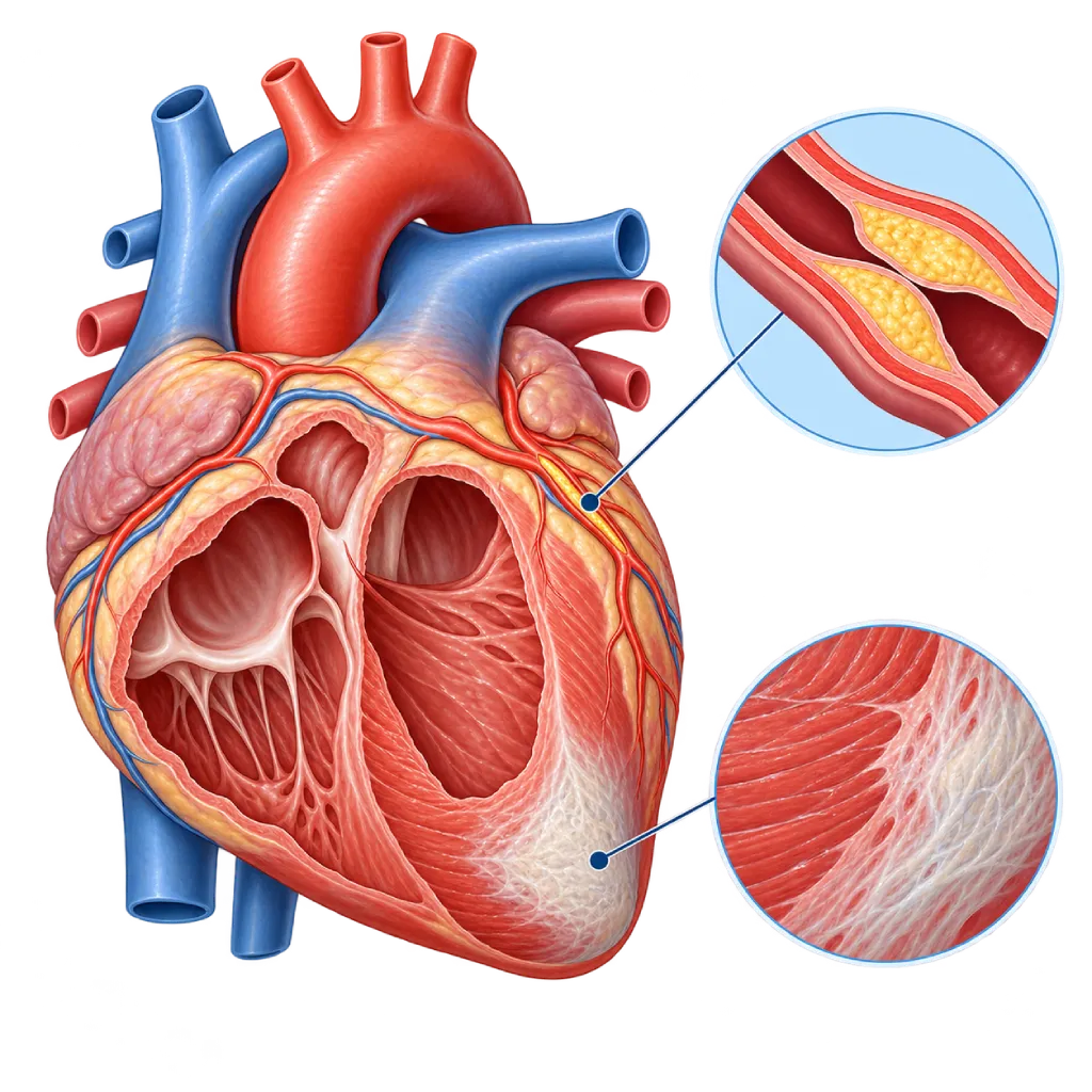
Ischemic cardiomyopathy is a weakening of the heart muscle and its pumping action caused by coronary artery disease, usually after a heart attack or because of long-term narrowing of several coronary arteries, and it can lead to heart failure.
SourcesSigns That Need Urgent Help
- Severe chest pain that does not go away after a few minutes of rest
- Severe shortness of breath at rest or being unable to lie down
- Coughing up frothy or pink sputum
- Fainting or nearly fainting
- A very fast heartbeat with severe dizziness or weakness
- Cold, pale skin and extreme weakness
If these signs appear, go to the nearest emergency department right away or call Heart Attack Center 1500 258.
01 / Understanding the condition
About Ischemic Cardiomyopathy
Ischemic cardiomyopathy is a condition in which the heart muscle weakens because of reduced blood flow caused by coronary artery disease. The term is generally used when the ejection fraction, the percentage of blood pumped out of the left ventricle with each heartbeat, falls to 40 percent or less, and the cause is a previous heart attack or severe narrowing of the coronary arteries. Ischemic cardiomyopathy is a common cause of heart failure with reduced ejection fraction, which is covered on the Heart Failure with Reduced Ejection Fraction page.
How the Heart Muscle Weakens
During a heart attack, part of the heart muscle dies and is replaced by scar tissue that cannot contract. If the damaged area is large, the left ventricle can widen and change shape to compensate, a process called remodeling. In addition, muscle that keeps receiving too little blood because of long-standing narrowing can weaken without dying. This is called living (viable) muscle, and some of its function can improve if blood flow is restored.
How It Differs from Other Types
In the 2023 European Society of Cardiology (ESC) guidelines on cardiomyopathies, weakening of the heart muscle that is fully explained by coronary artery disease is not classified as a cardiomyopathy, but the term ischemic cardiomyopathy is still commonly used by doctors and in research. This distinction matters because treatment also covers the underlying coronary disease. Weakening of the pump that is not caused by coronary disease is covered on the Nonischemic Cardiomyopathy page, while an overview of all types is covered on the Cardiomyopathy page.
Principles of Treatment
Treatment has three parts. First, heart failure medicines that have been shown in studies to reduce hospital stays and deaths. Second, control of coronary artery disease with medicines that protect the blood vessels, control of cholesterol, blood pressure, and blood sugar, and quitting smoking. Third, in certain patients, revascularization, meaning coronary bypass surgery or stent placement, as decided by the heart team. The STICH study showed that bypass surgery lowers the long-term risk of death in suitable patients, while the REVIVED study found no added benefit of stent placement compared with medicines alone in the group studied. Patients whose ejection fraction remains low are also assessed for an ICD.
02 / Understanding the condition
Symptoms to Recognize.
Common Symptoms
- Shortness of breath during activity, and later also when lying down
- Tiring easily and quickly running out of energy
- Swelling in the ankles and legs
- Pain or pressure in the chest during activity or stress
- Palpitations or a heartbeat that feels irregular
- Waking up at night short of breath
Symptoms in Older Adults
In older adults, especially those with diabetes, symptoms are often atypical and chest pain may not be felt.
- Weakness and reduced daily activity without a clear cause
- Reduced appetite or a feeling of fullness in the stomach
- Unusual confusion or sleepiness
- Often waking at night to urinate
- Shortness of breath when walking short distances, without chest pain
- Falling easily or feeling unsteady
03 / Understanding the condition
What Causes This Condition?
- A heart attack (myocardial infarction) that leaves scar tissue in the heart muscle
- Repeated heart attacks that enlarge the area of damaged heart muscle
- Severe narrowing in several coronary arteries, so the heart muscle lacks blood for a long time
- Narrowing at the start of the main coronary artery that supplies most of the left ventricle
- Atherosclerosis, the buildup of fatty plaque in the walls of the coronary arteries
04 / Understanding the condition
Who Is at Higher Risk?
Lifestyle
Smoking, a diet high in saturated fat and salt, too little physical activity, and excess weight speed up narrowing of the coronary arteries.
Medical
Hypertension, diabetes, high LDL cholesterol, and chronic kidney disease. A large heart attack or one that is treated late increases the risk of the heart muscle weakening.
Genetic
A family history of early coronary artery disease, meaning before age 55 in men or 65 in women, and a high lipoprotein(a) level.
Age
Risk increases with age, especially after 45 in men and after menopause in women.
Groups at Higher Risk
- Heart Attack Survivors: Especially if the heart attack was large, affected the front wall of the heart, or was treated late.
- People with Diabetes: Diabetes speeds up coronary narrowing and can make a heart attack go unnoticed, so muscle damage is discovered late.
- People with Narrowing in Several Coronary Arteries: Narrowing in several coronary arteries can reduce the blood supply to a large area of heart muscle.
- Current Smokers: Smoking damages the vessel walls, speeds up atherosclerosis, and increases the risk of another heart attack.
05 / Understanding the condition
How Is It Diagnosed?
Your doctor asks about any history of heart attack, chest pain, and risk factors, then checks for signs of heart failure. Echocardiography measures the ejection fraction and shows parts of the wall that do not move normally. To confirm coronary artery disease as the cause, the doctor assesses the coronary arteries with coronary angiography or, in certain patients, coronary CT. Imaging tests such as cardiac MRI, stress echocardiography, or nuclear imaging can show the extent of scar tissue and the parts of the muscle that are still alive. This information is brought together by the heart team, made up of cardiologists and cardiac surgeons, to choose the treatment. Research shows that the results of tests for living muscle do not always determine who will benefit from bypass surgery, so the decision also considers the pattern of narrowing, your symptoms, and your general condition.
Echocardiography
Measures the ejection fraction and shows parts of the heart wall that do not move normally, matching the area supplied by the affected coronary artery.
Coronary Angiography
Shows the location and severity of coronary narrowing, as the basis for choosing medicines alone, stent placement, or bypass surgery.
CT Coronary Angiography
A catheter-free option to look for coronary narrowing in certain patients, for example when the likelihood of coronary disease is still unclear.
Cardiac MRI
Shows the extent of scar tissue from a heart attack and assesses living muscle, while also helping assess the risk of rhythm disorders.
Myocardial Perfusion Imaging
Compares blood flow to the heart muscle at rest and under stress to assess areas with reduced flow and living muscle.
Electrocardiogram (ECG)
Looks for signs of a previous heart attack, conduction problems such as left bundle branch block, and rhythm disorders.
NT-proBNP and BNP Test
A blood test that reflects strain on the heart, to support the diagnosis and monitoring of heart failure.
06 / Understanding the condition
Treatment Options.
Treatment is decided by a doctor after an assessment. The categories below can help you follow the conversation with your doctor.
Main Heart Failure Medicines
An ARNI or ACE inhibitor, a beta blocker, a mineralocorticoid antagonist, and an SGLT2 inhibitor are chosen by your doctor, with doses increased gradually.
Coronary Disease Prevention Medicines
Antiplatelet medicines and cholesterol-lowering medicines such as statins to prevent another heart attack, with an LDL cholesterol target based on your level of risk.
Coronary Bypass Surgery
Considered for patients with narrowing in several arteries and a low ejection fraction who are suitable for surgery, because it has been shown to lower the long-term risk of death.
Percutaneous Coronary Intervention
Stent placement is chosen for certain patients after a heart team discussion, for example to relieve chest pain or when the surgical risk is high.
ICD and Cardiac Resynchronization Therapy
An ICD is considered if the ejection fraction remains 35 percent or less after at least three months of treatment and more than 6 weeks after a heart attack; CRT is for patients with certain conduction problems.
Cardiac Rehabilitation
Supervised exercise, education, and support to improve fitness and control risk factors.
Advanced Heart Failure Therapy
A left ventricular assist device (LVAD) or heart transplant is considered for selected patients.
07 / Understanding the condition
Complications to Watch For.
Heart Failure
A weak pump causes fluid to build up in the lungs and legs, and this can worsen at any time and require a hospital stay.
Ventricular Tachycardia and Sudden Cardiac Arrest
Scar tissue can become the source of a very fast ventricular rhythm. This risk is the basis for considering an ICD if the ejection fraction remains low.
Blood Clot in the Left Ventricle
Parts of the wall that do not move, especially at the tip of the heart, can form a clot that may travel to the brain and cause a stroke.
Mitral Valve Leakage
Changes in the shape of the left ventricle after a heart attack can pull on the mitral valve so it does not close tightly.
Repeat Heart Attack
The underlying coronary artery disease can still cause a new attack if its risk factors are not controlled.
08 / Understanding the condition
What Can You Do?
- Quit smoking and avoid secondhand smoke
- Control your blood pressure, blood sugar, and LDL cholesterol according to your doctor's targets
- Go to the emergency room right away for severe chest pain, because fast treatment of a heart attack limits muscle damage
- Take your coronary prevention and heart failure medicines regularly
- Join a cardiac rehabilitation program after a heart attack or surgery
- Limit salt and weigh yourself every morning
- Stay active as your doctor advises, for example by walking almost every day
09 / Understanding the condition
When Should You See a Doctor?
If you have had a heart attack or have coronary artery disease, see a cardiologist if any of the following symptoms appear. Severe chest pain that does not go away with rest needs emergency room care right away.
- Shortness of breath or tiring easily during activities that used to feel light
- Leg swelling or rapid weight gain over a few days
- Chest pain that occurs more often or with lighter activity
- Palpitations, dizziness, or nearly fainting
- You have never had your heart's pumping function checked after a heart attack
Don't Wait in an Emergency.
If symptoms are severe or come on suddenly, go to the nearest emergency department right away or call Heart Attack Center 1500 258, rather than waiting for an appointment.
10 / Common questions
Questions You May Have.
Frequently asked questions about ischemic cardiomyopathy.
Can damaged heart muscle recover?
Scar tissue from a heart attack cannot turn back into muscle that contracts. However, muscle that has only been short of blood without dying can improve if its blood flow is restored. Heart failure medicines can also improve the function and shape of the left ventricle in some patients. How much improvement is possible is assessed by your doctor based on imaging results and the course of your condition.
What is the difference between ischemic cardiomyopathy and coronary artery disease?
Coronary artery disease is narrowing of the blood vessels that supply the heart muscle, covered on the Coronary Artery Disease page. Ischemic cardiomyopathy is its effect on the heart muscle: a pump that has weakened because of damage or a long-term shortage of blood flow. Not everyone with coronary artery disease develops ischemic cardiomyopathy. Controlling coronary disease and getting fast treatment during a heart attack help prevent it.
Do I need bypass surgery?
Not every patient does. Bypass surgery is considered when there is narrowing in several arteries or in the main artery, the heart muscle can still be saved, and the surgical risk is acceptable. The STICH study showed a long-term survival benefit of bypass in suitable patients. The decision is made by the heart team together with you, weighing your symptoms, the pattern of narrowing, the function of other organs, and your personal preferences.
Why is stent placement not always recommended?
Stent placement opens a narrowing and is useful during a heart attack or to relieve chest pain. However, the REVIVED study in patients with a low ejection fraction and stable coronary disease found no reduction in deaths or hospital stays for heart failure compared with medicines alone. For this reason, a stent is chosen for a clear purpose, such as relieving symptoms, and decided together with the heart team.
When is an ICD needed?
An ICD is considered if the ejection fraction remains 35 percent or less despite at least three months of heart failure treatment, and more than 6 weeks have passed since the last heart attack. The device detects and stops dangerous ventricular rhythms. The ejection fraction is usually measured again after treatment and revascularization, because some patients improve and no longer need an ICD.
Can I exercise?
Yes, and regular exercise is recommended. A cardiac rehabilitation program helps you start exercising safely, under supervision, and gradually, while learning to recognize warning signs. Moderate-intensity exercise such as walking or stationary cycling is generally recommended. Avoid suddenly lifting very heavy weights, and stop exercising if you have chest pain, severe shortness of breath, or dizziness.
Sources and Medical Review
Translated from the Indonesian edition medically reviewed by dr. Siska S. Danny, Sp.JP(K).
Sources as cited in the Indonesian edition.
- McDonagh TA, Metra M, Adamo M, Gardner RS, Baumbach A, Böhm M, et al. 2021 ESC Guidelines for the diagnosis and treatment of acute and chronic heart failure. Eur Heart J. 2021;42(36):3599-726.
- Vrints C, Andreotti F, Koskinas KC, Rossello X, Adamo M, Ainslie J, et al. 2024 ESC Guidelines for the management of chronic coronary syndromes. Eur Heart J. 2024;45(36):3415-537.
- Velazquez EJ, Lee KL, Jones RH, Al-Khalidi HR, Hill JA, Panza JA, et al. Coronary-Artery Bypass Surgery in Patients with Ischemic Cardiomyopathy. N Engl J Med. 2016;374(16):1511-20.
- Perera D, Clayton T, O'Kane PD, Greenwood JP, Weerackody R, Ryan M, et al. Percutaneous Revascularization for Ischemic Left Ventricular Dysfunction. N Engl J Med. 2022;387(15):1351-60.
- Panza JA, Ellis AM, Al-Khalidi HR, Holly TA, Berman DS, Oh JK, et al. Myocardial Viability and Long-Term Outcomes in Ischemic Cardiomyopathy. N Engl J Med. 2019;381(8):739-48.
- Zeppenfeld K, Tfelt-Hansen J, de Riva M, Winkel BG, Behr ER, Blom NA, et al. 2022 ESC Guidelines for the management of patients with ventricular arrhythmias and the prevention of sudden cardiac death. Eur Heart J. 2022;43(40):3997-4126.
- Arbelo E, Protonotarios A, Gimeno JR, Arbustini E, Barriales-Villa R, Basso C, et al. 2023 ESC Guidelines for the management of cardiomyopathies. Eur Heart J. 2023;44(37):3503-626.
Doctors Who Treat This Condition

dr. Widya T. Suwatri, Sp.BTKV, Subsp. JD(K)
Adult Cardiac Surgery
Advanced Cardiovascular Surgical Care +1 center
Clinical focusMinimally Invasive Cardiac Surgery · Coronary Artery Bypass Surgery
View Profile
dr. Siska S. Danny, Sp.JP(K)
Interventional Cardiology
Coronary Intervention Procedures
Clinical focusPCI for Acute Coronary Syndrome · Primary Coronary Intervention
View Profile
dr. Sunu B. Raharjo, Sp.JP(K), Ph.D.
Arrhythmia and Electrophysiology
Arrhythmia and Device Center
Clinical focusClinical and Interventional Electrophysiology · Cardiac Arrhythmia Ablation
View Profile