
Quick answer
What Is ST-Elevation Myocardial Infarction (STEMI)?
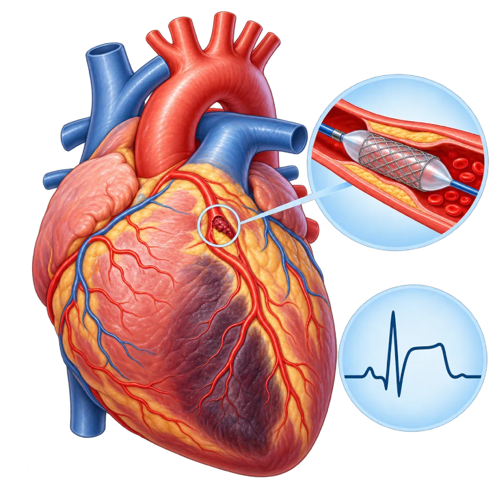
ST-elevation myocardial infarction (STEMI) is a heart attack caused by a complete blockage of a coronary artery, which shows up as a typical pattern on the ECG. The artery needs to be opened as soon as possible, usually with a catheter procedure.
Sources01 / Understanding the condition
About ST-Elevation Myocardial Infarction (STEMI)
ST-elevation myocardial infarction, or STEMI, is a type of heart attack identified by a typical pattern on the ECG recording: part of the line called the ST segment is raised higher than usual. This pattern usually means one coronary artery is completely blocked by a blood clot, so part of the heart muscle receives no blood flow at all. STEMI is an acute coronary syndrome, along with non-ST-elevation myocardial infarction and unstable angina. An overview of all types of heart attack is covered on the Myocardial Infarction / Heart Attack page.
What Happens in the Coronary Artery
Almost all STEMIs start with a cholesterol plaque in the wall of a coronary artery that cracks. A blood clot forms over the crack and closes the artery completely. The heart muscle beyond the blockage begins to die after about 20 to 40 minutes, starting from the inner layer and spreading through the full thickness of the wall over several hours. That is why doctors say "time is muscle". Less often, STEMI is caused by a coronary artery spasm, a tear in the artery wall, or a clot carried from elsewhere.
Opening the Artery as Soon as Possible
The main goal of STEMI treatment is reperfusion, which means restoring blood flow to the heart muscle. The 2023 ESC guidelines recommend primary percutaneous coronary intervention, which opens the artery with a balloon and stent through a catheter, if it can be done within 120 minutes of diagnosis. If this is not possible, for example because a hospital with catheterization facilities is too far away, the doctor gives clot-dissolving medicine (fibrinolytic therapy) and then refers the patient for coronary angiography. The benefit is greater the earlier the artery is opened, especially within the first 12 hours after symptoms start.
After the Emergency Phase
After the artery is opened, patients stay in the hospital for a few days with heart rhythm monitoring, because rhythm disorders often appear in the first 48 hours. If other arteries are also narrowed, the doctor may plan additional procedures, either during the hospital stay or a few weeks later. Long-term medicines, risk factor control, and cardiac rehabilitation help prevent another heart attack. Problems that can appear afterward are covered on the Post-Myocardial Infarction Complications page.
02 / Understanding the condition
Symptoms to Recognize.
Common Symptoms
- Pain, heaviness, or pressure in the center of the chest that persists and does not go away with rest
- Pain spreading to the left arm or both arms, the neck, jaw, or back
- Cold sweats and pale skin
- Shortness of breath
- Nausea or vomiting
- Dizziness, weakness, or feeling faint
- Intense anxiety or a feeling that something bad is about to happen
Symptoms in Adult Women
Chest pain is still the main complaint, but women more often have atypical accompanying symptoms, so they may seek help late.
- Shortness of breath or extreme tiredness that comes on suddenly
- Pain in the upper back, between the shoulder blades, neck, or jaw
- Nausea, vomiting, or discomfort in the pit of the stomach
- Pressure in the chest that is not very painful
- Dizziness or cold sweats without heavy activity
Symptoms in Older Adults
In older adults, chest pain may be mild or absent altogether.
- Sudden shortness of breath without clear chest pain
- New confusion or disorientation
- Severe weakness or suddenly being unable to stand
- Fainting or falling for no clear reason
03 / Understanding the condition
What Causes This Condition?
- A cholesterol plaque in a coronary artery that cracks and is then completely closed off by a blood clot
- Coronary artery disease that develops over many years without symptoms
- Severe, prolonged coronary artery spasm
- A spontaneous tear in the wall of a coronary artery, covered on the Spontaneous Coronary Artery Dissection page.
- A clot from a heart chamber or valve carried into a coronary artery (embolism)
- Sudden blockage of a previously placed stent, covered on the Stent Thrombosis page.
04 / Understanding the condition
Who Is at Higher Risk?
Lifestyle
Smoking is a strong risk factor for STEMI, especially at a young age. A diet high in saturated fat and salt, physical inactivity, and obesity add to the risk.
Medical
Hypertension, diabetes, high LDL cholesterol, and chronic kidney disease speed up plaque buildup. People who have had a stent or bypass surgery also remain at risk.
Genetic
A family history of early coronary artery disease, and familial hypercholesterolemia, which is very high LDL cholesterol from a young age due to inherited factors.
Age
Risk increases in men over 45 and women over 55. STEMI can also occur in people in their 30s and 40s, especially smokers.
Other
Cocaine or amphetamine use, severe emotional or physical stress, and air pollution can trigger an attack in people who already have plaque.
Groups at Higher Risk
- Young Smokers: Smoking is one of the main triggers of STEMI under age 50. The risk falls gradually after quitting.
- People with Diabetes: Diabetes speeds up hardening of the arteries and can make heart attack symptoms feel vague, so help may come late.
- People with Coronary Artery Disease: People who have had angina, a stent, or bypass surgery remain at risk because plaque can grow in other arteries.
- People with a Family History of Heart Disease: A history of early heart attack in a parent or sibling, including inherited high cholesterol, increases the risk from a young age.
05 / Understanding the condition
How Is It Diagnosed?
STEMI is diagnosed from symptoms and the ECG, without waiting for laboratory results. The ECG is recorded within 10 minutes of the patient first being seen by medical staff. If ST-segment elevation appears in two adjacent leads, or another equivalent pattern such as left bundle branch block with typical symptoms, the team immediately prepares to open the artery.
Blood is still drawn for troponin, kidney function, electrolytes, and blood clotting, but the results do not delay treatment. Coronary angiography shows the blocked artery and is followed directly by percutaneous coronary intervention. Echocardiography is done to assess pumping strength and look for complications. The doctor also rules out other conditions that can mimic STEMI, such as aortic dissection, pericarditis, or pulmonary embolism, because their treatment is very different.
Electrocardiogram (ECG)
Recorded within 10 minutes of the patient first being seen by medical staff. ST-segment elevation in two adjacent leads is the basis for the decision to open the artery.
Cardiac Troponin Test
Drawn on arrival to confirm heart muscle damage; in STEMI, treatment is started without waiting for the result.
Coronary Angiography
Shows the blocked artery and is followed directly by primary percutaneous coronary intervention when appropriate.
Echocardiography
Assesses pumping strength, parts of the wall that are not moving, valve leakage, and complications after the heart attack.
Laboratory Tests
Kidney function, electrolytes, complete blood count, blood sugar, and lipid profile for the safety of medicines and contrast dye and for prevention planning.
06 / Understanding the condition
Treatment Options.
Treatment is decided by a doctor after an assessment. The categories below can help you follow the conversation with your doctor.
Primary Percutaneous Coronary Intervention
Opens the artery with a balloon and stent through a catheter inserted at the wrist or groin. It is the main choice if it can be done within 120 minutes of diagnosis.
Fibrinolytic Therapy
Clot-dissolving medicine is given when a catheter procedure cannot be done in time, ideally within 10 minutes of diagnosis, followed by coronary angiography.
Antithrombotic Medicines
Two antiplatelet medicines and an injected anticoagulant to prevent new clots during and after the procedure; your doctor decides which ones and for how long.
Initial Care in the Emergency Room
Oxygen if levels are low, pain relief, and heart rhythm monitoring while preparing to open the artery.
Coronary Bypass Surgery
Rarely done as an emergency; considered when the artery cannot be opened with a catheter, the blockages affect many vessels, or there is a mechanical complication.
Long-Term Medicines and Rehabilitation
Statins, antiplatelet medicines, and other heart-protecting medicines based on pumping function, plus a cardiac rehabilitation program to restore fitness.
07 / Understanding the condition
Complications to Watch For.
Heart Rhythm Disorders
Often appear in the first 48 hours, ranging from a very slow heartbeat to ventricular fibrillation, which stops the heart and needs an immediate electric shock.
Acute Heart Failure
If a large area of muscle is damaged, the heart's pumping weakens, so fluid builds up in the lungs and the patient becomes very short of breath.
Cardiogenic Shock
Blood pressure drops because the heart's pumping is very weak, so the organs do not get enough blood. It requires intensive care and immediate opening of the artery.
Right Ventricular Infarction
A heart attack in the lower wall can involve the right ventricle, causing blood pressure to drop; treatment requires careful fluid management and specific medicines.
Mechanical Complications
A tear in the heart wall, the wall between the ventricles, or the muscle that supports the mitral valve in the first few days. These are rare but need surgery or an urgent procedure.
Blood Clot in the Left Ventricle
Can form after a large heart attack in the front wall of the heart and may break loose and travel to the brain, causing a stroke.
08 / Understanding the condition
What Can You Do?
- Quit smoking completely; the risk of a heart attack falls gradually after you stop.
- Control your blood pressure, blood sugar, and LDL cholesterol according to the targets your doctor sets.
- Be physically active for at least 150 minutes a week at moderate intensity.
- Choose a diet rich in vegetables, fruit, fish, and whole grains; limit salt and saturated fat.
- If you have had a stent, take antiplatelet medicines as advised and do not stop them on your own.
- Teach your family to recognize the signs of a heart attack, call an ambulance, and do chest compressions.
09 / Understanding the condition
When Should You See a Doctor?
STEMI cannot be treated at home. If you or someone near you has chest pain lasting more than 5 minutes, especially with cold sweats or shortness of breath, call an ambulance (119) or go to the nearest emergency room right away. Do not drive yourself and do not wait for the symptoms to ease.
After you go home from the hospital, contact your cardiologist or move your checkup forward if:
- Brief chest pain comes back during activity. If it lasts more than 5 minutes, go to the emergency room right away.
- You become more short of breath, your legs swell, or your weight rises quickly over a few days.
- You have palpitations or dizziness, or nearly faint.
- You notice medicine side effects, such as unusual bruising or bleeding.
10 / Common questions
Questions You May Have.
Frequently asked questions about STEMI and its treatment.
Is STEMI more dangerous than NSTEMI?
STEMI usually means a complete blockage, so damage happens quickly and the artery must be opened within hours. NSTEMI is usually a partial blockage, but that does not mean it is mild. In the long term, the risk with NSTEMI can be similar because patients are often older and have other diseases. Both are heart attacks that must be treated in the hospital, also covered on the Non-ST-Elevation Myocardial Infarction (NSTEMI) page.
Why don't doctors wait for troponin results in STEMI?
An ST-elevation pattern on the ECG with typical symptoms is enough to show that the artery is completely blocked. Troponin levels rise only after some time, and the test itself takes time. Waiting for the results means letting the heart muscle keep getting damaged. That is why blood is still drawn, but the team prepares to open the artery right away based on the ECG.
What is primary percutaneous coronary intervention?
It is an emergency procedure to open a blocked artery through a thin catheter inserted at the wrist or groin. The doctor passes a wire through the blockage, inflates a balloon, and then places a stent to keep the artery open. Patients are usually awake with local anesthesia. This procedure is covered on the Percutaneous Coronary Intervention (PCI) page.
How long will I stay in the hospital after STEMI?
If the artery is successfully opened and there are no complications, the stay is usually a few days. The 2023 ESC guidelines state that low-risk patients may be considered for earlier discharge after monitoring. If there is heart failure, a rhythm disorder, or another artery that needs treatment, the stay can be longer. Before going home, you will receive an explanation of your medicines, warning signs, and cardiac rehabilitation schedule.
Can STEMI happen at a young age?
Yes. In people in their 30s and 40s, STEMI is mainly linked to smoking, inherited high cholesterol, diabetes, or cocaine and amphetamine use. In young women, a heart attack can also be caused by a spontaneous tear in the wall of a coronary artery. Persistent chest pain at a young age still needs to be checked in the emergency room; do not brush it off as a minor ailment.
Will a stent cure me completely?
A stent opens the artery that caused the heart attack, but coronary artery disease in other arteries is still there. Plaque can keep growing if risk factors are not controlled. That is why long-term medicines, quitting smoking, a healthy diet, regular activity, and routine checkups are still needed to prevent another heart attack. Cardiac rehabilitation helps you build these habits.
Sources and Medical Review
Translated from the Indonesian edition medically reviewed by Dr. dr. Jajang Sinardja, Sp.JP(K).
Sources as cited in the Indonesian edition.
- Byrne RA, Rossello X, Coughlan JJ, et al. 2023 ESC Guidelines for the management of acute coronary syndromes. Eur Heart J. 2023;44(38):3720-3826.
- Thygesen K, Alpert JS, Jaffe AS, et al. Fourth universal definition of myocardial infarction (2018). Eur Heart J. 2019;40(3):237-269.
- Rao SV, O'Donoghue ML, Ruel M, et al. 2025 ACC/AHA/ACEP/NAEMSP/SCAI Guideline for the Management of Patients With Acute Coronary Syndromes: A Report of the American College of Cardiology/American Heart Association Joint Committee on Clinical Practice Guidelines. Circulation. 2025;151(13):e771-e862.
- Reed GW, Rossi JE, Cannon CP. Acute myocardial infarction. Lancet. 2017;389(10065):197-210.
- Damluji AA, van Diepen S, Katz JN, et al. Mechanical Complications of Acute Myocardial Infarction: A Scientific Statement From the American Heart Association. Circulation. 2021;144(2):e16-e35.
Doctors Who Treat This Condition

dr. Adrianus Kosasih, Sp.JP(K)
Interventional Cardiology
Coronary Intervention Procedures
Clinical focusPrimary Coronary Intervention · PCI for Acute Coronary Syndrome
View Profile
dr. Siska S. Danny, Sp.JP(K)
Interventional Cardiology
Coronary Intervention Procedures
Clinical focusPCI for Acute Coronary Syndrome · Primary Coronary Intervention
View Profile
Dr. dr. Jajang Sinardja, Sp.JP(K)
Interventional Cardiology
Coronary Intervention Procedures
Clinical focusAcute Coronary Syndrome · Primary Coronary Intervention
View Profile