
Quick answer
What Is Intravascular Ultrasound?
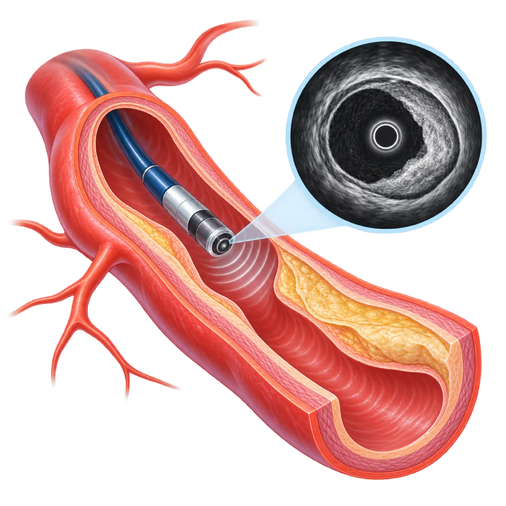
Intravascular ultrasound (IVUS) examines the coronary arteries from the inside with a thin catheter carrying a tiny ultrasound probe. The images show the vessel wall, plaque, and artery size, so the doctor can choose the stent size and make sure the stent is fully expanded. IVUS is done during cardiac catheterization and adds about 10 to 20 minutes.
Sources01 / Understanding the procedure
About Intravascular Ultrasound
What Is Intravascular Ultrasound
Coronary angiography shows the outline of the inside of a blood vessel filled with contrast dye, much like a silhouette, but it does not show the vessel wall itself. Intravascular ultrasound (IVUS) adds to this by looking from the inside. A very thin catheter with a tiny ultrasound probe at its tip is inserted into the coronary artery, where it sends out sound waves and picks up their echoes to create cross-sectional images of the vessel wall.
What It Can Show
IVUS shows the thickness and extent of plaque, whether there is calcium in the vessel wall, and the vessel diameter and length of the narrowed segment. If the calcium is thick and runs all the way around the vessel, the doctor may decide to shave it away first before placing a stent, which is covered on the Rotational Atherectomy (Rotablation) page. After a stent is placed, IVUS shows whether it is fully expanded, sits snugly against the wall, and covers all of the plaque without tears at its edges. IVUS also helps find the cause of renarrowing or a blood clot inside a previously placed stent.
When IVUS Is Used
The 2024 ESC guidelines for chronic coronary syndromes recommend imaging from inside the vessel, with IVUS or optical coherence tomography, to guide stent placement in complex narrowings. Examples include narrowings of the left main coronary artery, narrowings at branch points, long narrowings, or heavily calcified plaque. IVUS is also used to assess a moderate narrowing of the left main coronary artery that looks uncertain on angiography.
Benefits for Long-Term Results
Several large studies have shown that stent placement guided by IVUS reduces the need for repeat procedures and cardiac events compared with angiography guidance alone. A combined analysis in 2024 also found fewer heart attacks and fewer blood clots inside stents with intravascular imaging guidance. These benefits are seen mainly in complex narrowings.
IVUS and Optical Coherence Tomography
Both tests look from inside the vessel. IVUS uses sound waves, which penetrate deeper, and does not require an extra flush of contrast dye, making it suitable for large vessels, the origins of the coronary arteries, and patients with kidney problems. Optical coherence tomography uses light and gives more detailed images; it is covered on the Optical Coherence Tomography page. Your doctor chooses one of them based on your condition.
02 / Understanding the procedure
When Is This Procedure Considered?
A doctor may consider this procedure in the situations below. The decision is always made after a thorough assessment.
- Guiding Stent Placement in Complex NarrowingsFor example, in the left main coronary artery, at branch points, in long narrowings, or in calcified plaque.
- Confirming the Results of Stent PlacementAssesses whether the stent is fully expanded and sits snugly, so it can be corrected right away.
- Uncertain Narrowing of the Left Main Coronary ArteryMeasures the inner area of the vessel to assess whether treatment is needed.
- Finding the Cause of Problems with a Previous StentIn renarrowing or a blood clot inside a stent, to determine suitable treatment.
- Assessing Calcium Before Stent PlacementDetermines whether calcium needs to be shaved away first, for example with rotational atherectomy, before the stent is placed.
- Reducing Contrast Dye in Patients with Kidney ProblemsMeasurements from IVUS images help the doctor place a stent using less contrast dye.
Situations to Consider First
- Unstable ConditionWith very low blood pressure or a serious emergency, the main treatment takes priority.
- Very Severe or Tortuous NarrowingsThe IVUS catheter may not be able to pass yet, so the narrowing is first widened with a small balloon.
- Very Small VesselsThe catheter can block flow in small vessels, so the doctor weighs the benefits.
- Conditions That Prevent CatheterizationFor example, active bleeding or an uncontrolled infection, because IVUS requires catheterization.
03 / Before the procedure
How Should You Prepare?
IVUS is performed as part of cardiac catheterization or stent placement, so the preparation follows that procedure. The team will give you written instructions.
Tests before the procedure. Blood tests, including kidney function and blood clotting, as well as an ECG.
Medications you are taking. Bring a list of all your medications. Antiplatelet medications are usually continued, while other blood thinners and some diabetes medications, such as metformin, may need to be adjusted. Do not change any medication without your doctor's instructions.
Fasting. Follow the fasting instructions from the team, usually for a few hours before the procedure.
Allergies. Tell the team if you have ever had an allergy to contrast dye, medications, adhesive tape, or latex.
Companion. Arrange for someone to take you home, because you are advised not to drive after the procedure.
Unsure about the preparation instructions? Ask our team before the procedure.
04 / During the procedure
How Is the Procedure Done?
IVUS is performed in the catheterization laboratory by an interventional cardiologist under local anesthesia. The whole catheterization with IVUS usually takes about 1 hour, longer if it includes stent placement in a complicated narrowing. Taking the IVUS images itself adds about 10 to 20 minutes.
Access and Angiography
The doctor inserts a catheter through an artery in the wrist or groin, images the coronary arteries with contrast dye, and then decides which segment to examine. Blood thinners and medications that widen the blood vessels are given.
Inserting the IVUS Catheter
A very thin guide wire is passed across the narrowing, and then the IVUS catheter is advanced along the wire until it is past the segment to be examined.
Recording the Images
The catheter is pulled back slowly, usually with an automatic pullback device, while recording cross-sectional images along the vessel. This step usually causes no discomfort.
Deciding the Next Step and Checking Again
The doctor measures the vessel and plaque on the screen to choose the balloon and stent. After the stent is placed, IVUS is repeated; if the stent is not yet fully expanded, a balloon can be inflated again right away.
05 / Benefits and risks
Benefits and Risks Should Be Discussed Together.
Every medical procedure has benefits and risks. Your doctor will explain both for your situation before the procedure.
Benefits You Can Expect
- Direct View of the Vessel WallShows plaque, calcium, and vessel size that are not visible on angiography.
- Suitable Stent SizeThe stent diameter and length are chosen based on the directly measured vessel size.
- Better Stent Placement ResultsA stent that is not fully expanded or not sitting snugly can be corrected in the same procedure.
- Fewer Repeat EventsLarge studies show that IVUS guidance reduces repeat procedures and cardiac events.
- No Extra Contrast DyeImages are taken without a contrast flush, making it suitable for patients with kidney problems.
The benefits of a procedure are not a promise of a cure. Ask how the results will affect your care plan.
Risks You Should Know
- General Catheterization RisksBruising or bleeding at the puncture site, which is usually mild and treatable.
- Coronary Artery SpasmThe artery can narrow briefly as the catheter enters; this is prevented and treated with medication that widens the blood vessels.
- Chest DiscomfortSometimes occurs while the catheter is in a severe narrowing and goes away once the catheter is pulled back.
- Heart Rhythm DisturbancesRare, usually brief, and quickly resolved.
- Coronary Artery Wall InjuryVery rare, such as a tear or blood clot that needs prompt treatment, including a stent.
Risks can differ from person to person. A full explanation and consent are given after an individual assessment.
06 / After the procedure
What Is Recovery Like?
Recovery follows the catheterization or stent placement done at the same time. IVUS itself does not add to the recovery time.
After the procedure. If the wrist was used for access, a compression band is worn for a few hours. If the groin was used, you lie down for a few hours with your leg straight.
Hospital stay. After stent placement, patients usually stay in the hospital for one night. If only the examination was done without a stent, some patients can go home on the same day.
Medications. If a stent was placed, take your antiplatelet medication every day as prescribed and do not stop it without instructions from your cardiologist.
Activity. For a few days, avoid lifting heavy objects with the arm or leg used for access.
Warning signs to watch for. Go to the emergency room right away if you have:
- Chest pain, shortness of breath, or cold sweats
- Growing swelling or bleeding at the puncture site
- A hand or leg that feels cold, pale, numb, or painful
- A rash, itching, or shortness of breath after receiving contrast dye
Know the Signs That Need Prompt Care.
Follow the team's guidance on warning signs. If severe or sudden symptoms appear after the procedure, go to the nearest emergency department right away or call Heart Attack Center 1500 258.
07 / Team and support
Doctors and Procedure Support
The doctors below are listed for this procedure. Listed by name, not by ranking.

dr. Adrianus Kosasih, Sp.JP(K)
Interventional Cardiology
Coronary Intervention Procedures
Clinical focusPrimary Coronary Intervention · PCI for Acute Coronary Syndrome
View Profile
Dr. dr. Dafsah A. Juzar, Sp.JP(K)
Interventional Cardiology
Coronary Intervention Procedures +1 center
Clinical focusComplex Coronary Intervention · Cardiac Catheterization
View Profile
dr. Puti S. Saus, Sp.JP(K)
Interventional Cardiology
Coronary Intervention Procedures
Clinical focusAcute Coronary Syndrome · Primary Coronary Intervention
View ProfileRotational Atherectomy System
A system for treating calcified plaque in the coronary arteries. A small diamond-coated burr spins at high speed over a guidewire…
Intravascular Lithotripsy (IVL) System
A special balloon catheter that emits pulses of sonic pressure waves to crack calcium in the coronary artery wall. It is used…
Intracoronary Imaging System (IVUS / OCT)
Imaging from inside the coronary artery using ultrasound (IVUS) or near-infrared light (OCT). It is used during catheterization…
The availability of technology and test support is confirmed with the team. Technology is chosen according to clinical need.
08 / Common questions
Before You Plan the Procedure.
Frequently asked questions about IVUS; the decision to use it is made by your doctor when planning catheterization or stent placement.
How is IVUS different from coronary angiography?
Coronary angiography takes pictures of the outline of the contrast-filled vessel from the outside, so plaque in the vessel wall is not seen directly. IVUS looks from inside the vessel and shows the wall, plaque, calcium, and the true size of the vessel. That is why IVUS is used alongside angiography, especially when planning stent placement and checking its results.
How is IVUS different from OCT?
Both look from inside the blood vessel. IVUS uses sound waves, penetrates deeper so the full size of the vessel can be seen, and does not need an extra flush of contrast dye. OCT uses infrared light and gives much more detailed images, but requires contrast dye. Your doctor chooses based on the location of the narrowing, the size of the vessel, and your kidney function.
Does IVUS hurt?
You cannot feel the IVUS catheter moving inside the blood vessel. Taking the images does not require a contrast flush, so it usually does not cause a warm sensation. In a very severe narrowing, the catheter can briefly block flow and cause chest discomfort that goes away once the catheter is pulled back. Tell the team if you feel this.
Does every stent placement require IVUS?
Not always. Guidelines recommend intravascular imaging mainly for complex narrowings, for example in the left main coronary artery, at branch points, in long narrowings, or in heavily calcified plaque. For simple narrowings, your doctor may decide that angiography is enough. Ask your doctor whether IVUS or OCT is planned for you.
How much extra time does IVUS add?
Taking IVUS images of one vessel usually takes only a few minutes, and all the steps together usually add about 10 to 20 minutes to the catheterization. If IVUS is repeated after the stent is placed, the time increases slightly. The length of the whole procedure depends mainly on how complex the treated narrowing is.
Is IVUS safe for patients with kidney problems?
IVUS does not need a contrast flush to take images. Because of this, IVUS can help the doctor place a stent with less contrast dye, for example by using measurements from IVUS images to replace some contrast X-ray images. Kidney function is still checked before the procedure, IV fluids are given when needed, and it is checked again afterward.
Sources and Medical Review
Translated from the Indonesian edition medically reviewed by dr. Puti S. Saus, Sp.JP(K).
Sources as cited in the Indonesian edition.
- Vrints C, Andreotti F, Koskinas KC, et al. 2024 ESC Guidelines for the management of chronic coronary syndromes. Eur Heart J. 2024;45(36):3415-3537.
- Räber L, Mintz GS, Koskinas KC, et al. Clinical use of intracoronary imaging. Part 1: guidance and optimization of coronary interventions. An expert consensus document of the European Association of Percutaneous Cardiovascular Interventions. Eur Heart J. 2018;39(35):3281-3300.
- Hong SJ, Kim BK, Shin DH, et al. Effect of Intravascular Ultrasound-Guided vs Angiography-Guided Everolimus-Eluting Stent Implantation: The IVUS-XPL Randomized Clinical Trial. JAMA. 2015;314(20):2155-2163.
- Zhang J, Gao X, Kan J, et al. Intravascular Ultrasound Versus Angiography-Guided Drug-Eluting Stent Implantation: The ULTIMATE Trial. J Am Coll Cardiol. 2018;72(24):3126-3137.
- Lee JM, Choi KH, Song YB, et al. Intravascular Imaging-Guided or Angiography-Guided Complex PCI. N Engl J Med. 2023;388(18):1668-1679.
- Li X, Ge Z, Kan J, et al. Intravascular ultrasound-guided versus angiography-guided percutaneous coronary intervention in acute coronary syndromes (IVUS-ACS): a two-stage, multicentre, randomised trial. Lancet. 2024;403(10439):1855-1865.
- Stone GW, Christiansen EH, Ali ZA, et al. Intravascular imaging-guided coronary drug-eluting stent implantation: an updated network meta-analysis. Lancet. 2024;403(10429):824-837.