
Quick answer
What Is Ventricular Septal Defect (VSD) Closure?
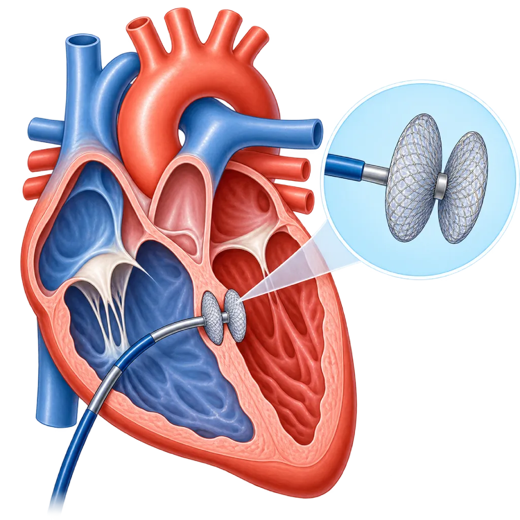
Ventricular septal defect (VSD) closure is a procedure to close a hole in the wall between the heart's right and left lower chambers. A large hole overworks the heart and lungs. It is closed with a surgical patch in about 3 to 4 hours or, for certain types, with a catheter device in about 2 to 3 hours.
Sources01 / Understanding the procedure
About Ventricular Septal Defect (VSD) Closure
What Is Ventricular Septal Defect Closure
A ventricular septal defect (VSD) is a hole in the wall (septum) that separates the right and left lower chambers of the heart (ventricles). VSD is one of the congenital heart defects commonly found in babies and children. Because the pressure in the left ventricle is much higher, blood flows forcefully through the hole into the right ventricle and on to the lungs. VSD closure aims to stop this excess flow. An overview of closing other holes in the heart is covered on the Heart Defect Closure page.
Not Every VSD Needs to Be Closed
Many small VSDs, especially those in the muscular part of the septum, get smaller or close on their own in the first years. A small VSD that does not strain the heart usually only needs monitoring with echocardiography. In contrast, a large VSD can make a baby breathless, sweaty and quickly tired during feeding, and slow to gain weight. If left untreated, the pressure in the lung blood vessels can rise and, over time, remain persistently high.
Surgical Closure
Surgery is a common way to close a large VSD in babies, usually within the first year of life. The surgeon opens the breastbone, temporarily stops the heart with the help of a heart-lung machine, then stitches a patch over the hole, usually through the right atrium without opening the ventricle. The long-term results are good in the majority of children. Congenital heart surgery in general is covered on the Congenital Heart Surgery page.
Catheter Closure
In older children and adults, VSDs in the muscular part of the septum and some perimembranous VSDs, meaning holes near the valves, can be closed with a device through a catheter. The requirement is that the hole is far enough from the aortic and tricuspid valves. This approach does not require a cut in the chest, but the heart rhythm needs to be monitored because the device sits close to the heart's electrical pathway.
Closure in Adults
In adults, closure is recommended when the left ventricle is enlarged because of the leak and the lung pressure is not yet too high. Closure is also considered if there has been an infection of the heart's inner lining, or if a leaflet of the aortic valve starts to be pulled into the hole, causing the valve to leak. If the lung pressure is suspected to be high, the doctor measures it with right heart catheterization, covered on the Right Heart Catheterization page.
02 / Understanding the procedure
When Is This Procedure Considered?
A doctor may consider this procedure in the situations below. The decision is always made after a thorough assessment.
- Large VSD with Heart Failure in BabiesThe baby is breathless, sweats during feeding, and has trouble gaining weight despite medication.
- Enlarged Left VentricleThe leak causes the left atrium and left ventricle to enlarge, even without symptoms.
- Lung Pressure Starting to RiseEarlier closure prevents the pressure in the lung blood vessels from remaining persistently high.
- History of EndocarditisA past infection of the heart's inner lining around the hole.
- Aortic Valve Pulled Into the HoleA leaflet of the aortic valve sags toward the hole, so the valve starts to leak.
Situations to Consider First
- Very High Lung PressureWhen blood has already reversed to flow from right to left (Eisenmenger syndrome), closure can be harmful.
- Small VSD Without Strain on the HeartRegular monitoring is enough, because it does not strain the heart and often closes on its own.
- Hole Too Close to a Valve for a CatheterA device could interfere with how the valve works, so the hole is better suited to surgical closure.
- Active InfectionA planned procedure is postponed until the infection, including a lung infection, is resolved.
03 / Before the procedure
How Should You Prepare?
Preparation depends on age and the closure method. The team will give you written instructions, and parents are involved at every step.
Tests before the procedure. Echocardiography, ECG, chest X-ray, and blood tests. If the lung pressure is suspected to be high, the doctor may request cardiac catheterization.
Nutrition and medicines. Babies with a large VSD often need medicines that remove excess fluid (diuretics) and high-calorie milk so they are strong enough for surgery.
Fasting. Follow the fasting schedule from the anesthesia team. The time limits for breast milk, formula, and water are explained separately.
General health. Tell the team if your child has a fever, cough, or cold. For older children, complete any needed dental treatment.
Preparing your child and family. Explain things to your child in simple words. Arrange for someone to stay with your child during the hospital stay, because after surgery your child will be cared for in the intensive care unit for a few days.
Unsure about the preparation instructions? Ask our team before the procedure.
04 / During the procedure
How Is the Procedure Done?
VSD surgery is done in the operating room under general anesthesia and takes about 3 to 4 hours. Catheter closure is done in the catheterization lab, also under general anesthesia, and takes about 2 to 3 hours.
Surgery with a Heart-Lung Machine
The surgeon opens the breastbone, connects the heart to a heart-lung machine, then temporarily stops the heart.
Stitching the Patch
The hole is closed with a patch, usually through the right atrium and the opening of the tricuspid valve. The result is checked with an echocardiogram through the esophagus before the chest is closed.
Catheter Closure
A catheter is inserted through a blood vessel in the groin. After the hole is measured with echocardiography and X-ray, the closure device is opened so that it clamps both sides of the hole.
Confirming the Result
The team checks for any residual leak, valve function, and heart rhythm before the procedure ends.
05 / Benefits and risks
Benefits and Risks Should Be Discussed Together.
Every medical procedure has benefits and risks. Your doctor will explain both for your situation before the procedure.
Benefits You Can Expect
- Less Strain on the Heart and LungsThe excess flow stops, so the enlarged left ventricle can shrink back.
- Better Growth in BabiesAfter a large hole is closed, babies usually feed more easily and gain weight.
- Preventing Damage to the Lung Blood VesselsTimely closure prevents the pressure in the lung blood vessels from becoming persistently high.
- Lower Risk of Heart InfectionThe forceful flow that can trigger endocarditis is gone once the hole is closed.
The benefits of a procedure are not a promise of a cure. Ask how the results will affect your care plan.
Risks You Should Know
- Rhythm Disorders and Heart BlockThe electrical pathway near the hole can be disrupted; a small proportion of patients need a pacemaker.
- Residual LeakA small leak may remain at the edge of the patch or device and often gets smaller on its own.
- Valve LeakageThe aortic or tricuspid valve may leak if it is affected by the device or patch.
- Risks of Open-Heart SurgeryBleeding, wound infection, and fluid around the heart, which are monitored in the intensive care unit.
- Device Displacement or Damage to Blood CellsRare in catheter closure and may require a further procedure.
Risks can differ from person to person. A full explanation and consent are given after an individual assessment.
06 / After the procedure
What Is Recovery Like?
After surgery, the child stays in the intensive care unit for about 1 to 3 days, then on the ward, with a total hospital stay of about 5 to 10 days. After catheter closure, most patients go home within 1 to 3 days.
Intensive care unit. Your child may still need a breathing machine for a short time, along with chest tubes and temporary pacing wires. These are removed gradually as your child recovers.
Rhythm monitoring. The heart rhythm is recorded regularly, because problems with the heart's electrical conduction can occur, including after going home.
Surgical wound. Keep the wound clean and dry. Avoid lifting your child by the armpits and avoid rough play for about 6 weeks so that the breastbone can heal.
Follow-up visits. Echocardiography and ECG are done regularly. For the first 6 months after closure, dental procedures may require preventive antibiotics.
Warning signs. Go to the emergency room right away if your child has:
- Fast breathing, shortness of breath, or bluish lips
- High fever, or a wound that is red, swollen, and oozing pus
- Fainting, severe weakness, or a very slow heartbeat
- Persistent vomiting, or refusing to drink or breastfeed
- Growing swelling or bleeding at the puncture site
Know the Signs That Need Prompt Care.
Follow the team's guidance on warning signs. If severe or sudden symptoms appear after the procedure, go to the nearest emergency department right away or call Heart Attack Center 1500 258.
07 / Team and support
Doctors and Procedure Support
The doctors below are listed for this procedure. Listed by name, not by ranking.

dr. Radityo Prakoso, Sp.JP(K)
Pediatrics & Congenital Heart Disease
Congenital and Structural Heart Center
Clinical focusPediatric Cardiology Consultation · Congenital Heart Disease Care
View Profile
dr. Salomo Purba, Sp.BTKV, Subsp.JPK(K)
Pediatric and Congenital Heart Surgery
Congenital and Structural Heart Center +2 centers
Clinical focusPediatric Heart Surgery · Congenital Heart Defect Surgery
View Profile
dr. Sri Diniharini, Sp.JP(K)
Echocardiography & Cardiovascular Imaging
Cardiovascular Diagnostic Center +1 center
Clinical focusAdvanced Echocardiography · Transesophageal Echocardiography
View Profile08 / Common questions
Before You Plan the Procedure.
Questions parents and patients often ask about ventricular septal defect (VSD) closure.
Can my child's VSD close on its own?
Yes, especially if the hole is small. Many small VSDs in the muscular part of the septum close on their own in the first few years, and some perimembranous VSDs also get smaller. Large VSDs rarely close on their own. The doctor will monitor your child's growth and the size of the hole with regular echocardiograms, then recommend closure if the hole strains the heart.
At what age does a large VSD need surgery?
A large VSD that causes heart failure or poor growth is usually operated on within the first year of life, often at about 3 to 6 months of age, and sooner if medicines no longer help. Timely closure prevents the pressure in the lung blood vessels from remaining persistently high. The doctor will decide the timing based on symptoms, weight, and echocardiogram results.
What is the difference between surgery and catheter closure?
Surgery uses a patch stitched in place with the help of a heart-lung machine and can be used for almost all types of VSD, including in babies. Catheter closure does not require a cut in the chest and has a shorter recovery, but it only suits certain VSDs that are far enough from the valves, usually in older children and adults.
Will my child need a pacemaker?
A small proportion of patients need one. The heart's electrical pathway lies close to perimembranous VSDs, so it can be disrupted by stitches or by a device. This disruption is often temporary. If electrical conduction does not recover, the doctor will implant a pacemaker. Because rhythm disorders can appear later, an ECG is still checked at follow-up visits.
When can my child return to school and play?
After catheter closure, children can usually return to school in about one week. After surgery, children can go back to school about 3 to 6 weeks later, and rough play is postponed until the breastbone has healed, about 6 weeks. Once recovered, the majority of children can play and exercise like their peers.
Does a closed VSD still need follow-up visits?
Yes. Regular follow-up with echocardiography and ECG checks for any residual leak, valve function, heart rhythm, and lung pressure. In children with good results, the time between visits gets longer over time. Patients who had surgery as children are still advised to have occasional check-ups as adults, especially before pregnancy or if new symptoms appear.
Sources and Medical Review
Translated from the Indonesian edition medically reviewed by dr. Radityo Prakoso, Sp.JP(K).
Sources as cited in the Indonesian edition.
- Baumgartner H, De Backer J, Babu-Narayan SV, et al. 2020 ESC Guidelines for the management of adult congenital heart disease. Eur Heart J. 2021;42(6):563-645.
- Stout KK, Daniels CJ, Aboulhosn JA, et al. 2018 AHA/ACC Guideline for the Management of Adults With Congenital Heart Disease: A Report of the American College of Cardiology/American Heart Association Task Force on Clinical Practice Guidelines. Circulation. 2019;139(14):e698-e800.
- Feltes TF, Bacha E, Beekman RH, et al. Indications for Cardiac Catheterization and Intervention in Pediatric Cardiac Disease: A Scientific Statement From the American Heart Association. Circulation. 2011;123(22):2607-2652.
- Yang J, Yang L, Yu S, et al. Transcatheter versus surgical closure of perimembranous ventricular septal defects in children: a randomized controlled trial. J Am Coll Cardiol. 2014;63(12):1159-1168.
- Penny DJ, Vick GW. Ventricular septal defect. Lancet. 2011;377(9771):1103-1112.