
Quick answer
What Is Cardiac Tamponade?
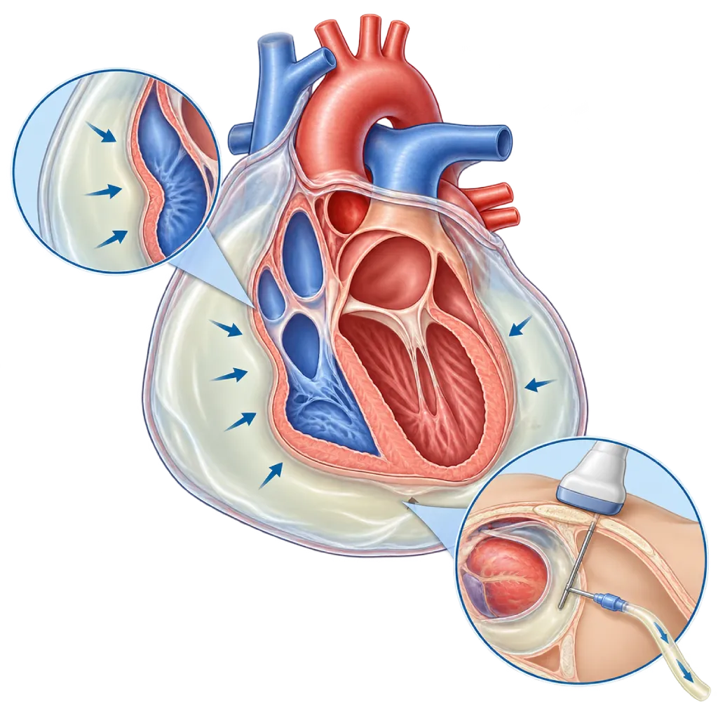
Cardiac tamponade is an emergency in which fluid or blood in the sac around the heart presses on the heart, so it cannot fill and pump blood adequately. The fluid needs to be removed right away.
Sources01 / Understanding the condition
About Cardiac Tamponade
The heart is wrapped in the pericardium, a thin two-layered sac that normally contains a small amount of lubricating fluid. In cardiac tamponade, fluid, blood, pus, or air inside this sac presses on the heart. This pressure prevents the heart chambers, especially the right atrium and right ventricle, from filling with enough blood. As a result, less blood is pumped to the rest of the body and blood pressure drops.
How Tamponade Develops
Tamponade usually starts with fluid in the sac around the heart, covered on the Pericardial Effusion page. What matters is not only the amount of fluid but also how fast it builds up. Bleeding into the sac within minutes to hours, for example after surgery or a catheter procedure on the heart, can cause tamponade even when the amount is relatively small. In contrast, fluid that builds up slowly over weeks can exceed one liter before it presses on the heart.
Causes to Recognize
Causes include complications of heart surgery, catheter procedures, or pacemaker implantation; cancer that has spread to the lining of the heart; TB; inflammation of the lining of the heart, covered on the Pericarditis page; severe kidney failure; and chest injury. Tamponade can also occur when a tear in the aorta extends into the sac around the heart, covered on the Aortic Dissection page, or when the heart wall ruptures after a heart attack, covered on the Post-Myocardial Infarction Complications page.
Signs Your Doctor Looks For
Doctors look for a combination of low blood pressure, bulging neck veins, and heart sounds that seem distant. Other signs are a fast heart rate and a drop in blood pressure that is larger than usual when breathing in, called pulsus paradoxus. Echocardiography confirms the diagnosis by showing the fluid and the compressed heart chambers.
Emergency Treatment
The main treatment is to remove the fluid as quickly as possible. Usually, the doctor uses a needle and a small tube inserted from below the breastbone, guided by echocardiography or X-ray, a procedure called pericardiocentesis. For bleeding from a tear in the aorta or heart wall, chest injury, pus-filled fluid, or fluid trapped in pockets, the fluid is removed through surgery. The 2015 ESC guidelines and a 2014 ESC position statement recommend a scoring system (triage) to decide how urgently the procedure is needed. After that, the doctor looks for and treats the cause so the fluid does not come back.
02 / Understanding the condition
Symptoms to Recognize.
Common Symptoms
With sudden bleeding, symptoms appear within minutes to hours. With fluid that builds up slowly, symptoms worsen over several days.
- Shortness of breath that keeps getting worse
- Rapid breathing and feeling more comfortable sitting up and leaning forward
- Chest pain or a feeling of pressure in the chest
- A fast, pounding heartbeat
- Weakness, dizziness, or light-headedness
- Restlessness or anxiety without a clear cause
- Loss of appetite and tiring easily in the slowly developing form
03 / Understanding the condition
What Causes This Condition?
- Bleeding into the sac around the heart after heart surgery, a catheter procedure, ablation, or pacemaker implantation
- Cancer that has spread to the lining of the heart, such as lung cancer, breast cancer, or lymphoma
- Tuberculosis (TB) and bacterial infections that produce fluid or pus
- Inflammation of the lining of the heart (pericarditis), although it rarely leads to tamponade
- A tear in the aorta (aortic dissection) or a tear in the heart wall after a heart attack
- Chest injury, for example from an accident or a stab wound
- Severe kidney failure (uremia) and autoimmune diseases
04 / Understanding the condition
Who Is at Higher Risk?
Medical
A large or growing pericardial effusion, cancer, TB, severe kidney failure, autoimmune diseases, and the use of blood thinners after a heart procedure.
Age
More common in adults and older adults, as cancer, heart procedures, and kidney failure become more common in these age groups.
Other
Heart surgery, a catheter procedure, ablation, or pacemaker implantation within the past few days to weeks, as well as chest injury.
Groups at Higher Risk
- Patients After Heart Surgery or Heart Procedures: Bleeding into the sac around the heart can occur hours to weeks after surgery, a catheter procedure, or pacemaker implantation.
- Patients with Cancer: Lung cancer, breast cancer, and lymphoma can spread to the lining of the heart and cause fluid to build up repeatedly.
- People with a Large Pericardial Effusion: A large, persistent amount of fluid can increase at any time until it presses on the heart, so regular checkups are needed.
- People with Kidney Failure: The buildup of fluid and metabolic waste in severe kidney failure can trigger an effusion that develops into tamponade.
- Chest Injury Victims: A hard blow or a stab wound to the chest can cause bleeding into the sac around the heart within a short time.
05 / Understanding the condition
How Is It Diagnosed?
Tamponade is diagnosed from physical signs and confirmed with imaging. In the emergency room, the doctor assesses blood pressure, heart rate, the neck veins, and heart sounds, and checks whether blood pressure drops excessively when the patient breathes in. Bedside echocardiography is the key test: it shows fluid around the heart, the right atrium and right ventricle being compressed as they fill, and widened large veins.
An ECG can show a fast heart rate, small electrical waves, or a pattern that alternates from beat to beat. A chest X-ray can show an enlarged heart shadow when fluid builds up slowly. CT is used when fluid is trapped in pockets after surgery or a tear in the aorta is suspected. After the fluid is removed, the fluid and blood are tested in the laboratory to look for the cause.
Echocardiography
The key test to see the fluid and the compressed right heart chambers, and also to guide fluid removal.
Physical Examination
Assesses blood pressure, the neck veins, heart sounds, and any drop in blood pressure when breathing in.
Electrocardiogram (ECG)
Looks for a fast heart rate, small electrical waves, or a pattern that alternates from beat to beat.
Chest X-Ray
Shows an enlarged heart shadow when fluid builds up slowly, as well as lung abnormalities.
Cardiac CT Scan
Used when fluid is trapped in pockets after heart surgery, or when a tear in the aorta is suspected.
Laboratory Tests
Tests the removed fluid and the blood to look for infection, TB, cancer cells, or kidney problems.
06 / Understanding the condition
Treatment Options.
Treatment is decided by a doctor after an assessment. The categories below can help you follow the conversation with your doctor.
Pericardiocentesis
Removal of fluid with a needle and a small tube guided by echocardiography or X-ray. The tube is usually left in place for a few days until the fluid decreases.
Surgical Fluid Removal
Chosen for bleeding from a tear in the aorta or heart wall, chest injury, pus-filled fluid, or fluid trapped in pockets after heart surgery.
Temporary Support in the Emergency Room
Intravenous fluids can help the heart fill while the procedure is being prepared. Medications that reduce heart filling, such as diuretics, are avoided.
Treating the Cause
For example, anti-TB medications, antibiotics, cancer treatment, dialysis, or anti-inflammatory medications for pericarditis, so the fluid does not come back.
Pericardial Window
Surgery to create an opening so fluid can keep draining out of the sac around the heart, considered when tamponade keeps coming back.
07 / Understanding the condition
Complications to Watch For.
Shock
Blood pressure falls so low that the organs do not get enough blood. Shock in general is covered on the Hypotensive Shock page.
Cardiac Arrest
If the pressure on the heart is not relieved quickly, the heart can stop pumping effectively. Removing the fluid quickly prevents this.
Organ Dysfunction
Reduced blood flow can affect the kidneys, liver, and brain, which usually recover after the fluid is removed.
Recurrent Tamponade
Fluid can build up again, especially with cancer or when the cause has not been treated, so a pericardial window operation is sometimes needed.
Procedure Complications
Fluid removal is generally safe when guided by imaging, but in rare cases it can cause bleeding, rhythm disorders, or puncture of nearby organs.
08 / Understanding the condition
What Can You Do?
- If you have a pericardial effusion, attend your echocardiography follow-up appointments on schedule
- After heart surgery or a heart procedure, report any new shortness of breath, weakness, or dizziness right away
- Complete your treatment for TB and pericarditis as your doctor advises
- Take your blood thinners as directed and have your blood levels checked on schedule
- Control your blood pressure to lower the risk of a tear in the aorta
- Wear a seat belt when traveling by car to reduce the risk of chest injury
09 / Understanding the condition
When Should You See a Doctor?
Cardiac tamponade is an emergency. If you or someone near you has worsening shortness of breath, weakness, near fainting, or cold and pale skin, call an ambulance (119) right away or ask to be taken to a nearby emergency room. Be especially alert if symptoms appear after heart surgery or a heart procedure, or in someone known to have fluid in the sac around the heart.
Make an appointment with a doctor if:
- You have been told you have a pericardial effusion and need follow-up
- You recently had a heart procedure and are tiring more quickly or feel slightly short of breath
- You have cancer, TB, or kidney failure and develop a feeling of fullness in your chest
10 / Common questions
Questions You May Have.
Frequently asked questions about cardiac tamponade.
What is the difference between cardiac tamponade and pericardial effusion?
A pericardial effusion means there is excess fluid in the sac around the heart, and in the majority of cases it does not press on the heart. Tamponade is when that fluid is already pressing on the heart, so its filling and pumping are impaired, with signs such as shortness of breath, weakness, and a drop in blood pressure. So tamponade is a severe form of effusion that requires the fluid to be removed right away.
How much fluid can cause tamponade?
There is no fixed amount, because how fast the fluid builds up matters more than how much there is. Bleeding that occurs within minutes to hours can press on the heart even when the amount is relatively small. In contrast, fluid that increases slowly over weeks can exceed one liter before causing severe symptoms. That is why doctors assess the effect of the fluid on the heart, not just the amount.
Is the fluid removal procedure painful?
Pericardiocentesis is done under local anesthesia, so you usually feel only a small prick and a sensation of pressure. The doctor inserts a thin needle from below the breastbone, guided by echocardiography or X-ray, and then places a small tube. Many patients feel their shortness of breath ease soon after the fluid drains. The tube is usually removed after a few days, once only a little fluid is coming out.
Can tamponade happen again?
Yes, especially if the cause has not been treated, such as cancer, TB, or kidney failure. That is why, after the fluid is removed, the doctor looks for and treats the cause, then monitors you with echocardiography. If the fluid keeps coming back, the doctor may consider a pericardial window operation so the fluid can drain out. Attend your follow-up appointments on schedule even if your symptoms have gone away.
Why can tamponade develop after heart surgery?
After surgery or a procedure on the heart, blood or fluid can seep into the sac around the heart, especially if the patient is taking blood thinners. The lining of the heart can also become inflamed a few days to weeks after surgery. That is why symptoms such as shortness of breath, weakness, or dizziness that appear after going home should be reported to your doctor right away or checked in the emergency room.
Can people with tamponade recover fully?
If the pressure from the fluid is relieved quickly, the heart usually recovers its function in a short time, because the heart muscle itself is not damaged. Long-term recovery depends more on the cause. Tamponade caused by inflammation or a heart procedure generally improves after treatment, while tamponade caused by cancer requires cancer treatment and closer monitoring.
Sources and Medical Review
Translated from the Indonesian edition medically reviewed by dr. Siska S. Danny, Sp.JP(K).
Sources as cited in the Indonesian edition.
- Adler Y, Charron P, Imazio M, Badano L, Barón-Esquivias G, Bogaert J, et al. 2015 ESC Guidelines for the diagnosis and management of pericardial diseases: The Task Force for the Diagnosis and Management of Pericardial Diseases of the European Society of Cardiology (ESC). Eur Heart J. 2015;36(42):2921-2964.
- Ristić AD, Imazio M, Adler Y, Anastasakis A, Badano LP, Brucato A, et al. Triage strategy for urgent management of cardiac tamponade: a position statement of the European Society of Cardiology Working Group on Myocardial and Pericardial Diseases. Eur Heart J. 2014;35(34):2279-2284.
- Adler Y, Ristić AD, Imazio M, Brucato A, Pankuweit S, Burazor I, et al. Cardiac tamponade. Nat Rev Dis Primers. 2023;9(1):36.
- Spodick DH. Acute Cardiac Tamponade. N Engl J Med. 2003;349(7):684-690.
- Schulz-Menger J, Collini V, Gröschel J, Adler Y, Brucato A, Christian V, et al. 2025 ESC Guidelines for the management of myocarditis and pericarditis. Eur Heart J. 2025;46(40):3952-4041.
- Imazio M, Adler Y. Management of pericardial effusion. Eur Heart J. 2013;34(16):1186-1197.
Doctors Who Treat This Condition

Dr. dr. Dafsah A. Juzar, Sp.JP(K)
Interventional Cardiology
Coronary Intervention Procedures +1 center
Clinical focusComplex Coronary Intervention · Cardiac Catheterization
View Profile
dr. Siska S. Danny, Sp.JP(K)
Interventional Cardiology
Coronary Intervention Procedures
Clinical focusPCI for Acute Coronary Syndrome · Primary Coronary Intervention
View Profile
dr. BRM Ario S. Kuncoro, Sp.JP(K)
Echocardiography & Cardiovascular Imaging
Cardiovascular Diagnostic Center +2 centers
Clinical focusAdvanced and 3D Echocardiography · Transesophageal Echocardiography
View Profile