
Quick answer
What Is Chest X-Ray?
A chest X-ray is an image of the chest that takes a few seconds of X-ray exposure. It shows the size and shape of the heart, lung congestion or fluid, pneumonia, and the position of devices like a pacemaker. Its radiation dose is very low, and it is often an initial test for breathlessness or chest pain.
Sources01 / Understanding the procedure
About Chest X-Ray
What Is a Chest X-Ray
A chest X-ray, also called a chest radiograph, is an imaging test that uses low-dose X-rays to take pictures of the heart, lungs, large blood vessels, and chest bones. It is quick, painless, and widely available, so it is often the initial test when a doctor assesses chest symptoms.
How It Works in Brief
X-rays are sent from behind the body toward an image detector plate in front of the chest. Air in the lungs lets the X-rays pass through, so it appears dark, while the heart, bones, and fluid block the X-rays, so they appear bright. From these differences between light and dark, the doctor assesses the shape and size of each part.
What Can Be Assessed
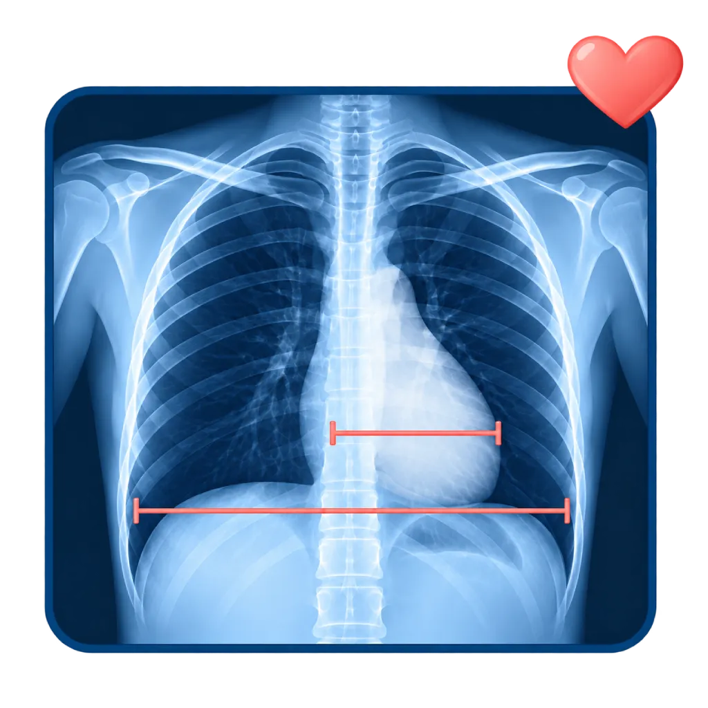
- Size and shape of the heart: if the width of the heart shadow is more than half the width of the chest cavity on an image taken from the back, the heart is probably enlarged.
- Congestion and fluid in the lungs: signs of heart failure, such as widened lung blood vessels and fluid in the lung tissue.
- Fluid in the pleural space: fluid between the lungs and the chest wall.
- Aorta and large vessels: widening of the aortic shadow or of the middle of the chest (mediastinum).
- Lungs: pneumonia, air outside the lung (pneumothorax), or other abnormalities.
- Devices in the chest: the position of pacemaker or ICD leads, catheters, and the wires that close the breastbone after surgery.
Chest X-Rays and the Heart
In shortness of breath or suspected heart failure, a chest X-ray helps show congestion in the lungs and look for other causes such as pneumonia. However, a normal chest X-ray does not rule out heart failure, and it is also not enough to rule out a tear in the wall of the aorta, so the doctor may add echocardiography, an NT-proBNP blood test, or a CT scan. After a pacemaker or ICD is implanted, a chest X-ray is used to check the position of the leads and make sure there is no air outside the lung.
02 / Understanding the procedure
When Is This Procedure Considered?
A doctor may consider this procedure in the situations below. The decision is always made after a thorough assessment.
- Shortness of BreathTo look for lung congestion, fluid, or pneumonia as the cause.
- Suspected Heart FailureTo assess heart size and signs of excess fluid in the lungs.
- Chest PainPart of the initial assessment to look for causes in the lungs, aorta, or bones.
- After Pacemaker, ICD, or Catheter PlacementTo check the position of the device and make sure there is no air outside the lung.
- Before and After Heart SurgeryTo assess the lungs and heart before surgery, and to monitor recovery afterward.
- Prolonged Cough or FeverTo look for a lung infection that can worsen heart disease.
Situations to Consider First
- PregnancyThe dose is very low, but tell the staff so the benefits and protection can be considered.
- Jewelry or Metal Objects on the ChestThese can block the image, so they need to be removed before the X-ray.
- Difficulty Holding Your Breath or Staying StillThe image may be blurred, so the X-ray may need to be repeated.
- Unable to StandThe X-ray is taken while you sit or lie down, with slightly different results.
03 / Before the procedure
How Should You Prepare?
A chest X-ray does not require fasting or any special preparation.
Clothing. You will be asked to change your top into an examination gown and remove jewelry, necklaces, or underwear with metal hooks.
Pregnancy. Tell the staff if you are or might be pregnant.
Devices in the chest. Let the staff know if you have a pacemaker, ICD, or another device in your chest.
Previous results. Bring previous chest X-ray results if you have them, so the doctor can compare any changes.
Unsure about the preparation instructions? Ask our team before the procedure.
04 / During the procedure
How Is the Procedure Done?
The whole examination is usually finished in about 10 minutes, while the X-ray exposure itself takes only a fraction of a second.
Positioning for the Back View
You stand with your chest against the plate, your hands on your hips, and your shoulders rolled forward so the shoulder blades do not cover the lungs.
Taking a Deep Breath
The staff ask you to take a deep breath and hold it for a few seconds while the image is taken, so the lungs are fully expanded and the image is not blurred.
Side View
If needed, a second image is taken from the side with both arms raised above your head.
Reading the Results
The images are examined by a doctor and then read together with your symptoms, ECG, and other tests. For patients who cannot stand, the image is taken with a portable machine at the bedside.
05 / Benefits and risks
Benefits and Risks Should Be Discussed Together.
Every medical procedure has benefits and risks. Your doctor will explain both for your situation before the procedure.
Benefits You Can Expect
- Fast and Widely AvailableThe images can be viewed within minutes.
- Assessing Heart Size and Fluid in the LungsHelps identify signs of heart failure.
- Looking for Other Causes of Shortness of BreathSuch as pneumonia, fluid in the pleural space, or air outside the lung.
- Checking Device PositionPacemaker and ICD leads and catheters can be confirmed to be in the right place.
- Very Low Radiation DoseEquivalent to a few days of natural background radiation.
The benefits of a procedure are not a promise of a cure. Ask how the results will affect your care plan.
Risks You Should Know
- Very Small Radiation ExposureThe risk is very low, and the dose is kept as low as possible.
- The X-Ray May Need to Be RepeatedIf you move or do not hold your breath, the image may be blurred.
- Not Every Abnormality Is VisibleThe heart's blood vessels, valves, and small abnormalities require other tests such as echocardiography or CT.
Risks can differ from person to person. A full explanation and consent are given after an individual assessment.
06 / After the procedure
What Is Recovery Like?
There is no recovery period. You can return to your activities right away, and no radiation remains in your body after the image is taken.
Results. The doctor will explain the results and decide whether further tests such as echocardiography or a CT scan are needed.
Warning signs. Go to the emergency room right away or contact the medical team if you experience:
- Shortness of breath that gets worse or comes on suddenly
- Chest pain that is severe or does not improve
- Coughing up blood
- Leg swelling with shortness of breath when lying down
Know the Signs That Need Prompt Care.
Follow the team's guidance on warning signs. If severe or sudden symptoms appear after the procedure, go to the nearest emergency department right away or call Heart Attack Center 1500 258.
07 / Team and support
Doctors and Procedure Support
The doctors below are listed for this procedure. Listed by name, not by ranking.

dr. Ika Ruriana, Sp.Rad
Radiology
Cardiovascular Diagnostic Center
Clinical focusDiagnostic Radiology · Radiography (X-Ray)
View Profile
dr. Santi R. Dewayanti, Sp.P
Pulmonology
Clinical focusLung and Respiratory Disease · Asthma and COPD
View Profile
dr. Siska S. Danny, Sp.JP(K)
Interventional Cardiology
Coronary Intervention Procedures
Clinical focusPCI for Acute Coronary Syndrome · Primary Coronary Intervention
View Profile08 / Common questions
Before You Plan the Procedure.
Frequently asked questions about chest X-rays. Your doctor will explain what the results mean for your condition.
Is a chest X-ray dangerous because of radiation?
The radiation dose of a chest X-ray is very low, roughly equivalent to the natural radiation you receive from the environment over a few days. The risk is very small compared with the benefit of the information obtained. No radiation remains in your body after the image is taken, so it is safe for you to be near your family.
Can pregnant women have a chest X-ray?
A chest X-ray can be done during pregnancy when it is truly needed, because the dose is very low and the X-rays are aimed at the chest, not the abdomen. The staff can provide additional shielding. Still, tell the staff if you are or might be pregnant so the benefits and protection can be considered.
What does an enlarged heart on an X-ray mean?
The heart is considered enlarged when its shadow is more than half the width of the chest cavity on an image taken from the back. This can be caused by thickened or dilated heart muscle, valve problems, or fluid around the heart. However, an image taken while lying down can make the heart look larger, so echocardiography is usually needed.
Does a normal chest X-ray mean my heart is healthy?
Not necessarily. A chest X-ray assesses the size of the heart and lungs, but it cannot show narrowing of the heart's blood vessels, pumping function, or the valves in detail. Heart failure can also occur with a normal heart size. That is why the doctor may request an ECG, echocardiography, or other tests based on your symptoms.
Why do I need a chest X-ray after a pacemaker is implanted?
A chest X-ray confirms that the pacemaker or ICD leads are in the right position inside the heart. It also checks whether air has leaked outside the lung because of the puncture during implantation, which is rare but needs prompt treatment. A repeat X-ray may be done at a follow-up visit if needed.
Why do I have to hold my breath during the X-ray?
Taking a deep breath fully expands the lungs so the lung tissue and heart shadow are clearly visible. Holding your breath for a few seconds prevents chest movement that could blur the image. If the image is blurred, the X-ray needs to be repeated and you will receive additional radiation, although it is still very small.
Sources and Medical Review
Translated from the Indonesian edition medically reviewed by dr. Santi R. Dewayanti, Sp.P.
Sources as cited in the Indonesian edition.
- McDonagh TA, Metra M, Adamo M, et al. 2021 ESC Guidelines for the diagnosis and treatment of acute and chronic heart failure. Eur Heart J. 2021;42(36):3599-3726.
- Mazzolai L, Teixido-Tura G, Lanzi S, et al. 2024 ESC Guidelines for the management of peripheral arterial and aortic diseases. Eur Heart J. 2024;45(36):3538-3700.
- Glikson M, Nielsen JC, Kronborg MB, et al. 2021 ESC Guidelines on cardiac pacing and cardiac resynchronization therapy. Eur Heart J. 2021;42(35):3427-3520.
- Mettler FA Jr, Huda W, Yoshizumi TT, Mahesh M. Effective doses in radiology and diagnostic nuclear medicine: a catalog. Radiology. 2008;248(1):254-263.