
Quick answer
What Is Myocardial Infarction with Non-Obstructive Coronary Arteries (MINOCA)?
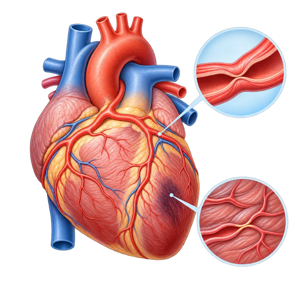
Myocardial infarction with non-obstructive coronary arteries (MINOCA) is a heart attack confirmed by a rise in troponin, but angiography finds no severe narrowing in the coronary arteries. The cause needs to be found so the right treatment can be given.
Sources01 / Understanding the condition
About Myocardial Infarction with Non-Obstructive Coronary Arteries (MINOCA)
Myocardial infarction with non-obstructive coronary arteries, or MINOCA, is a heart attack confirmed by a rise in troponin and signs of reduced blood flow, but coronary angiography finds no narrowing of 50 percent or more in the main coronary arteries. A systematic review found this condition in about 6 out of 100 people with a heart attack. MINOCA is more common in women and in younger people than heart attacks caused by a blockage.
The 2023 ESC guidelines treat MINOCA as a working diagnosis, meaning a starting point for finding the cause, not a final diagnosis. An overview of heart attacks is covered on the Myocardial Infarction / Heart Attack page.
Causes the Doctor Looks For
MINOCA has various causes. A small plaque can crack or erode and form a clot that then dissolves on its own, so the artery looks almost normal when examined. A coronary artery can go into spasm and temporarily shut off blood flow. The small blood vessels inside the heart muscle may not work properly. A clot from elsewhere can travel to a coronary artery. The artery wall can also tear without any injury, covered on the Spontaneous Coronary Artery Dissection page. Sometimes the heart's oxygen demand exceeds its supply, for example with severe anemia or a very fast heart rhythm.
Conditions That Mimic MINOCA
Some diseases cause similar symptoms and a similar rise in troponin, even though they are not heart attacks caused by reduced blood flow. Examples include inflammation of the heart muscle, covered on the Myocarditis page, and sudden weakening of the heart muscle often triggered by stress, covered on the Takotsubo Cardiomyopathy page. Telling them apart is important because their treatment differs, and this is where cardiac MRI plays a major role.
Not a Mild Condition
Finding no severe blockage often brings patients relief, but the risk of another cardiac event is still real. Research shows that the risk of death within a year in MINOCA is lower than in heart attacks caused by a blockage, but it is far from zero. Some patients also feel confused or anxious after being told their arteries are clear, and the doctor's explanation of the cause helps answer those doubts. For this reason, further tests, treatment based on the cause, and regular follow-up are still needed.
02 / Understanding the condition
Symptoms to Recognize.
Common Symptoms
The symptoms are the same as in other heart attacks; MINOCA can only be identified after angiography.
- Pain, pressure, or heaviness in the chest
- Pain spreading to the arm, neck, jaw, or back
- Shortness of breath
- Cold sweat
- Nausea or discomfort in the upper abdomen
- Palpitations or dizziness
Symptoms in Adult Women
MINOCA is more common in women. Symptoms can be typical or vague.
- Chest pain at rest, at night, or in the early morning, which can signal artery spasm
- Shortness of breath or extreme tiredness
- Pain in the upper back, neck, jaw, or shoulder
- Nausea or discomfort in the upper abdomen
- Palpitations with a cold sweat
03 / Understanding the condition
What Causes This Condition?
- A small plaque that cracks or erodes and forms a clot that dissolves on its own
- Spasm of a large coronary artery that temporarily shuts off blood flow
- Poor function of the small blood vessels inside the heart muscle (microvascular)
- A spontaneous tear in the coronary artery wall, covered on the Spontaneous Coronary Artery Dissection page.
- A blood clot that travels to a coronary artery, for example in atrial fibrillation or a blood clotting disorder
- Oxygen supply that does not meet the heart's needs, for example with severe anemia, very low blood pressure, or a very fast rhythm
04 / Understanding the condition
Who Is at Higher Risk?
Lifestyle
Smoking triggers coronary artery spasm and damages small blood vessels. Using cocaine, amphetamines, or other stimulants can also trigger spasm.
Medical
Blood clotting disorders such as antiphospholipid syndrome, atrial fibrillation, autoimmune disease, and a history of vasospastic angina. High blood pressure and high cholesterol still play a role in MINOCA caused by plaque.
Age
On average, people with MINOCA are younger than people who have a heart attack caused by a coronary artery blockage.
Other
Female sex, severe emotional or physical stress, and pregnancy and the period after childbirth, which are linked to spontaneous coronary artery tears.
Groups at Higher Risk
- Young and Middle-Aged Women: MINOCA and spontaneous coronary artery tears are more common in women, including during pregnancy or after giving birth.
- Smokers: Smoking triggers coronary artery spasm and damages the small blood vessels in the heart muscle.
- People with Blood Clotting Disorders: Conditions such as antiphospholipid syndrome make clots form more easily and travel to the coronary arteries.
- Cocaine or Stimulant Users: These substances trigger coronary artery spasm and suddenly increase the heart's oxygen demand.
05 / Understanding the condition
How Is It Diagnosed?
MINOCA is diagnosed in stages. First, the doctor confirms a heart attack according to the universal definition of myocardial infarction: troponin rises or falls with levels above the normal limit, together with symptoms, ECG changes, or signs on imaging. Second, coronary angiography finds no narrowing of 50 percent or more. Third, there is no other clear cause, such as pulmonary embolism or a severe infection.
After that, the search for the cause begins. The 2023 ESC guidelines recommend cardiac MRI for patients with MINOCA whose cause is still unclear, to distinguish infarction from myocarditis or Takotsubo cardiomyopathy. Imaging from inside the artery with IVUS or OCT during catheterization can find a cracked plaque or a small tear. A spasm provocation test may be done in selected patients. Echocardiography and blood tests help look for the source of a clot or a blood clotting disorder.
Cardiac Troponin Test
Proves heart muscle damage. The rising or falling pattern of troponin helps confirm that the injury is recent.
Coronary Angiography
Shows no narrowing of 50 percent or more in the main coronary arteries, which is required for the working diagnosis of MINOCA.
Cardiac MRI
Recommended by the 2023 ESC guidelines when the cause is unclear, to distinguish infarction from myocarditis or Takotsubo cardiomyopathy and to assess the extent of injury.
Optical Coherence Tomography (OCT)
Imaging from inside the artery during catheterization in selected patients, to find a cracked or eroded plaque or a small tear that is not visible on angiography.
Echocardiography
Assesses pumping strength and heart wall motion, and looks for a source of clots inside the heart.
A coronary artery spasm provocation test in the catheterization lab may be done in selected patients suspected of having artery spasm.
06 / Understanding the condition
Treatment Options.
Treatment is decided by a doctor after an assessment. The categories below can help you follow the conversation with your doctor.
Initial Care as for a Heart Attack
Until the cause is known, the patient is treated and monitored like any other heart attack, following acute coronary syndrome guidelines.
Antiplatelet Drugs and Statins
Given if the cause is a cracked or eroded plaque; the doctor decides the type and duration.
Vasodilator Medicines
Calcium channel blockers and nitrates for coronary artery spasm. Some types of beta blockers are avoided if spasm is the cause.
Anticoagulant Medicines
Given if the cause is a clot that traveled from the heart, for example in atrial fibrillation, or a blood clotting disorder.
Treating a Coronary Artery Tear
Usually with medicines and monitoring, because the tear often heals on its own; a stent is used only in certain situations.
Cardiac Rehabilitation
Supervised exercise, education, and risk factor control to restore fitness and self-confidence.
07 / Understanding the condition
Complications to Watch For.
Repeat Heart Attack
Can happen if the cause, such as spasm or a cracked plaque, has not been found and treated.
Heart Rhythm Disorders
Injured heart muscle can trigger an irregular rhythm, especially in the early phase after the attack.
Heart Failure
If the muscle injury is extensive, the heart's pumping strength decreases, causing shortness of breath and swelling.
Stroke
In MINOCA caused by a traveling clot, for example from atrial fibrillation, a similar clot can block a blood vessel in the brain.
08 / Understanding the condition
What Can You Do?
- Quit smoking, because smoking triggers coronary artery spasm.
- Avoid cocaine, amphetamines, and other stimulants.
- Make sure the cause of your MINOCA has been investigated, including with cardiac MRI if your doctor recommends it.
- Take the medicines that match the cause found, and do not stop them on your own.
- Control your blood pressure, cholesterol, and blood sugar to the targets set by your doctor.
- Manage severe stress, for example through counseling or a cardiac rehabilitation program.
09 / Understanding the condition
When Should You See a Doctor?
When symptoms like those of a heart attack appear, you cannot tell whether there is a blockage or not, so call an ambulance right away or go to the nearest emergency room if chest pain lasts more than 5 minutes. After you have been diagnosed with MINOCA, make a follow-up appointment with a cardiologist if:
- The cause of your MINOCA is still unclear and you have not had a cardiac MRI.
- Brief chest pain comes back, especially at rest, at night, or in the early morning.
- You feel palpitations, tire easily, or get short of breath with light activity.
- You are planning a pregnancy after having had a coronary artery tear.
10 / Common questions
Questions You May Have.
Frequently asked questions about heart attack without coronary artery blockage (MINOCA).
If my arteries are clear, why is it called a heart attack?
A heart attack is defined by damage to the heart muscle caused by reduced blood flow, not by whether there is a large blockage. In MINOCA, troponin rises and there are signs of reduced blood flow, but the cause does not show up as narrowing on angiography. For example, the clot has already dissolved, the artery went into spasm and then returned to normal, or the problem lies in small blood vessels that cannot be seen.
What is the difference between MINOCA and myocarditis or Takotsubo cardiomyopathy?
All three can cause chest pain, a rise in troponin, and an angiogram without severe blockage. In MINOCA, the muscle injury is caused by a problem with blood flow. In myocarditis, the cause is inflammation of the heart muscle. In Takotsubo cardiomyopathy, the heart muscle weakens temporarily, often after severe stress. Cardiac MRI helps tell them apart, and treatment differs for each condition.
Why do I need a cardiac MRI after a normal angiogram?
Angiography only shows the inside of the large arteries. Cardiac MRI looks directly at the heart muscle, so it can show injury patterns typical of infarction, myocarditis, or Takotsubo cardiomyopathy. The 2023 ESC guidelines recommend cardiac MRI when the cause of MINOCA is unclear. The results guide the choice of the right medicines and prevent unnecessary treatment.
Do I still need to take heart medicines?
Usually yes, but the type depends on the cause. If the cause is a cracked plaque, antiplatelet drugs and statins are usually given. If the cause is spasm, vasodilator medicines play a bigger role. If the cause is a traveling clot, anticoagulants may be needed. That is why it is important to confirm the cause. Do not stop your medicines on your own without talking to your doctor.
Can MINOCA come back?
Yes, especially if the cause has not been found or treated. Research shows that the risk of another cardiac event in MINOCA is lower than after a heart attack caused by a blockage, but it is still real. Regular follow-up, medicines matched to the cause, quitting smoking, and cardiac rehabilitation help lower that risk. Go to the emergency room right away if chest pain comes back and lasts more than 5 minutes.
Sources and Medical Review
Translated from the Indonesian edition medically reviewed by Dr. med. dr. Denio A. Ridjab, Sp.JP(K).
Sources as cited in the Indonesian edition.
- Byrne RA, Rossello X, Coughlan JJ, et al. 2023 ESC Guidelines for the management of acute coronary syndromes. Eur Heart J. 2023;44(38):3720-3826.
- Tamis-Holland JE, Jneid H, Reynolds HR, et al. Contemporary Diagnosis and Management of Patients With Myocardial Infarction in the Absence of Obstructive Coronary Artery Disease: A Scientific Statement From the American Heart Association. Circulation. 2019;139(18):e891-e908.
- Agewall S, Beltrame JF, Reynolds HR, et al. ESC working group position paper on myocardial infarction with non-obstructive coronary arteries. Eur Heart J. 2017;38(3):143-153.
- Thygesen K, Alpert JS, Jaffe AS, et al. Fourth universal definition of myocardial infarction (2018). Eur Heart J. 2019;40(3):237-269.
- Pasupathy S, Air T, Dreyer RP, Tavella R, Beltrame JF. Systematic review of patients presenting with suspected myocardial infarction and nonobstructive coronary arteries. Circulation. 2015;131(10):861-870.
- Reynolds HR, Maehara A, Kwong RY, et al. Coronary Optical Coherence Tomography and Cardiac Magnetic Resonance Imaging to Determine Underlying Causes of Myocardial Infarction With Nonobstructive Coronary Arteries in Women. Circulation. 2021;143(7):624-640.
Doctors Who Treat This Condition

dr. Puti S. Saus, Sp.JP(K)
Interventional Cardiology
Coronary Intervention Procedures
Clinical focusAcute Coronary Syndrome · Primary Coronary Intervention
View Profile
Dr. dr. Jajang Sinardja, Sp.JP(K)
Interventional Cardiology
Coronary Intervention Procedures
Clinical focusAcute Coronary Syndrome · Primary Coronary Intervention
View Profile
Dr. med. dr. Denio A. Ridjab, Sp.JP(K)
Interventional Cardiology
Coronary Intervention Procedures +2 centers
Clinical focusInterventional Cardiology · Coronary Artery Disease
View Profile