
Quick answer
What Is Cardiac MRI?
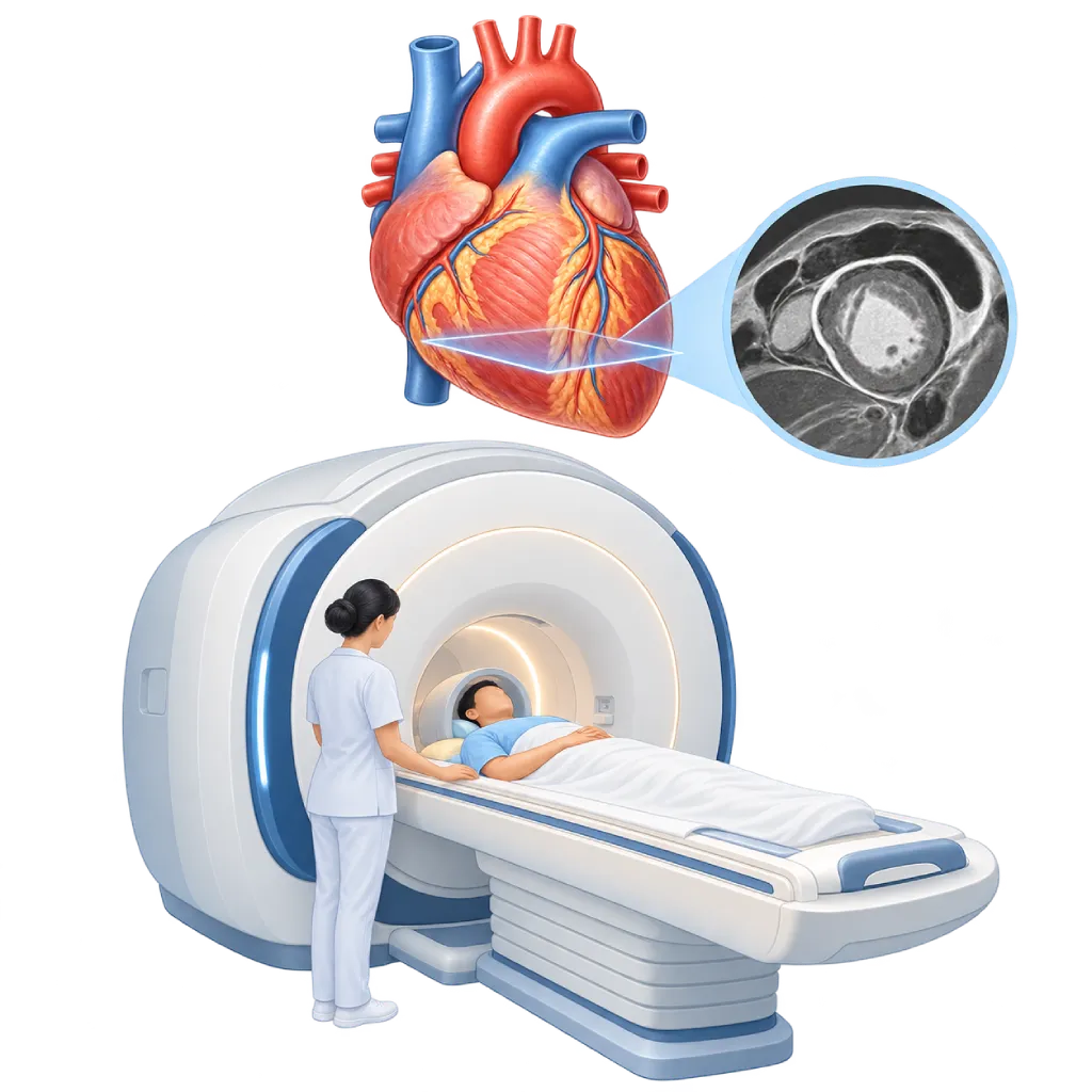
Cardiac MRI is a test that uses a strong magnetic field and radio waves to create moving images of the heart, without X-rays. It measures the heart chambers and pumping strength, and shows inflammation, scar tissue, or buildup of substances in the heart muscle. The test usually takes 30 to 60 minutes.
Sources01 / Understanding the procedure
About Cardiac MRI
What Is Cardiac MRI
Cardiac MRI, also called CMR or cardiovascular magnetic resonance, is an imaging test that uses a strong magnetic field and radio waves. Signals from body tissues are processed by a computer into moving images of the heart from various angles. This test does not use X-rays, and it is usually considered when the doctor needs information that echocardiography or other tests have not provided.
What Is Assessed
Cardiac MRI measures the size of the heart chambers, wall thickness, and ejection fraction, which is the percentage of blood pumped out each time the heart beats. Its measurements are consistent, so they are easy to compare over time. This test also assesses blood flow through the valves, the aorta, and the blood vessels of the lungs, as well as the condition of the sac around the heart.
Seeing the Heart Muscle Tissue
A distinctive feature of cardiac MRI is its ability to tell different types of tissue apart. After gadolinium contrast is injected, areas of muscle that have become scar tissue or are inflamed hold on to the contrast longer and appear bright. This pattern of brightness helps distinguish damage from a heart attack from myocarditis, which is inflammation of the heart muscle, or from cardiomyopathy. Special mapping techniques can show swelling, iron buildup, or protein buildup such as in amyloidosis.
When It Is Considered
Cardiomyopathy and heart failure guidelines recommend cardiac MRI to look for the cause of weakened or thickened heart muscle, to assess myocarditis, and to assess scar tissue before deciding on certain procedures or devices. Cardiac MRI is also used to monitor congenital heart abnormalities in adults and diseases of the aorta. In some patients, MRI with a stress medication can assess blood flow to the heart muscle.
Limitations
The test takes longer than CT and requires holding your breath several times. The narrow space inside the scanner can make some people anxious, and a very irregular heart rhythm can reduce image quality. Many newer pacemakers and defibrillators can be scanned with special settings, but some devices and metal objects in the body are not safe near a magnetic field.
MRI or Other Tests
Echocardiography is usually the first test to assess the structure and function of the heart, which is covered on the Echocardiography page. CT is more suitable for looking at the coronary arteries and calcium, while MRI is used more often to assess the heart muscle tissue. The doctor chooses a test based on the question to be answered.
02 / Understanding the procedure
When Is This Procedure Considered?
A doctor may consider this procedure in the situations below. The decision is always made after a thorough assessment.
- Weak Heart Muscle Without a Clear CauseHelps find the cause, such as scar tissue, inflammation, or cardiomyopathy.
- Suspected MyocarditisShows swelling and inflammation of the heart muscle, for example after a viral infection.
- Hypertrophic CardiomyopathyMeasures wall thickness and the extent of scar tissue to assess risk.
- Suspected Buildup of Substances in the Heart MuscleFor example, amyloidosis, sarcoidosis, or iron overload.
- Congenital Heart Disease in AdultsMonitors the size and function of the right ventricle and blood flow after surgery.
- Assessing Scar Tissue After a Heart AttackHelps estimate which parts of the muscle can still recover if blood flow is restored.
Situations to Consider First
- Implanted Devices That Are Not MRI-SafeFor example, some older pacemakers, certain clips on brain blood vessels, or ear implants.
- Metal Fragments in the Eye or BodyMetal can move or heat up in the magnetic field.
- Severe Kidney ImpairmentThe doctor weighs the use of gadolinium contrast, or a technique without contrast is chosen.
- PregnancyGadolinium contrast is avoided; MRI without contrast is done when the benefit is clear.
- Severe Fear of Enclosed SpacesCan be eased with a mild sedative, or the doctor may choose another test.
03 / Before the procedure
How Should You Prepare?
The team will ask about your medical history and any devices in your body before your test is scheduled.
Metal and implanted devices. Tell the team if you have a pacemaker, defibrillator, brain blood vessel clip, ear implant, or drug pump, or if you have ever been hit by metal fragments. Bring your device ID card if you have one.
Personal items. Jewelry, watches, bank cards, hearing aids, and other metal objects are removed before entering the MRI room. You will be given an examination gown.
Food and medications. You can usually eat and take your medications as usual. If an MRI with a stress medication is planned, you may be asked to avoid coffee, tea, and chocolate for at least 12 hours beforehand.
Kidneys, pregnancy, and anxiety. Let the team know if you have kidney disease, might be pregnant, or are afraid of enclosed spaces.
Unsure about the preparation instructions? Ask our team before the procedure.
04 / During the procedure
How Is the Procedure Done?
Cardiac MRI is done in the MRI room without anesthesia and usually takes 30 to 60 minutes, sometimes longer if a stress medication is used.
Safety Check
The staff make sure no metal is brought in. A small IV line is placed in your arm if contrast is needed, and ECG electrodes are attached to your chest to synchronize the images with your heartbeat.
Entering the Scanner
You lie on a table that moves into the tunnel of the MRI scanner. A signal-receiving pad is placed on your chest, and you are given ear protection because the scanner makes loud knocking sounds. You can still talk to the staff through a speaker.
Recording the Images
Images are taken in several sequences. For the majority of sequences, you are asked to hold your breath for about 10 to 20 seconds. Lying still helps make the images clear.
Gadolinium Contrast
If needed, contrast is injected through the IV, and more images are taken a few minutes later to look for scar tissue or inflammation. You may feel a brief cold sensation in your arm during the injection.
05 / Benefits and risks
Benefits and Risks Should Be Discussed Together.
Every medical procedure has benefits and risks. Your doctor will explain both for your situation before the procedure.
Benefits You Can Expect
- No X-RaysThere is no radiation exposure, so it can be repeated for monitoring.
- Distinguishes Types of TissueShows scar tissue, inflammation, and buildup of substances in the heart muscle.
- Consistent MeasurementsChamber size and pumping strength can be compared over time.
- Helps Guide Treatment ChoicesThe pattern of scar tissue helps determine the choice of medication, procedure, or device.
- Views from Many AnglesImages can be taken from various angles without being blocked by bone or lungs.
The benefits of a procedure are not a promise of a cure. Ask how the results will affect your care plan.
Risks You Should Know
- Anxiety in an Enclosed SpaceCan be reduced with explanation, music, or a mild sedative.
- Loud Scanner NoiseEar protection is provided to protect your hearing.
- Reaction to Gadolinium ContrastRare; usually mild, such as nausea, dizziness, or a rash.
- Contrast Risk in Severe Kidney ImpairmentKidney function is assessed first and the type of contrast is chosen carefully.
- Interference with Implanted DevicesPrevented with a safety check and special pacemaker settings.
- Effects of Stress MedicationDuring an MRI with a stress medication, you may briefly feel breathless, have palpitations, or feel warm.
Risks can differ from person to person. A full explanation and consent are given after an individual assessment.
06 / After the procedure
What Is Recovery Like?
After the test, the IV is removed and you can go home and resume your usual activities, unless you were given a sedative.
Test results. Cardiac MRI images need to be processed and measured carefully, so reading them takes longer than for simpler tests. The doctor explains the results during your consultation.
If you have a pacemaker. The team restores and checks your device settings before you go home.
Warning signs to watch for. Contact the medical team right away or go to the emergency room if you experience:
- Itching, a rash, or facial swelling after contrast
- Shortness of breath or chest pain
- Swelling or pain at the IV site
Know the Signs That Need Prompt Care.
Follow the team's guidance on warning signs. If severe or sudden symptoms appear after the procedure, go to the nearest emergency department right away or call Heart Attack Center 1500 258.
07 / Team and support
Doctors and Procedure Support
The doctors below are listed for this procedure. Listed by name, not by ranking.

dr. BRM Ario S. Kuncoro, Sp.JP(K)
Echocardiography & Cardiovascular Imaging
Cardiovascular Diagnostic Center +2 centers
Clinical focusAdvanced and 3D Echocardiography · Transesophageal Echocardiography
View Profile
Dr. med. dr. Denio A. Ridjab, Sp.JP(K)
Interventional Cardiology
Coronary Intervention Procedures +2 centers
Clinical focusInterventional Cardiology · Coronary Artery Disease
View Profile
dr. Siska S. Danny, Sp.JP(K)
Interventional Cardiology
Coronary Intervention Procedures
Clinical focusPCI for Acute Coronary Syndrome · Primary Coronary Intervention
View Profile08 / Common questions
Before You Plan the Procedure.
Frequently asked questions about cardiac MRI, from safety to how it differs from other tests.
Is cardiac MRI safe?
Cardiac MRI does not use X-rays, so there is no radiation exposure. The main risk comes from the strong magnetic field acting on metal and devices in the body, which is why a safety check is done before entering the MRI room. Gadolinium contrast is generally safe; allergic reactions are rare, and in severe kidney impairment the doctor weighs its use.
Can I have an MRI if I have a pacemaker?
Many newer pacemakers and defibrillators are designed to allow MRI under certain conditions. In many cases, older devices can also be scanned with special settings and close monitoring. The team needs to know the brand and type of your device and its leads, and then adjusts the device before and after the test. Bring your device ID card.
What if I am afraid of enclosed spaces?
Tell the team from the start. The staff explain each step, you can talk to them through a speaker, and you hold a call button to get their attention if needed. Closing your eyes or listening to music can help. If your anxiety is severe, the doctor can give a mild sedative or choose another suitable test.
Why does the test take quite a long time?
Cardiac MRI consists of several image sequences, each assessing something different, such as heart movement, blood flow, and muscle tissue. The majority of sequences are taken while you hold your breath for a few seconds, and some can only be taken a few minutes after the contrast is injected. That is why the whole test usually takes 30 to 60 minutes.
How is cardiac MRI different from a cardiac CT scan?
A cardiac CT scan uses X-rays and is mainly used to look at the coronary arteries, calcium, and heart structures quickly. Cardiac MRI does not use X-rays, takes longer, and is used more often to assess the heart muscle, such as scar tissue, inflammation, or buildup of substances. The doctor chooses a test based on the question to be answered.
How is cardiac MRI different from echocardiography?
Echocardiography uses sound waves, is quick, and can be done at the bedside, so it is usually the first test. Cardiac MRI provides images that are not blocked by bone or lungs, consistent measurements, and information about the heart muscle tissue. MRI is usually considered when echocardiography results have not fully answered the doctor's questions.
Sources and Medical Review
Translated from the Indonesian edition medically reviewed by dr. BRM Ario S. Kuncoro, Sp.JP(K).
Sources as cited in the Indonesian edition.
- Leiner T, Bogaert J, Friedrich MG, et al. SCMR Position Paper (2020) on clinical indications for cardiovascular magnetic resonance. J Cardiovasc Magn Reson. 2020;22(1):76.
- Kramer CM, Barkhausen J, Bucciarelli-Ducci C, et al. Standardized cardiovascular magnetic resonance imaging (CMR) protocols: 2020 update. J Cardiovasc Magn Reson. 2020;22(1):17.
- Arbelo E, Protonotarios A, Gimeno JR, et al. 2023 ESC Guidelines for the management of cardiomyopathies. Eur Heart J. 2023;44(37):3503-3626.
- Ferreira VM, Schulz-Menger J, Holmvang G, et al. Cardiovascular Magnetic Resonance in Nonischemic Myocardial Inflammation: Expert Recommendations. J Am Coll Cardiol. 2018;72(24):3158-3176.
- Indik JH, Gimbel JR, Abe H, et al. 2017 HRS expert consensus statement on magnetic resonance imaging and radiation exposure in patients with cardiovascular implantable electronic devices. Heart Rhythm. 2017;14(7):e97-e153.
- Weinreb JC, Rodby RA, Yee J, et al. Use of Intravenous Gadolinium-based Contrast Media in Patients with Kidney Disease: Consensus Statements from the American College of Radiology and the National Kidney Foundation. Radiology. 2021;298(1):28-35.