
Quick answer
What Is Aneurysm?
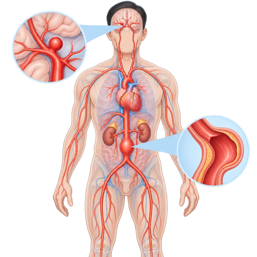
An aneurysm is a localized widening of a weakened artery wall, which makes the vessel bulge like a balloon. Aneurysms can form in the aorta, the brain, or the leg arteries, and most of them cause no symptoms until they grow, block blood flow, or rupture.
SourcesSigns That Need Urgent Help
- Sudden, very severe pain in the chest, back, or abdomen
- A very severe, sudden headache unlike any you have had before
- Fainting, severe weakness, and pale, cold, sweaty skin
- Stiff neck, vomiting, seizures, or reduced consciousness
- Sudden severe leg pain, with a pale, cold leg that is hard to move
If these signs appear, go to the nearest emergency department right away or call Heart Attack Center 1500 258.
01 / Understanding the condition
About Aneurysm
An aneurysm is a localized widening of an artery, a vessel that carries blood from the heart to the rest of the body. The weakened part of the artery wall can no longer withstand blood pressure well, so it bulges like a balloon. Doctors usually describe an artery as having an aneurysm when its diameter has reached about one and a half times its normal size or more.
Types of Aneurysm by Location
Aneurysms are named after where they occur. An aortic aneurysm occurs in the aorta, the main artery leaving the heart. When it is in the chest it is called a thoracic aortic aneurysm, and when it is in the abdomen it is called an abdominal aortic aneurysm. A widening that extends from the chest to the abdomen is called a thoracoabdominal aneurysm. A peripheral artery aneurysm forms in an artery outside the aorta and the brain, for example behind the knee or in the groin. A cerebral aneurysm forms in an artery inside the head and is treated by neurologists and neurosurgeons. Each type is covered in more detail on its own page listed below.
Why the Artery Wall Weakens
The artery wall is made of layers of muscle and elastic fibers. With age, smoking, high blood pressure, and plaque buildup (atherosclerosis) can damage these layers little by little. In some people, the weakness of the wall is present from birth, for example in connective tissue disorders such as Marfan syndrome, or runs in the family. Inflammation of the blood vessels, infection, and injury can also be causes, although less often.
Why Monitoring Matters
The majority of aneurysms grow slowly and cause no complaints for years. For this reason, aneurysms are often found by chance during an ultrasound, CT scan, or MRI done for another reason. Problems arise when an aneurysm keeps growing. An aortic aneurysm can tear or rupture and cause bleeding inside the body. An aneurysm behind the knee more often forms blood clots that block blood flow to the leg. A ruptured cerebral aneurysm causes bleeding around the brain.
An aneurysm found early can be monitored with regular imaging tests. Your doctor weighs its size, location, growth rate, symptoms, and your overall health to decide whether the aneurysm only needs monitoring or should be repaired with surgery or a procedure done from inside the blood vessel (endovascular).
02 / Understanding the condition
Types and Related Conditions
The following conditions belong to the Aneurysm group. Open each one for a full explanation.
03 / Understanding the condition
Symptoms to Recognize.
Common Symptoms
The majority of aneurysms cause no symptoms. The following complaints depend on the location and usually appear when the aneurysm grows or presses on nearby tissue.
- A pulsing feeling in the abdomen, especially around the navel
- Persistent dull pain in the chest, back, abdomen, or flank
- A pulsating lump behind the knee or in the groin
- Hoarseness, coughing, or difficulty swallowing with an aneurysm in the chest
- Pain around one eye or a new headache that does not go away
- Double vision or a drooping eyelid on one side
- Legs that feel cold, tingle, or hurt when walking
04 / Understanding the condition
What Causes This Condition?
- Plaque buildup and hardening of the artery walls (atherosclerosis), which weakens the layers of the wall
- High blood pressure that has gone uncontrolled for a long time, putting constant strain on the artery walls
- Inherited connective tissue disorders, such as Marfan syndrome, Loeys-Dietz syndrome, or vascular Ehlers-Danlos syndrome
- Congenital weakness at the branch points of brain arteries, which can run in families
- Inflammation of the blood vessels (vasculitis) or infection of the artery wall
- Injury to an artery, for example from an accident or a catheter puncture
05 / Understanding the condition
Who Is at Higher Risk?
Lifestyle
Smoking is strongly linked to aortic aneurysms and cerebral aneurysms, and it also speeds up their growth. Heavy alcohol use is linked to cerebral aneurysms.
Medical
High blood pressure, atherosclerosis, and polycystic kidney disease increase the risk. Having one aneurysm also raises the chance of having another one elsewhere.
Genetic
A history of aneurysm or aortic dissection in a parent or sibling, as well as inherited connective tissue disorders such as Marfan syndrome.
Age
The risk rises with age, especially after 60 for aortic aneurysms. Cerebral aneurysms are usually found in middle-aged adults.
Other
Men are more likely to have abdominal aortic aneurysms and aneurysms behind the knee, while cerebral aneurysms are more common in women.
Groups at Higher Risk
- Men Aged 65 and Older Who Have Ever Smoked: A group at higher risk of abdominal aortic aneurysm; guidelines recommend screening with an abdominal ultrasound at least once.
- People with Hypertension: High blood pressure that stays uncontrolled for a long time strains the artery walls, both in the aorta and in the brain.
- Close Relatives of People with Aneurysms: Parents, siblings, or children of people with an aortic aneurysm or a cerebral aneurysm are at higher risk.
- People with Connective Tissue Disorders: For example Marfan syndrome, Loeys-Dietz syndrome, or vascular Ehlers-Danlos syndrome, which make the artery walls more fragile from a young age.
- People with Polycystic Kidney Disease: This inherited kidney disorder is linked to cerebral aneurysms, so your doctor may recommend an examination of the blood vessels in the brain.
06 / Understanding the condition
How Is It Diagnosed?
Because they often cause no symptoms, aneurysms are frequently found during tests for something else or during screening of people at risk. The evaluation begins with questions about your symptoms, smoking habits, and family history, followed by a physical examination. Your doctor then chooses imaging based on the suspected location. Ultrasound is used for the abdominal aorta and leg arteries because it is quick and involves no radiation. Echocardiography assesses the root of the aorta near the heart. CT angiography shows the size and shape of the aneurysm in detail and is the basis for planning a procedure. MRI or MR angiography is used for blood vessels in the brain and for monitoring without X-rays.
If one aneurysm has been found, your doctor may recommend checking other locations, because more than one aneurysm can occur. For aneurysms linked to heredity, close family members may also be advised to get checked.
Physical Examination
Your doctor asks about your symptoms and family history, measures your blood pressure, feels for pulses in the abdomen, groin, and behind the knees, and checks nerve function if needed.
Vascular Ultrasound
Radiation-free sound waves used to measure the width of the abdominal aorta and leg arteries; used for screening and monitoring.
Echocardiography
Ultrasound of the heart to assess the aortic root, the ascending aorta, and the aortic valve.
CT angiography: a CT scan with contrast dye to measure an aneurysm in the chest, abdomen, legs, or head in detail and to plan a procedure.
MRI or MR angiography: imaging without X-rays to assess aneurysms in the brain and to monitor the aorta regularly.
Aortic Angiography
Examination of the aorta with a catheter and contrast dye, usually when planning or performing a repair procedure.
07 / Understanding the condition
Treatment Options.
Treatment is decided by a doctor after an assessment. The categories below can help you follow the conversation with your doctor.
Regular Monitoring
Small aneurysms without symptoms usually only need repeat checks with ultrasound, CT, or MRI at intervals set by your doctor based on their size and location.
Risk Factor Control
Quitting smoking, controlling blood pressure and cholesterol, and taking medicines as prescribed by your doctor to reduce strain on the artery walls and lower the risk of heart attack and stroke.
Endovascular Repair
A stent graft, a fabric tube with a metal frame, is placed from inside the vessel through an artery in the groin to seal off an aortic or peripheral artery aneurysm.
Open Surgery
The widened section of the artery is replaced with an artificial tube, or a bypass is created around it. This option is considered based on the shape of the aneurysm and your condition.
Cerebral Aneurysm Treatment
A brain aneurysm can be sealed from inside the vessel with fine metal coils (coiling) or clipped during surgery (clipping) by a neurosurgeon or a neurointerventional team.
08 / Understanding the condition
Complications to Watch For.
Aneurysm Rupture
The thinned wall can rupture and cause bleeding inside the body or around the brain. This is an emergency and needs immediate treatment.
Tear in the Aortic Wall
The inner layer of the aorta can tear so that blood flows between the layers of the wall. Covered on the Aortic Dissection page.
Blood Clots and Blockage
Clots can form inside the aneurysm and then block the artery or break off into smaller vessels, for example in the leg.
Pressure on Nearby Tissues
A large aneurysm can press on nearby nerves, airways, the esophagus, or veins and cause symptoms.
Subarachnoid Hemorrhage
A ruptured cerebral aneurysm causes bleeding in the space around the brain, a type of stroke covered on the Hemorrhagic Stroke page.
09 / Understanding the condition
What Can You Do?
- Quit smoking and avoid secondhand smoke around you
- Keep your blood pressure within the target agreed with your doctor
- Manage your cholesterol through a balanced diet and medicines if prescribed
- Stay physically active regularly, within your ability and your doctor's advice
- If a close family member has an aneurysm, ask whether you need screening
- If you already know you have an aneurysm, keep your follow-up visits and repeat imaging appointments
10 / Understanding the condition
When Should You See a Doctor?
Aneurysms often cause no symptoms, so early checkups are helpful for people at risk. If you have sudden severe pain in the chest, back, abdomen, or head, if you faint, or if your leg suddenly turns pale and cold, go to the nearest emergency room right away. Make an appointment with a doctor if you:
- Are a man aged 65 or older who has ever smoked, for abdominal aortic screening
- Have a parent or sibling with an aneurysm or aortic dissection
- Have a connective tissue disorder such as Marfan syndrome, or a bicuspid aortic valve
- Feel a pulsing in your abdomen or a pulsating lump behind your knee or in your groin
- Have persistent hoarseness, an unusual headache, or double vision
- Have been diagnosed with an aneurysm and have not had your scheduled follow-up scan
Don't Wait in an Emergency.
If symptoms are severe or come on suddenly, go to the nearest emergency department right away or call Heart Attack Center 1500 258, rather than waiting for an appointment.
11 / Common questions
Questions You May Have.
Frequently asked questions about aneurysms.
Is an aneurysm the same as a stroke?
No. An aneurysm is a widening in the wall of an artery. If a cerebral aneurysm ruptures, blood leaks into the space around the brain and causes a hemorrhagic stroke. However, the majority of small cerebral aneurysms never rupture, and a stroke can also happen without an aneurysm, for example when a blood vessel in the brain is blocked. Aneurysms in the abdominal aorta or the legs do not directly cause a stroke.
Do all aneurysms need surgery?
No. Many small aneurysms only need monitoring with regular imaging while risk factors are controlled. Repair is considered when an aneurysm reaches a certain size, grows quickly, causes symptoms, or forms clots. The size threshold differs for each location, for example the abdominal aorta, the thoracic aorta, or the artery behind the knee. Your doctor also weighs your age, general health, and preferences.
Can an aneurysm shrink or go away on its own?
Aneurysms generally do not shrink on their own, because an artery wall that has widened does not return to its original shape. What can be done is to slow its growth and lower the risk of complications, mainly by quitting smoking and controlling blood pressure. After repair with a stent graft, the aneurysm sac outside the stent may shrink over time, and this is monitored with follow-up scans.
Can aneurysms run in families?
Yes. Some thoracic aortic aneurysms are linked to genetic disorders, such as Marfan syndrome or Loeys-Dietz syndrome, and some occur in several family members without a clear syndrome. Cerebral aneurysms are also found more often when two or more close relatives have had one. If your family has a history like this, talk with your doctor about whether you and your family need to be checked.
Can people with an aneurysm exercise?
Usually yes. Moderate activities such as walking, leisurely cycling, or swimming help keep your blood pressure and blood vessels healthy. What usually needs to be avoided is heavy weightlifting or exercise that makes you hold your breath and strain, because it can raise blood pressure suddenly. Safe activity limits differ for each person, so ask your doctor based on the size and location of your aneurysm.
What is the difference between an aneurysm and an aortic dissection?
An aneurysm is a widening of the artery wall that usually develops slowly. An aortic dissection is a tear in the inner layer of the aorta that lets blood enter and flow between the layers of the wall. A dissection happens suddenly and is an emergency. A widened aorta is more prone to dissection, but dissection can also occur in an aorta that is not widened. Dissection is covered on the Aortic Dissection page.
Sources and Medical Review
Translated from the Indonesian edition medically reviewed by dr. Suko Adiarto, Sp.JP(K), Ph.D..
Sources as cited in the Indonesian edition.
- Mazzolai L, Teixido-Tura G, Lanzi S, et al. 2024 ESC Guidelines for the management of peripheral arterial and aortic diseases. Eur Heart J. 2024;45(36):3538-3700.
- Isselbacher EM, Preventza O, Hamilton Black J, et al. 2022 ACC/AHA Guideline for the Diagnosis and Management of Aortic Disease: A Report of the American Heart Association/American College of Cardiology Joint Committee on Clinical Practice Guidelines. Circulation. 2022;146(24):e334-e482.
- Wanhainen A, Van Herzeele I, Bastos Goncalves F, et al. European Society for Vascular Surgery (ESVS) 2024 Clinical Practice Guidelines on the Management of Abdominal Aorto-Iliac Artery Aneurysms. Eur J Vasc Endovasc Surg. 2024;67(2):192-331.
- Farber A, Angle N, Avgerinos E, et al. The Society for Vascular Surgery clinical practice guidelines on popliteal artery aneurysms. J Vasc Surg. 2022;75(1S):109S-120S.
- Etminan N, de Sousa DA, Tiseo C, et al. European Stroke Organisation (ESO) guidelines on management of unruptured intracranial aneurysms. Eur Stroke J. 2022;7(3):LXXXI-CVI.
- Hoh BL, Ko NU, Amin-Hanjani S, et al. 2023 Guideline for the Management of Patients With Aneurysmal Subarachnoid Hemorrhage: A Guideline From the American Heart Association/American Stroke Association. Stroke. 2023;54(7):e314-e370.
Doctors Who Treat This Condition

dr. Suko Adiarto, Sp.JP(K), Ph.D.
Interventional Cardiology
Vascular Diagnostic and Therapeutic Center +1 center
Clinical focusCoronary Intervention · Peripheral Vascular Intervention
View Profile
dr. Widya T. Suwatri, Sp.BTKV, Subsp. JD(K)
Adult Cardiac Surgery
Advanced Cardiovascular Surgical Care +1 center
Clinical focusMinimally Invasive Cardiac Surgery · Coronary Artery Bypass Surgery
View Profile