
Quick answer
What Is Vascular Ultrasound?
Vascular ultrasound, often called Doppler ultrasound, is a test of the blood vessels that uses sound waves to see the vessel walls and blood flow. It looks for narrowed neck or leg arteries, clots in the veins, leaking vein valves, and widening of the abdominal aorta. One area usually takes about 30 minutes, without radiation.
Sources01 / Understanding the procedure
About Vascular Ultrasound
What Is Vascular Ultrasound
Vascular ultrasound is a test of the blood vessels using high-frequency sound waves. A probe placed on the skin sends out sound waves and picks up the echoes that bounce back from the vessel walls and from moving blood cells. This test is also often called Doppler ultrasound. It uses no radiation, needles, or contrast agents, so it is safe to repeat for monitoring.
Three Types of Images
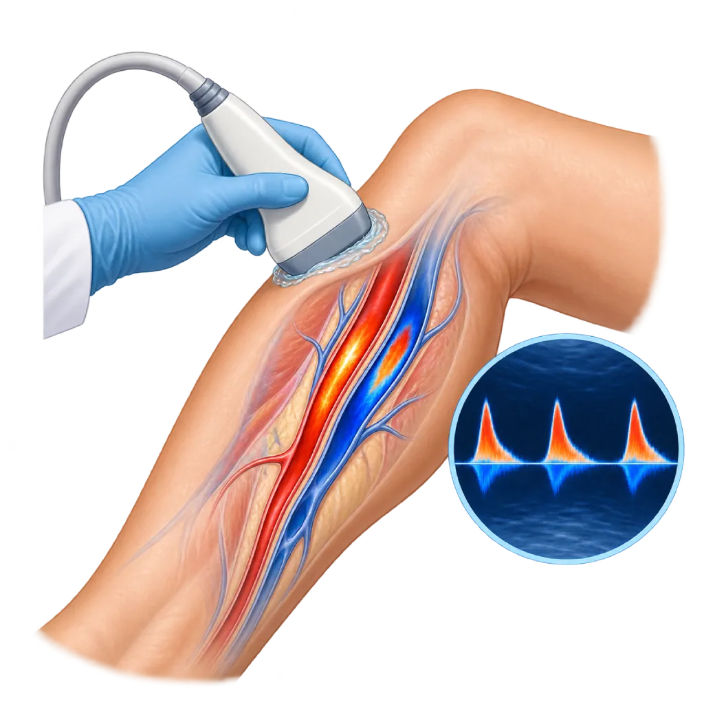
Black-and-white images show the vessel walls, plaque, clots, and vessel size. Color Doppler shows the direction of blood flow, while the Doppler graph measures its speed. Flow that is much faster at one point indicates a narrowing. The combination of images and Doppler is called duplex ultrasound, which is covered in more detail on the Duplex Ultrasound page.
Blood Vessels in the Neck
Carotid artery ultrasound assesses plaque and narrowing in the main blood vessels that carry blood to the brain. This test is recommended after a stroke or a mild stroke that improves within a short time, and may be considered in certain people, for example when a whooshing sound (bruit) is heard in the neck. A severe narrowing may need a procedure to prevent stroke.
Blood Vessels in the Legs and Arms
In the veins, ultrasound looks for blood clots in deep vein thrombosis and assesses leaking vein valves in varicose veins or legs that often swell. In the arteries, ultrasound finds the location and length of a narrowing or blockage in peripheral artery disease, and is often combined with blood pressure measurements at the ankles and arms. Ultrasound of the arm vessels is also used before creating dialysis access, to choose a suitable artery and vein.
Blood Vessels in the Abdomen
Abdominal aortic ultrasound measures the diameter of the aorta to detect an aneurysm, which is a widening that can grow without symptoms, and to monitor its size. Ultrasound can also assess the kidney arteries when a narrowing that causes high blood pressure is suspected. An abdominal scan usually requires fasting so that gas in the intestines does not block the images.
After Vascular Ultrasound
Vascular ultrasound is often the first test for blood vessel disease. When a more complete map of the blood vessels is needed before a procedure, the doctor may follow up with CT angiography, MR angiography, or catheter angiography. Ultrasound is also used to monitor stents, bypass grafts, and dialysis access after a procedure.
02 / Understanding the procedure
When Is This Procedure Considered?
A doctor may consider this procedure in the situations below. The decision is always made after a thorough assessment.
- After a Stroke or Mild StrokeLooks for narrowing of the carotid arteries in the neck that can trigger another stroke.
- Sudden Leg Swelling and PainLooks for blood clots in the deep veins of the legs or arms.
- Varicose Veins or Frequently Swollen LegsAssesses leaking vein valves before deciding on treatment.
- Calf Pain When WalkingLooks for narrowing of the leg arteries in peripheral artery disease.
- Suspected Widening of the Abdominal AortaMeasures the diameter of the aorta in people at risk, such as older men who smoke.
- Monitoring After a ProcedureRegularly checks stents, bypass grafts, or dialysis access.
Situations to Consider First
- No Absolute ContraindicationsVascular ultrasound can be done in almost everyone, including during pregnancy.
- Open Wounds in the Examination AreaThe area is avoided or examined with a sterile cover.
- Severe Pain When PressedProbe pressure is adjusted, and the doctor may choose another method.
- Excess Intestinal GasCan block images of the abdominal aorta; the test is repeated after fasting.
03 / Before the procedure
How Should You Prepare?
Preparation depends on the area being examined. The team will give you instructions when your test is scheduled.
Neck, legs, and arms. No fasting is needed. You may eat, drink, and take your medications as usual.
Abdomen. You usually need to fast for about 6 to 8 hours beforehand, for example from the night before for a morning test, so that gas in the intestines does not block the images. A little plain water is usually allowed.
Clothing. Wear loose clothing that is easy to open over the area being examined. Remove necklaces for a neck examination.
Bring your records. Bring previous blood vessel test results, records of procedures such as stents or surgery, and a list of your medications.
Unsure about the preparation instructions? Ask our team before the procedure.
04 / During the procedure
How Is the Procedure Done?
Vascular ultrasound is done in an examination room without anesthesia. One area usually takes about 30 minutes, and longer if several areas are examined at once or both legs are assessed.
Body Position
You lie on your back for examinations of the neck, abdomen, and leg arteries. To assess the leg vein valves, you may be asked to stand or lie with your head raised so the veins fill with blood.
Gel and Probe
Gel is applied to your skin, then a staff member moves the probe along the blood vessel being examined. You may hear a pulsing sound from the Doppler, which is the sound of blood flow.
Compression and Maneuvers
In the veins, the probe is pressed gently to see whether the vein collapses; a vein that does not collapse indicates a clot. To assess the valves, the calf is squeezed and then released, or you are asked to bear down briefly.
Measurements
Flow speed, degree of narrowing, and vessel diameter are measured and recorded. For the leg arteries, blood pressure at the ankles and arms may be measured at the same time.
05 / Benefits and risks
Benefits and Risks Should Be Discussed Together.
Every medical procedure has benefits and risks. Your doctor will explain both for your situation before the procedure.
Benefits You Can Expect
- No Radiation or NeedlesOnly gel and a probe on the skin, so it is safe to repeat for monitoring.
- Seeing Blood Flow DirectlyThe direction and speed of blood flow appear on the screen during the test.
- Many Areas in One VisitThe neck, legs, arms, and abdomen can be examined as needed.
- An Initial Test Before ProceduresHelps decide whether CT angiography or catheter angiography is needed.
- Safe During PregnancyCan be used to look for clots in the leg veins during pregnancy.
The benefits of a procedure are not a promise of a cure. Ask how the results will affect your care plan.
Risks You Should Know
- Discomfort When PressedEspecially if the leg is swollen or painful; probe pressure is adjusted.
- Obstructed ImagesIntestinal gas, a large body size, calcified plaque, or wounds can limit the images.
- Results Depend on the ExaminerAccuracy depends on the examiner's experience and a standardized protocol.
- Further Tests May Be NeededIf the results are unclear, the doctor may request CT angiography or catheter angiography.
Risks can differ from person to person. A full explanation and consent are given after an individual assessment.
06 / After the procedure
What Is Recovery Like?
After the gel is wiped off, you can go home right away and resume your usual activities. If your abdomen was examined, you may eat again.
Test results. The doctor assesses the recorded images and measurements, then explains the results during your consultation. Findings such as a clot in a deep vein may need treatment on the same day.
Repeat tests. Vascular ultrasound is repeated periodically to monitor carotid narrowing, the size of an aortic aneurysm, or stents and bypass grafts, on the schedule set by your doctor.
Warning signs to watch for. Go to the emergency room right away if you experience:
- A drooping face, weakness on one side of the body, or slurred speech
- Sudden shortness of breath or chest pain
- A leg that suddenly becomes very swollen, painful, pale, or cold
- Severe abdominal or back pain that comes on suddenly
Know the Signs That Need Prompt Care.
Follow the team's guidance on warning signs. If severe or sudden symptoms appear after the procedure, go to the nearest emergency department right away or call Heart Attack Center 1500 258.
07 / Team and support
Doctors and Procedure Support
The doctors below are listed for this procedure. Listed by name, not by ranking.

dr. Emanoel Oepangat, Sp.JP(K)
Interventional Cardiology
Vascular Diagnostic and Therapeutic Center +1 center
Clinical focusCoronary Intervention · Peripheral Vascular Intervention
View Profile
dr. Ika Ruriana, Sp.Rad
Radiology
Cardiovascular Diagnostic Center
Clinical focusDiagnostic Radiology · Radiography (X-Ray)
View Profile
dr. Indah A. Putri, Sp.N, Subsp, NIOO(K), MSc
Neurovascular, Interventional, Otology, Imaging, and Ophthalmology
Clinical focusStroke and Cerebrovascular Disease · Neurosonology
View ProfileDoppler Vascular Ultrasound
An ultrasound test of the blood vessels that shows the vessel wall and the blood flow at the same time. Doctors use it to look…
The availability of technology and test support is confirmed with the team. Technology is chosen according to clinical need.
08 / Common questions
Before You Plan the Procedure.
Frequently asked questions about vascular ultrasound, from preparation to how it relates to other tests.
What is the difference between vascular ultrasound, Doppler ultrasound, and duplex ultrasound?
The three terms are often used interchangeably. Vascular ultrasound is the general name for ultrasound of the blood vessels. Doppler is the part of the test that assesses blood flow, while duplex means combining black-and-white images and Doppler in one test. In practice, blood vessel ultrasound almost always uses the duplex technique. What matters more is the area examined, such as the neck, legs, or abdomen.
Does vascular ultrasound hurt?
Usually not. You only feel the cool gel and the probe moving across your skin. During a vein examination, the probe is pressed slightly to see whether the vein collapses, and this can feel uncomfortable if your leg is swollen or painful. Tell the staff if it hurts so the pressure can be adjusted.
Do I need to fast?
Only for examinations of the blood vessels in the abdomen, such as the aorta or kidney arteries. Fasting for about 6 to 8 hours helps reduce intestinal gas that can block the images. For the neck, legs, and arms, you do not need to fast and may take your medications as usual. Follow the team's instructions when your test is scheduled.
Who needs a neck artery ultrasound?
Carotid artery ultrasound is recommended after a stroke or a mild stroke, to look for narrowing that can trigger another stroke. It may also be considered if the doctor hears a whooshing sound in the neck or before certain heart surgeries. Guidelines do not recommend routine carotid ultrasound for everyone without symptoms, so the doctor weighs its benefit for you.
Is vascular ultrasound safe during pregnancy?
It is safe. Ultrasound uses sound waves without radiation or contrast, so it can be used during pregnancy. During pregnancy, the risk of blood clots in the leg veins increases, and vein ultrasound is the first test if a leg suddenly becomes swollen and painful. Tell the staff how far along your pregnancy is so the examination position can be adjusted.
Can ultrasound replace angiography?
For many purposes, ultrasound is enough for diagnosis and monitoring. However, if a procedure such as balloon angioplasty, a stent, or surgery is planned, the doctor often needs a more complete map of the blood vessels with CT angiography, MR angiography, or catheter angiography. Ultrasound images can also be blocked by heavy calcification or intestinal gas. The doctor decides on the appropriate follow-up test.
Sources and Medical Review
Translated from the Indonesian edition medically reviewed by dr. Emanoel Oepangat, Sp.JP(K).
Sources as cited in the Indonesian edition.
- Mazzolai L, Teixido-Tura G, Lanzi S, et al. 2024 ESC Guidelines for the management of peripheral arterial and aortic diseases. Eur Heart J. 2024;45(36):3538-3700.
- Naylor R, Rantner B, Ancetti S, et al. European Society for Vascular Surgery (ESVS) 2023 Clinical Practice Guidelines on the Management of Atherosclerotic Carotid and Vertebral Artery Disease. Eur J Vasc Endovasc Surg. 2023;65(1):7-111.
- Kakkos SK, Gohel M, Baekgaard N, et al. European Society for Vascular Surgery (ESVS) 2021 Clinical Practice Guidelines on the Management of Venous Thrombosis. Eur J Vasc Endovasc Surg. 2021;61(1):9-82.
- De Maeseneer MG, Kakkos SK, Aherne T, et al. European Society for Vascular Surgery (ESVS) 2022 Clinical Practice Guidelines on the Management of Chronic Venous Disease of the Lower Limbs. Eur J Vasc Endovasc Surg. 2022;63(2):184-267.
- Wanhainen A, Van Herzeele I, Bastos Goncalves F, et al. European Society for Vascular Surgery (ESVS) 2024 Clinical Practice Guidelines on the Management of Abdominal Aorto-Iliac Artery Aneurysms. Eur J Vasc Endovasc Surg. 2024;67(2):192-331.
- Needleman L, Cronan JJ, Lilly MP, et al. Ultrasound for Lower Extremity Deep Venous Thrombosis: Multidisciplinary Recommendations From the Society of Radiologists in Ultrasound Consensus Conference. Circulation. 2018;137(14):1505-1515.