
Quick answer
What Is Atrioventricular Block?
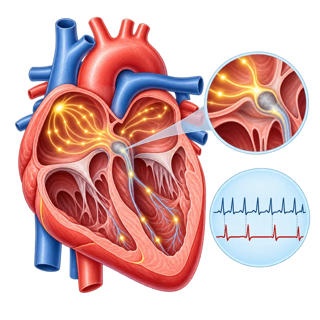
Atrioventricular block is a blockage of the electrical signal from the upper chambers of the heart (atria) to the lower chambers (ventricles). Its degree varies, from mild slowing with no symptoms to complete block, which makes the heartbeat very slow and usually requires a pacemaker.
SourcesSigns That Need Urgent Help
- Sudden fainting without warning
- Very slow pulse with weakness, paleness, or cold sweats
- Chest pain or severe shortness of breath
- Sudden confusion or difficulty waking up
- A brief seizure with loss of consciousness
If these signs appear, go to the nearest emergency department right away or call Heart Attack Center 1500 258.
01 / Understanding the condition
About Atrioventricular Block
The heart's electrical signal starts in the sinus node, spreads through the upper chambers (atria), then passes through the atrioventricular node (AV node) before entering the lower chambers (ventricles). The AV node works like a gate that adds a short pause so the atria can finish filling the ventricles with blood. In atrioventricular block, or AV block, the signal at this gate or in the pathway below it is slowed or stopped. The disturbance can be permanent or come and go, and its effect depends on the degree and location of the block.
Degrees of AV Block
First-degree block means every signal still reaches the ventricles, only more slowly. It shows up on an ECG and usually causes no symptoms. Second-degree block means some signals are not conducted. Mobitz type I, or Wenckebach, is often found in fit young people or during sleep and is usually harmless. Mobitz type II usually occurs lower in the pathway and is more likely to progress to complete block. Third-degree block, or complete block, means no signals from the atria are conducted. The ventricles then beat on their own at a slow rate, often 30 to 45 beats per minute.
Causes
In adults, common causes are aging and hardening of the tissue in the heart's electrical system. Other causes include heart attack, medicines that slow conduction, heart valve surgery or procedures such as catheter-based aortic valve replacement (TAVI), inflammation of the heart muscle, sarcoidosis, and high potassium levels. There is also congenital AV block, for example in babies born to mothers with lupus.
Why the Degree Matters
The degree and location of the block determine treatment. First-degree block and Mobitz I without symptoms usually only need monitoring. Mobitz II block, high-grade block, and complete block usually require a permanent pacemaker even before symptoms appear, unless the cause is temporary and can be corrected. In younger people with no clear cause, the doctor also looks for an underlying heart muscle disease.
AV block is one source of a slow heartbeat. An overview of slow heartbeats from various causes is covered on the Bradycardia page.
02 / Understanding the condition
Symptoms to Recognize.
Common Symptoms
- No symptoms in first-degree block and some second-degree block
- Tiring easily and reduced ability to do physical activity
- Dizziness or lightheadedness
- Shortness of breath when walking or climbing stairs
- A heartbeat that feels slow or occasionally seems to skip a beat
- Chest discomfort
Symptoms in Older Adults
AV block due to aging is common in older adults, and the symptoms are often vague.
- Frequent falls without a clear reason
- Confusion or forgetfulness that comes and goes
- Weakness and sleeping a lot during the day
Symptoms in Newborns
Congenital AV block is often already detected during pregnancy, especially in mothers with lupus or anti-Ro antibodies.
- Slow fetal heart rate during prenatal checkups
- Slow heart rate in the baby at birth
- Difficulty feeding and tiring easily
- Fast breathing or appearing limp
03 / Understanding the condition
What Causes This Condition?
- Aging and hardening of the tissue in the heart's electrical conduction system
- Heart attack, especially one affecting the lower wall of the heart
- Medicines that slow the AV node, such as beta blockers, digoxin, or antiarrhythmics
- Valve surgery, catheter-based aortic valve replacement (TAVI), or ablation near the AV node
- Inflammation or disease of the heart muscle, such as myocarditis, sarcoidosis, and amyloidosis
- Blood potassium levels that are too high
- Congenital AV block, for example in babies born to mothers with lupus
- High vagal nerve tone in athletes or during sleep, usually causing first-degree block or Mobitz I
04 / Understanding the condition
Who Is at Higher Risk?
Age
Degeneration of the conduction tissue increases after age 60 to 70, so AV block and the need for a pacemaker are more common in older adults.
Medical
Coronary artery disease, a history of heart attack, cardiomyopathy, aortic valve disease, sarcoidosis, amyloidosis, and chronic kidney disease with high potassium.
Genetic
A small proportion of conduction disorders run in families, for example lamin A/C gene abnormalities or myotonic muscular dystrophy.
Other
A history of heart valve surgery, TAVI, or ablation, as well as taking several medicines that slow conduction at the same time.
Groups at Higher Risk
- Older Adults: Aging of the conduction tissue makes AV block and the need for a pacemaker more common after age 70.
- Patients After Aortic Valve Procedures: TAVI or aortic valve surgery can press on the conduction pathway near the valve, causing new block.
- People with Coronary Artery Disease: A heart attack can disrupt blood flow to the AV node; this type of block is often temporary.
- People Taking Heart Rate Medicines: Beta blockers, certain calcium channel blockers, and digoxin can slow conduction through the AV node.
05 / Understanding the condition
How Is It Diagnosed?
AV block is often first seen on a routine ECG. The ECG shows the relationship between the electrical waves of the atria and the ventricles, so the doctor can determine the degree of block. The width of the QRS complex and changes when the heart rate increases give clues as to whether the block is in the AV node or lower in the pathway.
If the block comes and goes, a Holter monitor or long-term recorder is used to capture it. The doctor also looks for correctable causes: medicines that slow conduction, high potassium, heart attack, or inflammation of the heart muscle. Echocardiography assesses pumping function and the valves. In people under 60 with no clear cause, the doctor may recommend a cardiac MRI to look for sarcoidosis or other heart muscle diseases.
Electrocardiogram (ECG)
Determines the degree of block from the relationship between the atrial (P) and ventricular (QRS) waves, as well as the width of the QRS complex.
Holter Monitoring
Captures block that comes and goes and matches it with symptoms such as dizziness or fainting.
Implantable Loop Recorder (ILR)
An implanted recorder for rare, unexplained fainting that records the heart rhythm for up to about three years.
Echocardiography
Assesses pumping function and the valves and looks for a heart muscle disease underlying the block.
Cardiac MRI
Looks for sarcoidosis, inflammation, or other heart muscle diseases, especially in people under 60 with no clear cause.
Blood Electrolyte Test
Measures potassium that is too high, a correctable cause of block.
Electrophysiology Study
In certain cases, measures the location of the block within the conduction pathway when the ECG and other recordings are not yet clear.
06 / Understanding the condition
Treatment Options.
Treatment is decided by a doctor after an assessment. The categories below can help you follow the conversation with your doctor.
Regular Monitoring
First-degree block and Mobitz I without symptoms usually only need monitoring with regular ECGs.
Treating Correctable Causes
Adjusting medicines, correcting potassium, or treating a heart attack or inflammation, because some blocks improve afterward.
Temporary Pacing
Used in the intensive care unit when severe block appears suddenly, while waiting for the block to improve or for a permanent device to be implanted.
Permanent Pacemaker
Recommended for Mobitz II block, high-grade block, and complete block that is not caused by something temporary, with or without symptoms.
Physiologic Pacing or Resynchronization
If the ventricles are expected to be paced very often or pumping function is reduced, the doctor may choose conduction system pacing or a resynchronization device (CRT).
07 / Understanding the condition
Complications to Watch For.
Sudden Fainting (Stokes-Adams Attack)
In high-grade block, the ventricles can stop beating for a few seconds, so you faint without warning and risk injury.
Heart Failure
A very slow heartbeat over a long time reduces the amount of blood pumped, which can cause shortness of breath and swelling.
Cardiac Arrest
Untreated complete block can cause the ventricles to stop beating or trigger dangerous ventricular arrhythmias.
Pacemaker Complications
Infection, lead displacement, or reduced pumping function from long-term right ventricular pacing can occur, so the device needs regular checkups.
08 / Understanding the condition
What Can You Do?
- Control your blood pressure, blood sugar, and cholesterol, and stop smoking to prevent coronary artery disease
- Show all your medicines and supplements to your doctor, especially medicines that regulate heart rate
- Have follow-up ECGs on schedule if you have first-degree block or Mobitz I
- If you have kidney disease, follow the advice on limiting potassium
- After TAVI or valve surgery, follow the rhythm monitoring schedule you are given
- Women with lupus or anti-Ro antibodies should seek medical advice before pregnancy
09 / Understanding the condition
When Should You See a Doctor?
If your ECG shows AV block, discuss the results with a cardiologist even if you feel well, especially if it is described as second- or third-degree block. Also make an appointment if you tire easily, feel dizzy, get short of breath with light activity, or often feel as if your heart skips a beat.
Go to the emergency room right away if:
- You faint, especially suddenly and without warning
- Your pulse is very slow, with weakness, paleness, or cold sweats
- You develop chest pain, severe shortness of breath, or sudden confusion
- Your heart rate slows noticeably after valve surgery or TAVI
Don't Wait in an Emergency.
If symptoms are severe or come on suddenly, go to the nearest emergency department right away or call Heart Attack Center 1500 258, rather than waiting for an appointment.
10 / Common questions
Questions You May Have.
Frequently asked questions about atrioventricular block, or heart block.
Is first-degree AV block dangerous?
Usually not. In first-degree block, every signal still reaches the ventricles, only slightly more slowly. This condition is often found in healthy people, athletes, or older adults, and it usually causes no symptoms. Your doctor may recommend regular ECGs to watch how it develops, especially if you have another heart condition or take medicines that slow conduction.
Does complete heart block always require a pacemaker?
Almost always, unless the cause is temporary and can be corrected, for example a medicine effect, high potassium, or certain heart attacks that improve within a few days. In persistent complete block, a pacemaker is recommended even before symptoms appear, because of the risk of sudden fainting and cardiac arrest. Your doctor will confirm the cause first before deciding on a permanent device.
What do Mobitz I and Mobitz II mean?
Both are second-degree block, in which some signals from the atria are not conducted to the ventricles. In Mobitz I, the signal delay gets longer and longer until one beat is dropped; it usually occurs in the AV node and is often harmless. In Mobitz II, beats are dropped suddenly without prior slowing, usually lower in the pathway, and this type is more likely to progress to complete block.
Why can AV block appear after TAVI or valve surgery?
The heart's electrical conduction pathway lies very close to the aortic valve. When a valve is implanted or repaired, the surrounding tissue can be compressed or swell, blocking the signal. Some blocks improve within a few days, while others persist and require a pacemaker. That is why the heart rhythm is monitored for several days after the procedure and at later follow-up visits.
Can people with AV block exercise?
It depends on the degree of block and its treatment. In first-degree block or Mobitz I without symptoms, exercise is usually safe. In high-grade block, exercise is postponed until treatment is complete, usually after the pacemaker is implanted and the wound has healed. After that, the majority of sports can be resumed while avoiding hard impacts to the device area. Ask your doctor about safe limits.
Sources and Medical Review
Translated from the Indonesian edition medically reviewed by dr. Sunu B. Raharjo, Sp.JP(K), Ph.D..
Sources as cited in the Indonesian edition.
- Kusumoto FM, Schoenfeld MH, Barrett C, Edgerton JR, Ellenbogen KA, Gold MR, et al. 2018 ACC/AHA/HRS Guideline on the Evaluation and Management of Patients With Bradycardia and Cardiac Conduction Delay: A Report of the American College of Cardiology/American Heart Association Task Force on Clinical Practice Guidelines and the Heart Rhythm Society. Circulation. 2019;140(8):e382-e482.
- Glikson M, Nielsen JC, Kronborg MB, Michowitz Y, Auricchio A, Barbash IM, et al. 2021 ESC Guidelines on cardiac pacing and cardiac resynchronization therapy. Eur Heart J. 2021;42(35):3427-3520.
- Chung MK, Patton KK, Lau CP, Dal Forno ARJ, Al-Khatib SM, Arora V, et al. 2023 HRS/APHRS/LAHRS guideline on cardiac physiologic pacing for the avoidance and mitigation of heart failure. Heart Rhythm. 2023;20(9):e17-e91.
- Rodés-Cabau J, Ellenbogen KA, Krahn AD, Latib A, Mack M, Mittal S, et al. Management of Conduction Disturbances Associated With Transcatheter Aortic Valve Replacement: JACC Scientific Expert Panel. J Am Coll Cardiol. 2019;74(8):1086-1106.
- Brignole M, Moya A, de Lange FJ, Deharo JC, Elliott PM, Fanciulli A, et al. 2018 ESC Guidelines for the diagnosis and management of syncope. Eur Heart J. 2018;39(21):1883-1948.
Doctors Who Treat This Condition

Dr. dr. Faris Basalamah, Sp.JP(K)
Interventional Cardiology
Coronary Intervention Procedures +1 center
Clinical focusComplex Coronary Intervention · Cardiac Electrophysiology
View Profile
Dr. dr. Dicky A. Hanafy, Sp.JP(K)
Arrhythmia and Electrophysiology
Arrhythmia and Device Center +1 center
Clinical focusElectrophysiology and Arrhythmia Ablation · Pacemaker and ICD Implantation
View Profile
dr. Sunu B. Raharjo, Sp.JP(K), Ph.D.
Arrhythmia and Electrophysiology
Arrhythmia and Device Center
Clinical focusClinical and Interventional Electrophysiology · Cardiac Arrhythmia Ablation
View Profile