
Quick answer
What Is Cardiomegaly?
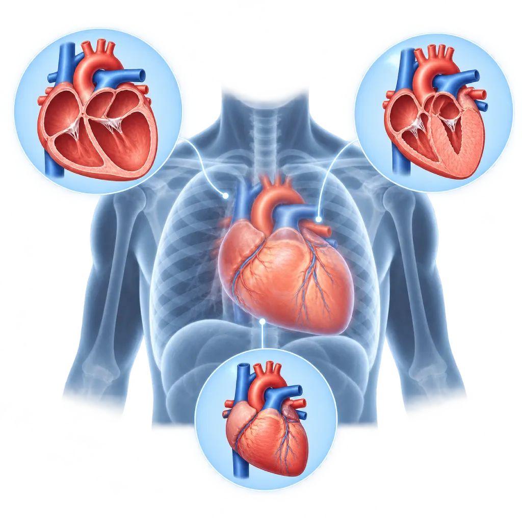
Cardiomegaly means the heart is larger than normal, usually seen on a chest X-ray or echocardiography. It is not a disease on its own, but a sign of another condition, such as long-standing hypertension, valve disease, cardiomyopathy, or heart failure.
SourcesSigns That Need Urgent Help
- Severe shortness of breath at rest or being unable to lie down
- Coughing up frothy or pink sputum
- Chest pain lasting more than a few minutes
- Fainting or near-fainting
- A very fast heartbeat with dizziness or weakness
- Weakness on one side of the body, an uneven face, or slurred speech
If these signs appear, go to the nearest emergency department right away or call Heart Attack Center 1500 258.
01 / Understanding the condition
About Cardiomegaly
Cardiomegaly is the term for a heart that is larger than normal. The term describes a test finding, not the name of a disease, and in everyday language people often call it a swollen heart. The heart can enlarge because its chambers widen (dilation), its walls thicken (hypertrophy), or both, as a result of working harder over a long time or because the heart muscle has weakened. Enlargement can affect a single chamber, for example the left atrium in mitral valve disease, or several chambers at once. What determines the treatment and course of the condition is the cause of the enlargement, not the size of the heart alone.
How Cardiomegaly Is Found
Cardiomegaly is often first seen on a chest X-ray, when the width of the heart shadow is more than half the width of the chest cavity. This measurement is called the cardiothoracic ratio. An X-ray only gives an estimate: the position of the image, a breath that is not deep enough, fat around the heart, or fluid in the sac around the heart can make the heart look larger. For this reason, echocardiography is used to confirm which chamber is enlarged, measure wall thickness, and assess pumping strength and the valves.
Causes to Look For
Common causes are high blood pressure that has gone uncontrolled for a long time, coronary artery disease or a previous heart attack, valve disease, and cardiomyopathy, a disease of the heart muscle covered on the Cardiomyopathy page. Other causes include congenital heart disease, high pressure in the lung blood vessels, a heart rhythm that is too fast for a long time, severe anemia, thyroid disorders, heavy drinking, and certain chemotherapy drugs. Mild enlargement can also occur during pregnancy and in trained athletes, covered on the Athlete's Heart page.
Why It Needs to Be Checked
A heart that is enlarged because of disease may work less efficiently. Over time, heart failure, rhythm disorders, or blood clots in the heart chambers can develop. Thickening of the left ventricular wall due to hypertension also signals a higher risk of stroke and heart failure. The good news is that many causes of cardiomegaly can be controlled. In some patients, the heart can shrink back after the cause is treated, for example when blood pressure is controlled, a valve is repaired, or heart failure medications are taken regularly. Heart failure is covered on the Heart Failure page.
02 / Understanding the condition
Symptoms to Recognize.
Common Symptoms
Cardiomegaly itself often causes no symptoms. Symptoms usually come from the underlying disease.
- Shortness of breath during activity or when lying down
- Tiring easily and reduced ability to be active
- Swelling in the legs, ankles, or abdomen
- Palpitations or an irregular heartbeat
- Dizziness or near-fainting
- Coughing when lying down at night
Symptoms in Infants
In babies, an enlarged heart is often related to congenital heart disease or inflammation of the heart muscle.
- Fast breathing or visible effort to breathe
- Sweating or tiring quickly during feeding
- Difficulty gaining weight
- Fussiness and looking pale
03 / Understanding the condition
What Causes This Condition?
- Hypertension that has gone uncontrolled for a long time, causing the heart walls to thicken
- Coronary artery disease or a previous heart attack that weakens the heart muscle
- Heart valve disease that strains the heart chambers, for example a leaking or narrowed valve
- Cardiomyopathy, including from inherited factors, alcohol, or after giving birth
- Congenital heart disease and high pressure in the lung blood vessels
- Fluid in the sac around the heart that makes the heart shadow look large on an X-ray
- Severe anemia, thyroid disorders, or a fast heart rhythm that lasts a long time
- Adaptation in trained athletes and during pregnancy, which is generally harmless
04 / Understanding the condition
Who Is at Higher Risk?
Lifestyle
Heavy drinking, smoking, using stimulants such as methamphetamine, excess body weight, and inactivity increase the likelihood of the heart enlarging.
Medical
Long-standing uncontrolled hypertension, diabetes, coronary artery disease, valve disease, chronic kidney disease, sleep apnea, and a history of chemotherapy or chest radiotherapy.
Genetic
A family history of cardiomyopathy or sudden death at a young age may signal an inherited heart muscle disease.
Age
Causes such as long-standing hypertension, coronary disease, and degenerative valve disease become more common with age.
Other
Pregnancy and the few months after giving birth can be linked to weakening of the heart muscle, covered on the Peripartum Cardiomyopathy page.
Groups at Higher Risk
- People with Long-Standing Hypertension: Years of high blood pressure make the left ventricular wall thicken, so regular ECG and echocardiography checks are useful.
- Heart Attack Survivors: Damaged heart muscle can make the left ventricle widen slowly, especially if treatment is not taken regularly.
- Families of People with Cardiomyopathy: Close relatives are advised to have regular ECG and echocardiography checks even without symptoms.
- Cancer Survivors Who Have Had Chemotherapy or Radiotherapy: Some cancer drugs and chest radiation can weaken the heart muscle, covered on the Cancer Therapy-Related Heart Disease page.
05 / Understanding the condition
How Is It Diagnosed?
Testing starts by confirming whether the heart is truly enlarged. A chest X-ray gives an initial estimate, but the result is affected by the position of the image and the depth of breath, so echocardiography is used to measure each chamber, wall thickness, pumping strength, and the valves, and to look for fluid in the sac around the heart. Your doctor also asks about symptoms and any history of high blood pressure, diabetes, heart attack, alcohol use, chemotherapy, and heart disease in the family. An ECG looks for signs of wall thickening or rhythm disorders, while blood tests, including NT-proBNP, help assess the strain on the heart and other causes such as anemia or thyroid disorders. If the cause is still unclear, cardiac MRI can show scar tissue, inflammation, or buildup of substances in the heart muscle, and the coronary arteries are examined if coronary artery disease is suspected. For cardiomyopathy that is suspected to be inherited, genetic testing and screening of family members may be recommended.
Chest X-Ray
Estimates heart size using the cardiothoracic ratio and looks for fluid in the lungs. The position of the image and the depth of breath affect the result.
Echocardiography
Confirms which chamber is enlarged, measures wall thickness, pumping strength, and the valves, and looks for fluid in the sac around the heart.
Electrocardiogram (ECG)
Looks for signs of wall thickening, rhythm disorders, and a previous heart attack.
NT-proBNP and BNP Test
Helps assess whether heart enlargement comes with heart failure and monitors treatment results.
Laboratory Tests
Complete blood count, kidney function, blood sugar, cholesterol, and thyroid function to look for causes and aggravating factors.
Cardiac MRI
Measures the heart chambers in detail and looks for scar tissue, inflammation, or buildup of substances in the heart muscle to determine the cause.
CT Coronary Angiography
Looks for narrowing of the coronary arteries as a cause of weakened heart muscle, sometimes followed by catheter angiography.
06 / Understanding the condition
Treatment Options.
Treatment is decided by a doctor after an assessment. The categories below can help you follow the conversation with your doctor.
Treating the Cause
For example, controlling blood pressure, improving coronary blood flow, repairing or replacing a valve, stopping alcohol, or treating anemia and thyroid disorders.
Heart Failure Medications
If the heart's pumping is weakened, your doctor prescribes several classes of medication that have been shown to improve symptoms and life expectancy, then adjusts them gradually. The type and dose are decided by your doctor.
Diuretics
Water pills to reduce shortness of breath and swelling caused by excess fluid in the body.
Valve Repair or Replacement
Done through surgery or a catheter procedure if the heart enlargement is caused by severe valve disease.
Special Pacemaker or ICD
Cardiac resynchronization therapy helps both ventricles contract together in certain patients, and an ICD prevents sudden cardiac arrest in high-risk patients.
Healthy Lifestyle and Rehabilitation
Limiting salt, staying active as advised, quitting smoking, and joining cardiac rehabilitation help improve your ability to be active.
07 / Understanding the condition
Complications to Watch For.
Heart Failure
A heart enlarged by disease can become weaker or stiffer, causing shortness of breath, tiredness, and swelling.
Heart Rhythm Disorders
Widened atria make atrial fibrillation more likely, while enlarged and scarred ventricles can trigger ventricular rhythm disorders.
Blood Clots and Stroke
Slow blood flow in widened heart chambers or during atrial fibrillation can form clots that travel to the brain.
Functional Valve Regurgitation
Widening of the ventricles can pull on the mitral or tricuspid valve so it does not close tightly and blood flows backward.
Sudden Cardiac Arrest
In some heart muscle diseases with very weak pumping, the risk of dangerous rhythm disorders rises. In certain patients, this risk can be reduced with an ICD.
08 / Understanding the condition
What Can You Do?
- Keep your blood pressure at your doctor's target and check it regularly
- Avoid heavy drinking, smoking, and stimulants
- Manage your weight, blood sugar, and cholesterol
- Take your heart medications regularly and attend follow-up visits as scheduled
- Treat anemia, thyroid disorders, and sleep apnea if present
- Have your heart checked if there is a family history of cardiomyopathy or you have had chemotherapy
- Limit salt if you have hypertension or heart failure
09 / Understanding the condition
When Should You See a Doctor?
If a chest X-ray report says your heart is enlarged, take the result to a cardiologist so it can be confirmed with echocardiography and the cause can be found, even if you have no symptoms. Go to the emergency room right away if you have severe shortness of breath at rest, chest pain, or fainting.
- A chest X-ray or health checkup reports an enlarged heart
- Shortness of breath during activity or when lying down, or waking up at night short of breath
- Swelling in the legs or abdomen, or rapid weight gain
- Palpitations or an irregular heartbeat
- High blood pressure that is not yet under control
- A close relative with cardiomyopathy or sudden death at a young age
Don't Wait in an Emergency.
If symptoms are severe or come on suddenly, go to the nearest emergency department right away or call Heart Attack Center 1500 258, rather than waiting for an appointment.
10 / Common questions
Questions You May Have.
Frequently asked questions about cardiomegaly, or an enlarged heart.
Is cardiomegaly the same as heart failure?
No. Cardiomegaly means the heart is larger than normal, while heart failure means the heart cannot pump or fill with blood as well as the body needs. The two often occur together, but the heart can enlarge without heart failure, for example in athletes, and heart failure can occur with a normal-sized heart. Echocardiography and blood tests help your doctor assess both.
Can an enlarged heart return to normal?
In some people, yes. The heart can shrink after the cause is treated, for example when blood pressure is controlled, a valve is repaired, a fast rhythm is brought under control, or alcohol is stopped. In heart failure with weak pumping, medication taken regularly can improve heart size and pumping strength within a few months. How much it improves depends on the cause and how long the enlargement has lasted.
Why does an X-ray show a large heart when echocardiography is normal?
A chest X-ray only shows the heart's shadow. An image taken while lying down or from front to back, a breath that is not deep enough, fat around the heart, or a certain chest shape can make the heart look larger. Echocardiography measures the heart's chambers and walls directly, so its results are more reliable for assessing heart size.
Can high blood pressure make the heart enlarge?
Yes. Long-term high blood pressure makes the left ventricle work harder, so its wall thickens. If it is not controlled, the heart muscle can become stiff, then weaken, and its chambers widen. Controlling your blood pressure to your doctor's target can reduce wall thickening and lower the risk of heart failure, stroke, and rhythm disorders.
Can people with cardiomegaly exercise?
Generally yes, and regular physical activity benefits the majority of stable heart conditions. The type and intensity of exercise depend on the cause of the enlargement, pumping strength, and whether there are rhythm disorders. Talk with your doctor before you start, or join a supervised cardiac rehabilitation program. Stop exercising if you have chest pain, dizziness, or severe shortness of breath.
Does an athlete's large heart count as cardiomegaly?
Athletes who train hard can have a larger heart as a normal adaptation, with pumping and filling that remain good. This is harmless and is covered on the Athlete's Heart page. However, enlargement in athletes still needs to be assessed by a doctor if it comes with symptoms, unusual ECG results, or a family history of heart disease.
Sources and Medical Review
Translated from the Indonesian edition medically reviewed by dr. Sri Diniharini, Sp.JP(K).
Sources as cited in the Indonesian edition.
- McDonagh TA, Metra M, Adamo M, Gardner RS, Baumbach A, Böhm M, et al. 2021 ESC Guidelines for the diagnosis and treatment of acute and chronic heart failure. Eur Heart J. 2021;42(36):3599-726.
- Heidenreich PA, Bozkurt B, Aguilar D, Allen LA, Byun JJ, Colvin MM, et al. 2022 AHA/ACC/HFSA Guideline for the Management of Heart Failure: A Report of the American College of Cardiology/American Heart Association Joint Committee on Clinical Practice Guidelines. Circulation. 2022;145(18):e895-e1032.
- Arbelo E, Protonotarios A, Gimeno JR, Arbustini E, Barriales-Villa R, Basso C, et al. 2023 ESC Guidelines for the management of cardiomyopathies. Eur Heart J. 2023;44(37):3503-626.
- McEvoy JW, McCarthy CP, Bruno RM, Brouwers S, Canavan MD, Ceconi C, et al. 2024 ESC Guidelines for the management of elevated blood pressure and hypertension. Eur Heart J. 2024;45(38):3912-4018.
- Lang RM, Badano LP, Mor-Avi V, Afilalo J, Armstrong A, Ernande L, et al. Recommendations for cardiac chamber quantification by echocardiography in adults: an update from the American Society of Echocardiography and the European Association of Cardiovascular Imaging. J Am Soc Echocardiogr. 2015;28(1):1-39.
- Truszkiewicz K, Poręba R, Gać P. Radiological Cardiothoracic Ratio in Evidence-Based Medicine. J Clin Med. 2021;10(9):2016.
Doctors Who Treat This Condition

dr. Siska S. Danny, Sp.JP(K)
Interventional Cardiology
Coronary Intervention Procedures
Clinical focusPCI for Acute Coronary Syndrome · Primary Coronary Intervention
View Profile
dr. Sri Diniharini, Sp.JP(K)
Echocardiography & Cardiovascular Imaging
Cardiovascular Diagnostic Center +1 center
Clinical focusAdvanced Echocardiography · Transesophageal Echocardiography
View Profile
Dr. med. dr. Denio A. Ridjab, Sp.JP(K)
Interventional Cardiology
Coronary Intervention Procedures +2 centers
Clinical focusInterventional Cardiology · Coronary Artery Disease
View Profile