
Quick answer
What Is Heart Failure with Reduced Ejection Fraction?
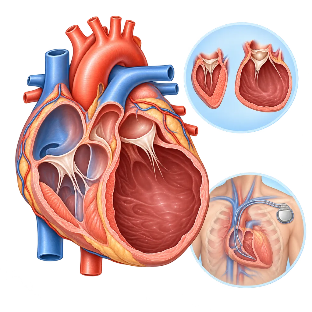
Heart failure with reduced ejection fraction is heart failure in which the pumping of the left ventricle has weakened, so only 40% or less of the blood in the left ventricle is pumped out with each beat. This type has medicine and device options proven to extend life expectancy.
SourcesSigns That Need Urgent Help
- Severe shortness of breath at rest or being unable to lie down
- Frothy white or pink phlegm
- Fainting or nearly fainting
- A very fast heartbeat with dizziness or severe weakness
- Chest pain that does not go away with rest
- Cold, pale skin and confusion
- Repeated ICD shocks, or a shock accompanied by feeling unwell
If these signs appear, go to the nearest emergency department right away or call Heart Attack Center 1500 258.
01 / Understanding the condition
About Heart Failure with Reduced Ejection Fraction
Heart failure with reduced ejection fraction, known internationally as HFrEF, is a type of heart failure that occurs because the muscle of the left ventricle has weakened and cannot contract strongly. Ejection fraction is the percentage of blood in the left ventricle that is pumped out each time the heart beats. In a healthy heart the value is 50% or more, while in this type it is 40% or less.
A weak muscle is usually followed by enlargement of the left ventricle. An enlarged heart pumps less and less efficiently, the mitral valve can start to leak, and the body holds on to salt and water. This change in shape is called remodeling, and the majority of heart failure medicines work by slowing it. The symptoms are the same as heart failure in general: shortness of breath, tiring easily, and swelling in the legs. How severe the symptoms are does not always match the ejection fraction number, so even people with mild symptoms still need full treatment.
Common Causes
The main cause is coronary artery disease, especially after a heart attack, covered on the Ischemic Cardiomyopathy page. Other causes are dilated cardiomyopathy, including inherited forms, long-standing hypertension, valve disease, excessive alcohol use, side effects of cancer treatment, myocarditis, and a heart rate that stays too fast for a long time.
Four Main Classes of Medicine
The ESC 2021 and AHA/ACC/HFSA 2022 guidelines recommend four classes of medicine for almost everyone with this type: an ACE inhibitor or ARNI, a beta blocker, a mineralocorticoid receptor antagonist (MRA), and an SGLT2 inhibitor. Each has been proven to reduce hospital stays and deaths, and the benefits add up when they are used together. Your doctor starts them and raises the doses gradually while monitoring blood pressure, heart rate, kidney function, and potassium. Diuretics are added to relieve shortness of breath and swelling.
Pumping Function Can Improve
In some patients, the ejection fraction rises above 40% after several months of treatment. This is called heart failure with improved ejection fraction. Even with improvement, medicines are generally continued, because research shows that stopping them can cause pumping function to fall again. If the ejection fraction stays at 35% or less, your doctor will discuss devices such as an ICD, which prevents sudden death from dangerous ventricular rhythms, or resynchronization therapy, which coordinates the contraction of both ventricles.
02 / Understanding the condition
Symptoms to Recognize.
Common Symptoms
- Shortness of breath when climbing stairs or walking fast
- Shortness of breath when lying down, or waking at night short of breath
- Tiring easily and wearing out quickly during activity
- Swelling in the ankles and legs
- Weight gain due to fluid buildup
- Palpitations or an irregular heartbeat
- Cold hands and feet
- Loss of appetite and feeling full quickly
03 / Understanding the condition
What Causes This Condition?
- Coronary artery disease and damage from a past heart attack
- Dilated cardiomyopathy, including forms that run in families
- Hypertension that has gone uncontrolled for a long time
- Heart valve disease, such as a leaking mitral valve or a narrowed aortic valve
- Excessive alcohol use over a long period
- Side effects of certain chemotherapy or chest radiotherapy
- Inflammation of the heart muscle (myocarditis) or peripartum cardiomyopathy
- A heart rate that stays too fast for a long time, for example in atrial fibrillation
04 / Understanding the condition
Who Is at Higher Risk?
Medical
A history of heart attack, coronary artery disease, hypertension, diabetes, and valve disease. Certain cancer treatments can also weaken the heart muscle.
Lifestyle
Smoking, excessive alcohol, and substance use such as cocaine or methamphetamine can damage the heart muscle.
Genetic
Some dilated cardiomyopathies are inherited. A family history of cardiomyopathy or sudden death at a young age should be shared with your doctor.
Age
The risk increases with age, and this type is more common in men than in women.
Groups at Higher Risk
- Heart Attack Survivors: Damaged muscle can cause the left ventricle to enlarge and the pump to weaken, so the ejection fraction needs to be assessed after a heart attack.
- Relatives of People with Dilated Cardiomyopathy: First-degree relatives are advised to have ECG and echocardiography screening, and genetic testing if needed.
- Patients Receiving Chemotherapy or Chest Radiotherapy: Some cancer treatments can affect the heart muscle; monitoring is done before, during, and after treatment.
- Heavy Drinkers: Large amounts of alcohol over many years can weaken the heart muscle; stopping drinking can improve its function.
05 / Understanding the condition
How Is It Diagnosed?
Diagnosis starts with your symptoms, a physical examination, an ECG, and an NT-proBNP or BNP test. Echocardiography confirms an ejection fraction of 40% or less, while also assessing the size of the ventricles, wall motion, and the valves. The next step is to look for the cause, because treating the cause can improve pumping function.
If coronary artery disease is suspected, your doctor will recommend coronary angiography or CT coronary angiography. Cardiac MRI helps distinguish scarring from a past heart attack from other cardiomyopathies, inflammation, or buildup of substances in the heart muscle. Blood tests assess kidney function, potassium, complete blood count, thyroid, and iron levels. The ECG is also used to measure the width of the QRS complex, which determines whether resynchronization therapy will help. Echocardiography is repeated after about three months of medication as the basis for deciding on an ICD.
Echocardiography
Confirms an ejection fraction of 40% or less and assesses ventricle size, wall motion, and the valves. Repeated after treatment is underway to assess improvement and the need for a device.
Electrocardiogram (ECG)
Looks for signs of a past heart attack and rhythm problems, and measures the width of the QRS complex, which determines the benefit of resynchronization therapy.
NT-proBNP and BNP Test
Supports the diagnosis and helps monitor the response to treatment.
Cardiac MRI
Distinguishes scarring from a past heart attack from other cardiomyopathies, inflammation, or buildup of substances in the heart muscle.
Coronary Angiography
Looks for narrowing of the coronary arteries as a cause and determines whether an artery needs to be opened.
Laboratory Tests
Kidney function, potassium, complete blood count, thyroid, and iron levels to look for causes and monitor the safety of medicines.
06 / Understanding the condition
Treatment Options.
Treatment is decided by a doctor after an assessment. The categories below can help you follow the conversation with your doctor.
Four Main Classes of Medicine
An ACE inhibitor or ARNI, a beta blocker, an MRA, and an SGLT2 inhibitor to reduce hospital stays and deaths. Your doctor raises the doses gradually; do not stop or change your medicines on your own.
Diuretics
Water pills to relieve shortness of breath and swelling. The dose is readjusted as excess fluid decreases or increases.
Additional Medicines as Needed
In certain situations, your doctor may add a medicine to slow the heart rate, digoxin, a combination of hydralazine and nitrates, or intravenous iron if you are iron deficient.
ICD and Resynchronization Therapy
An ICD is considered if the ejection fraction remains at 35% or less after about three months of full medication. CRT is helpful when the ventricles do not contract in sync, seen as a wide QRS complex.
Treating the Cause
Coronary revascularization in selected patients, valve repair including catheter-based mitral valve repair, and ablation if atrial fibrillation is making the condition worse.
Rehabilitation and Self-Care
Supervised exercise training, salt restriction, daily weighing, and regular checkups to adjust medicines.
Advanced Heart Failure Therapy
A mechanical heart pump (LVAD) or heart transplant for people who still have severe symptoms despite full treatment.
07 / Understanding the condition
Complications to Watch For.
Dangerous Ventricular Rhythms
A very low ejection fraction increases the risk of ventricular rhythms that can cause cardiac arrest. In certain patients, an ICD is implanted to prevent this.
Atrial Fibrillation and Stroke
Enlarged atria are prone to atrial fibrillation, which worsens heart failure and raises the risk of stroke.
Mitral Valve Leakage
An enlarged left ventricle pulls on the mitral valve leaflets so the valve does not close tightly and shortness of breath increases.
Impaired Kidney Function
Reduced blood flow strains the kidneys, and some medicines affect the kidneys and potassium, so routine blood tests are needed.
Acute Worsening
At any time, symptoms can worsen quickly and require hospital care, covered on the Acute Heart Failure page.
Advanced Heart Failure
If the pump keeps weakening despite full treatment, symptoms become severe and further options such as a mechanical heart pump or a transplant are needed.
08 / Understanding the condition
What Can You Do?
- Take your medicines as prescribed and come to checkups for gradual dose adjustments.
- Weigh yourself every morning; contact your doctor if you gain 2 kg or more in 3 days.
- Limit salt and avoid alcohol.
- Ask your doctor before using NSAID pain relievers, herbal remedies, or supplements.
- Join cardiac rehabilitation and stay active within your limits.
- Control your blood pressure, blood sugar, and cholesterol.
- Get influenza and pneumococcal vaccines as recommended by your doctor.
09 / Understanding the condition
When Should You See a Doctor?
See a cardiologist if shortness of breath, tiredness, or swelling appears for the first time, especially if you have had a heart attack or have family members with cardiomyopathy. If you have already been diagnosed, contact your doctor early if any of the following occur.
- You gain 2 kg or more in 3 days.
- Shortness of breath or swelling is worse than usual.
- You feel dizzy when standing up or your blood pressure is much lower than usual.
- You receive a shock from your ICD.
- Go to the emergency room right away if you have severe shortness of breath at rest, fainting, or chest pain.
Don't Wait in an Emergency.
If symptoms are severe or come on suddenly, go to the nearest emergency department right away or call Heart Attack Center 1500 258, rather than waiting for an appointment.
10 / Common questions
Questions You May Have.
Frequently asked questions about heart failure with reduced ejection fraction.
Can my ejection fraction go back up?
Yes. In some patients, the ejection fraction rises above 40% after several months of full medication, especially when the cause can be treated, for example a heart rate that is too fast, alcohol, or inflammation of the heart muscle. This is called improved ejection fraction. Treatment is still continued, because the TRED-HF study showed that pumping function can fall again if medicines are stopped.
Why do I have to take several medicines at once?
Each class of medicine works on a different pathway: some calm the hormones that hold on to salt and water, some slow the heart rate and protect the heart muscle, and some help the kidneys get rid of salt. Their benefits add up. A combined analysis of studies estimated that the four-class combination can add several years of life expectancy compared with older treatment. If you have side effects, tell your doctor instead of stopping on your own.
When do I need an ICD or CRT?
An ICD is considered if the ejection fraction remains at 35% or less after about three months of full treatment, to prevent sudden death from dangerous ventricular rhythms. CRT, a pacing device with leads to both ventricles, is helpful when the ECG shows the ventricles are not contracting in sync, seen as a wide QRS complex. The decision is made with your doctor, weighing your age, other illnesses, and your wishes.
My blood pressure dropped after taking my medicines. Is that dangerous?
Many heart failure medicines lower blood pressure, and a lower number than before is often still safe as long as you do not feel dizzy or weak. If you feel dizzy when standing up, nearly faint, or your blood pressure is much lower than usual, contact your doctor. Your doctor can readjust the timing or dose of your medicines. Do not stop your medicines on your own, because this can lead to worsening.
What is the difference between heart failure with reduced ejection fraction and dilated cardiomyopathy?
Dilated cardiomyopathy is one of the causes: a heart muscle disease that makes the left ventricle enlarged and weak without coronary or valve disease severe enough to explain it. Heart failure with reduced ejection fraction is a syndrome that can arise from many causes, including dilated cardiomyopathy and a past heart attack. That disease is covered on the Dilated Cardiomyopathy page.
Sources and Medical Review
Translated from the Indonesian edition medically reviewed by Dr. dr. Dudy A. Hanafy, Sp.BTKV, Subsp. JD(K), MARS.
Sources as cited in the Indonesian edition.
- McDonagh TA, Metra M, Adamo M, Gardner RS, Baumbach A, Böhm M, et al. 2021 ESC Guidelines for the diagnosis and treatment of acute and chronic heart failure. Eur Heart J. 2021;42(36):3599-3726.
- McDonagh TA, Metra M, Adamo M, Gardner RS, Baumbach A, Böhm M, et al. 2023 Focused Update of the 2021 ESC Guidelines for the diagnosis and treatment of acute and chronic heart failure. Eur Heart J. 2023;44(37):3627-3639.
- Heidenreich PA, Bozkurt B, Aguilar D, Allen LA, Byun JJ, Colvin MM, et al. 2022 AHA/ACC/HFSA Guideline for the Management of Heart Failure: A Report of the American College of Cardiology/American Heart Association Joint Committee on Clinical Practice Guidelines. Circulation. 2022;145(18):e895-e1032.
- McMurray JJV, Solomon SD, Inzucchi SE, Køber L, Kosiborod MN, Martinez FA, et al. Dapagliflozin in patients with heart failure and reduced ejection fraction. N Engl J Med. 2019;381(21):1995-2008.
- Vaduganathan M, Claggett BL, Jhund PS, Cunningham JW, Pedro Ferreira J, Zannad F, et al. Estimating lifetime benefits of comprehensive disease-modifying pharmacological therapies in patients with heart failure with reduced ejection fraction: a comparative analysis of three randomised controlled trials. Lancet. 2020;396(10244):121-128.
- Murphy SP, Ibrahim NE, Januzzi JL Jr. Heart failure with reduced ejection fraction: a review. JAMA. 2020;324(5):488-504.
- Halliday BP, Wassall R, Lota AS, Khalique Z, Gregson J, Newsome S, et al. Withdrawal of pharmacological treatment for heart failure in patients with recovered dilated cardiomyopathy (TRED-HF): an open-label, pilot, randomised trial. Lancet. 2019;393(10166):61-73.
Doctors Who Treat This Condition

dr. Siska S. Danny, Sp.JP(K)
Interventional Cardiology
Coronary Intervention Procedures
Clinical focusPCI for Acute Coronary Syndrome · Primary Coronary Intervention
View Profile
dr. Sunu B. Raharjo, Sp.JP(K), Ph.D.
Arrhythmia and Electrophysiology
Arrhythmia and Device Center
Clinical focusClinical and Interventional Electrophysiology · Cardiac Arrhythmia Ablation
View Profile
Dr. dr. Dudy A. Hanafy, Sp.BTKV, Subsp. JD(K), MARS
Adult Cardiac Surgery
Advanced Cardiovascular Surgical Care +1 center
Clinical focusMinimally Invasive Cardiac Surgery · Coronary Artery Bypass Surgery
View Profile