
Quick answer
What Is Mitral Valve Repair?
Mitral valve repair is a procedure to make a leaking mitral valve close tightly again while keeping the original valve. The repair can be done through heart surgery, usually 3 to 5 hours, or through a catheter from a vein in the groin using a clip, about 1 to 3 hours. The heart team chooses the suitable approach.
Sources01 / Understanding the procedure
About Mitral Valve Repair
What Is Mitral Valve Repair
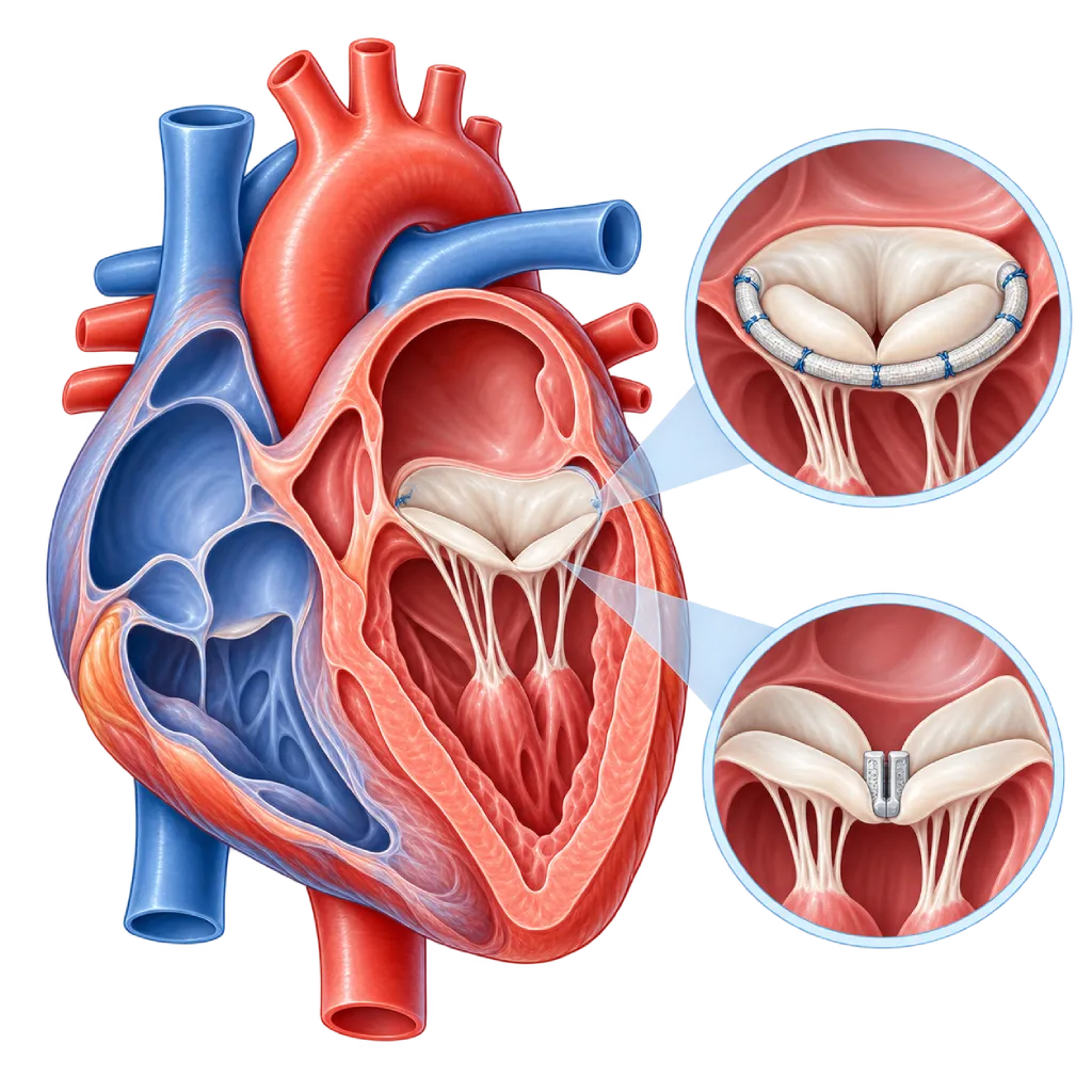
The mitral valve sits between the left atrium and the left ventricle of the heart. It has two leaflets held by fine cords (chordae) so that it closes tightly each time the heart pumps. If the valve does not close tightly, some blood flows back into the left atrium and the lungs. This condition is called mitral regurgitation or a leaking mitral valve, and it can cause shortness of breath, tiring easily, and an irregular heart rhythm.
Mitral valve repair is a procedure to correct this leak while keeping the original valve, rather than replacing it with an artificial valve.
Two Types of Leakage
In primary regurgitation, the valve itself is damaged, for example the leaflets are too floppy and bulge backward (prolapse) or the supporting cords have ruptured. In secondary regurgitation, the valve is relatively normal, but the left ventricle or left atrium is enlarged, for example in heart failure, so the two leaflets are pulled apart and cannot meet. The type of leakage helps determine how it is treated.
Repair Through Surgery
For severe primary regurgitation, guidelines recommend surgical repair when the result is expected to last a long time. The surgeon may trim the excess part of the leaflets, place artificial supporting cords, and sew a ring around the valve to support its circumference. Compared with replacement, repair preserves your own valve tissue and usually does not require lifelong blood thinners. In certain patients, the operation can be done through a small incision on the side of the chest, covered on the Minimally Invasive Heart Surgery page.
Repair Through a Catheter
In transcatheter edge-to-edge repair (TEER), a catheter is inserted from a vein in the groin, passed through the wall between the right and left atria, and then a small clip grasps the two leaflets at the point of the leak. The valve then works as if it has two small openings that close more tightly.
This approach is considered for patients with primary regurgitation who are at high risk for surgery, and for some patients with secondary regurgitation who still have symptoms despite heart failure medications given according to guidelines.
The Role of the Heart Team
The choice between surgery, catheter repair, or valve replacement is made by the heart team together with you. The team weighs the type and severity of the leak, the shape of the valve on transesophageal echocardiography, your age, other illnesses, and surgical risk. Valve replacement, covered on the Heart Valve Surgery page, is chosen when the valve cannot be repaired well, for example in severe rheumatic damage.
02 / Understanding the procedure
When Is This Procedure Considered?
A doctor may consider this procedure in the situations below. The decision is always made after a thorough assessment.
- Severe Mitral Regurgitation with SymptomsShortness of breath, tiring easily, or swelling caused by blood flowing backward through the mitral valve.
- Left Ventricle Starting to Enlarge or WeakenReduced pumping function or a widening left ventricle due to a severe leak, even before symptoms are felt.
- Prolapse or Ruptured Valve CordsA leaflet bulges backward or its supporting cords rupture, causing a severe leak.
- New Atrial Fibrillation or Pulmonary HypertensionAn irregular rhythm or increased blood pressure in the lungs caused by a severe leak.
- Secondary Regurgitation in Heart FailureSymptoms persist despite guideline-based heart failure medications; catheter repair may be considered.
Situations to Consider First
- Valve Cannot Be Repaired WellFor example, severe calcification or extensive rheumatic damage; valve replacement is more suitable.
- Active Valve InfectionActive endocarditis is treated with antibiotics and, if needed, surgery, not with a clip.
- Mitral Valve Already NarrowedA clip can make the valve opening smaller, so it is not suitable if the valve is already narrow.
- Blood Clot in the HeartA clot in the left atrium or left ventricle needs to be treated first because it can break loose during the procedure.
- Very Advanced Heart FailureWhen the heart's pumping is very weak, repairing the valve alone is unlikely to help; other options are discussed.
03 / Before the procedure
How Should You Prepare?
Mitral valve repair is usually a planned procedure. The heart team explains whether you will have surgery or catheter repair, and your preparation is adjusted accordingly.
Transesophageal echocardiography. A small probe is passed through the mouth into the esophagus to clearly see the shape of the valve leaflets and the point of the leak. This test helps determine whether the valve can be repaired and how.
Other tests. Blood tests, an ECG, a chest X-ray, and examination of the coronary arteries with coronary angiography or CT, especially at certain ages or when there are risk factors for coronary artery disease.
Teeth and sources of infection. Infections of the teeth, gums, or skin should be treated first because germs can attach to the ring or clip.
Medications. Bring a complete list of your medications. Blood thinners may need to be stopped or switched several days beforehand, while heart failure medications are usually continued. Follow your doctor's instructions.
Fasting and consent. Follow the fasting instructions from the anesthesia team. Before you sign the consent form, your doctor explains the benefits, the risks, and the possibility that the valve may need to be replaced if the repair is not adequate.
Unsure about the preparation instructions? Ask our team before the procedure.
04 / During the procedure
How Is the Procedure Done?
Surgical repair is performed by the cardiac surgery team in the operating room under general anesthesia with the help of a heart-lung machine, usually taking 3 to 5 hours. Catheter repair is performed in the cardiac catheterization lab, usually under general anesthesia, and takes about 1 to 3 hours.
Monitoring with TEE
In both approaches, a transesophageal echocardiography (TEE) probe is placed after you are asleep. Images from this probe guide the procedure and assess the result of the repair.
Surgery: Repairing the Leaflets and Cords
After the chest is opened and the heart is stopped temporarily with the help of the heart-lung machine, the surgeon opens the left atrium, trims the valve leaflets or places artificial supporting cords, and then sews a ring around the valve.
Catheter: Placing the Clip
The catheter is inserted through a vein in the groin and passed through the wall between the two atria. The clip is guided to the point of the leak, grasps both leaflets, and is then released from the catheter. If needed, more than one clip is placed.
Assessing the Result
Before the procedure ends, any remaining leak and the valve opening are assessed with TEE. If the surgical result is not adequate, the surgeon can repeat the repair or replace the valve during the same operation.
05 / Benefits and risks
Benefits and Risks Should Be Discussed Together.
Every medical procedure has benefits and risks. Your doctor will explain both for your situation before the procedure.
Benefits You Can Expect
- Your Own Valve Is PreservedYour own valve tissue stays in use, so lifelong blood thinners are usually not needed.
- Symptoms Can ImproveShortness of breath and tiring easily can ease once the leak becomes smaller.
- Protecting Heart Pumping FunctionRepair at the right time helps prevent the left ventricle from enlarging and weakening further.
- An Option Without Opening the ChestFor patients at high risk for surgery, the leak can be reduced through a catheter.
The benefits of a procedure are not a promise of a cure. Ask how the results will affect your care plan.
Risks You Should Know
- Remaining or Recurrent LeakThe valve may leak again over time, so some patients need a repeat procedure.
- BleedingAt the surgical wound or the puncture site in the groin; some cases need a transfusion or an additional procedure.
- Heart Rhythm DisordersMainly atrial fibrillation after surgery, which can usually be treated with medication.
- StrokeClots or air can travel to the brain during the procedure; the team takes preventive measures.
- Problems with the Clip or Valve OpeningThe clip can detach from one leaflet, or the valve opening can become too narrow; both are rare.
- Infection and Other ComplicationsWound or valve infection, kidney problems, and, in a small number of patients, death.
Risks can differ from person to person. A full explanation and consent are given after an individual assessment.
06 / After the procedure
What Is Recovery Like?
The length of recovery depends on the repair method and the condition of your heart before the procedure.
After surgery. You usually stay 1 to 2 days in the ICU, with a total hospital stay of about 5 to 7 days. The breastbone needs about 6 to 8 weeks to heal, so avoid lifting heavy objects during that time.
After catheter repair. Many patients are walking the next day and go home within 1 to 3 days. Avoid strenuous activity for about one week.
Medications. A blood thinner or antiplatelet medication may be given in the first months as advised by your doctor. Heart failure medications are continued, especially for secondary regurgitation.
Follow-up visits and exercise. Follow-up echocardiograms are done periodically to assess any remaining leak. A cardiac rehabilitation program helps you regain your fitness safely.
Protecting the valve from infection. Rings and clips are made of man-made materials. Keep your teeth clean and tell your dentist about the procedure you have had.
Warning signs. Go to the emergency room right away if you have:
- Shortness of breath that returns or gets worse, especially when lying down
- Chest pain or an irregular, pounding heartbeat
- Facial drooping, weakness on one side of the body, or slurred speech
- Fever, or a surgical wound that is red, swollen, or oozing pus
- Bleeding or swelling that grows larger in the groin
Know the Signs That Need Prompt Care.
Follow the team's guidance on warning signs. If severe or sudden symptoms appear after the procedure, go to the nearest emergency department right away or call Heart Attack Center 1500 258.
07 / Team and support
Doctors and Procedure Support
The doctors below are listed for this procedure. Listed by name, not by ranking.

Dr. dr. Dafsah A. Juzar, Sp.JP(K)
Interventional Cardiology
Coronary Intervention Procedures +1 center
Clinical focusComplex Coronary Intervention · Cardiac Catheterization
View Profile
Dr. dr. Dudy A. Hanafy, Sp.BTKV, Subsp. JD(K), MARS
Adult Cardiac Surgery
Advanced Cardiovascular Surgical Care +1 center
Clinical focusMinimally Invasive Cardiac Surgery · Coronary Artery Bypass Surgery
View Profile
dr. Sri Diniharini, Sp.JP(K)
Echocardiography & Cardiovascular Imaging
Cardiovascular Diagnostic Center +1 center
Clinical focusAdvanced Echocardiography · Transesophageal Echocardiography
View ProfileOperating Theatre (Cardiac Operating Room)
An operating room dedicated to heart and blood vessel surgery, where the surgical, anesthesia, perfusion, and nursing teams work…
Minimally Invasive Cardiac Surgery (MICS) Technology
A way of performing heart surgery through a small incision between the ribs or through part of the breastbone, using long-shafted…
Live Surgery Facility
Cameras and an audio-video link in the operating room that allow an operation to be broadcast for education and discussion among…
4D Echocardiography
An ultrasound scan of the heart that records three-dimensional images in real time to assess pumping function, the valves, and…
The availability of technology and test support is confirmed with the team. Technology is chosen according to clinical need.
08 / Common questions
Before You Plan the Procedure.
Questions patients and families often ask about mitral valve repair.
What is the difference between mitral valve repair and replacement?
In repair, your own valve is kept and reshaped so that it closes tightly. In replacement, the valve is removed and replaced with a mechanical or biological valve. When the valve can be repaired well, repair is usually preferred because left ventricular function is better preserved and lifelong blood thinners are usually not needed. Replacement is covered on the Heart Valve Surgery page.
Who is suitable for catheter repair?
Catheter repair is considered for patients with primary regurgitation who are at too high a risk for surgery, for example because of older age or other illnesses. It may also be considered for some heart failure patients with secondary regurgitation who still have symptoms despite guideline-based medications. The shape of the valve on transesophageal echocardiography also helps determine suitability.
Can the results of the repair last a long time?
In primary regurgitation repaired through surgery, many patients do not need repeat surgery for many years, and long-term studies have recorded results lasting up to two decades in many patients. Catheter repair usually leaves a small remaining leak that needs to be monitored. That is why follow-up echocardiograms are still done periodically.
Is the procedure painful?
Both approaches are done under general anesthesia, so you do not feel anything during the procedure. After surgery, pain in the chest wound is normal for a few days to weeks and is controlled with medication. After catheter repair, there is usually only some soreness in the groin and mild throat discomfort from the TEE probe for one to two days.
Will I need to take blood thinners?
After repair, a blood thinner or antiplatelet medication is often given in the first months until the ring or clip is covered by tissue. After that, many patients no longer need it, unless there is another reason such as atrial fibrillation. The type and duration are decided by your doctor based on your condition and bleeding risk. Do not stop any medication without consulting your doctor.
When can I return to my usual activities?
After catheter repair, many patients return to light activities within a few days and avoid strenuous activity for about one week. After surgery through the breastbone, driving and heavy lifting are postponed for about 6 to 8 weeks, and full recovery may take several months. Your doctor decides the timing at your follow-up visit.
Sources and Medical Review
Translated from the Indonesian edition medically reviewed by Dr. dr. Dafsah A. Juzar, Sp.JP(K).
Sources as cited in the Indonesian edition.
- Praz F, Borger MA, Lanz J, et al. 2025 ESC/EACTS Guidelines for the management of valvular heart disease. Eur Heart J. 2025;46(44):4635-4736.
- Vahanian A, Beyersdorf F, Praz F, et al. 2021 ESC/EACTS Guidelines for the management of valvular heart disease. Eur Heart J. 2022;43(7):561-632.
- Otto CM, Nishimura RA, Bonow RO, et al. 2020 ACC/AHA Guideline for the Management of Patients With Valvular Heart Disease: A Report of the American College of Cardiology/American Heart Association Joint Committee on Clinical Practice Guidelines. Circulation. 2021;143(5):e72-e227.
- Lazam S, Vanoverschelde JL, Tribouilloy C, et al. Twenty-year outcome after mitral repair versus replacement for severe degenerative mitral regurgitation. Circulation. 2017;135(5):410-422.
- Feldman T, Foster E, Glower DD, et al. Percutaneous repair or surgery for mitral regurgitation. N Engl J Med. 2011;364(15):1395-1406.
- Stone GW, Lindenfeld J, Abraham WT, et al. Transcatheter mitral-valve repair in patients with heart failure. N Engl J Med. 2018;379(24):2307-2318.
- Obadia JF, Messika-Zeitoun D, Leurent G, et al. Percutaneous repair or medical treatment for secondary mitral regurgitation. N Engl J Med. 2018;379(24):2297-2306.