
Quick answer
What Is Pulmonary Stenosis?
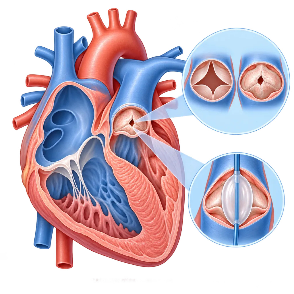
Pulmonary stenosis is a narrowing of the pulmonary valve, the outlet that carries blood from the heart's right ventricle to the lungs. It is usually present from birth. Mild forms often cause no symptoms, while severe forms need to be widened, usually with a balloon through a catheter.
SourcesSigns That Need Urgent Help
- A baby who looks increasingly blue, limp, or hard to wake
- Fainting during or after activity
- Severe chest pain during activity
- Severe shortness of breath at rest
- Swelling in the legs or a swollen belly from fluid
If these signs appear, go to the nearest emergency department right away or call Heart Attack Center 1500 258.
01 / Understanding the condition
About Pulmonary Stenosis
The pulmonary valve sits between the right ventricle of the heart and the pulmonary artery, the vessel that carries blood to the lungs to pick up oxygen. In pulmonary stenosis, this valve is narrowed because its leaflets are thickened, fused at the edges, or abnormally shaped. The right ventricle has to pump harder, so its muscle thickens. Pulmonary stenosis is a type of valve narrowing covered on the Heart Valve Stenosis page.
A Condition Present from Birth
The majority of pulmonary stenosis cases are congenital heart disease, meaning they form while the baby develops in the womb. It is a fairly common type of congenital heart disease, and it can occur on its own or as part of another defect such as tetralogy of Fallot. Some cases are linked to genetic syndromes, such as Noonan syndrome. In adults, acquired narrowing is rare, for example due to carcinoid syndrome. An overview of congenital heart defects is covered on the Congenital Heart Disease page.
From Mild to Critical
The severity of the narrowing is judged by the pressure difference across the valve on echocardiography. Mild stenosis usually does not get worse, causes no symptoms, and only needs monitoring. Moderate stenosis can get worse as a child grows, while severe stenosis exhausts the right ventricle and causes shortness of breath or tiring easily. Newborns can have a critical form: the valve is almost closed, so blood flow to the lungs depends on a fetal blood vessel called the ductus arteriosus. The baby looks bluish and needs immediate treatment.
Usually Treated Through a Catheter
If the narrowing is moderate to severe or causes symptoms, the valve is usually widened with a balloon through a catheter inserted into a blood vessel in the groin (pulmonary balloon valvuloplasty). The results usually last a long time. Surgery is chosen if the valve is very thick and stiff, there is narrowing below or above the valve, or there is another heart defect that needs to be repaired at the same time. After the procedure, the valve sometimes becomes leaky, so heart checkups are still needed into adulthood.
The good news is that the majority of people with pulmonary stenosis, whether it is mild or has been successfully widened, can grow up, go to school, work, and exercise like everyone else. The key is regular checkups so that changes in the valve or right ventricle are detected early.
02 / Understanding the condition
Symptoms to Recognize.
Common Symptoms
These symptoms are more common in teenagers and adults with moderate to severe stenosis.
- Often no symptoms, with the condition found from a heart murmur during an examination
- Shortness of breath during activity
- Tiring easily and reduced exercise capacity
- Chest pain during activity in severe narrowing
- Dizziness or nearly fainting during strenuous activity
- Palpitations
Symptoms in Newborns
The critical form in newborns needs prompt treatment by a pediatric heart team.
- Bluish skin, lips, or nails from the first days of life
- Fast breathing or visible difficulty breathing
- Poor feeding or tiring quickly while feeding
- Low oxygen levels on newborn pulse oximetry screening
Symptoms in Children
- A heart murmur heard during a routine checkup
- Tiring more quickly than peers during play
- Shortness of breath when running or exercising
- Dizziness or fainting during strenuous activity in severe narrowing
03 / Understanding the condition
What Causes This Condition?
- Abnormal formation of the pulmonary valve while the baby develops in the womb
- Part of another congenital heart defect, such as tetralogy of Fallot
- Certain genetic syndromes, such as Noonan syndrome, which often come with a thick, stiff valve
- Rubella infection during pregnancy, which is linked to narrowing of the branches of the pulmonary artery in the baby
- Narrowing of an artificial conduit or valve after congenital heart surgery
- Rare acquired causes in adults, such as carcinoid syndrome
04 / Understanding the condition
Who Is at Higher Risk?
Genetic
A family history of congenital heart disease and genetic syndromes such as Noonan syndrome increase the chance of a baby being born with pulmonary stenosis.
Other
Rubella infection during pregnancy, uncontrolled diabetes during pregnancy, and some medicines taken by pregnant women are linked to congenital heart disease in babies.
Medical
In adults, a history of congenital heart surgery with an artificial conduit or valve in the right ventricular outflow tract, as well as carcinoid syndrome, can cause narrowing.
Groups at Higher Risk
- Babies with a Family History of Congenital Heart Disease: If a parent or sibling has congenital heart disease, the chance of a baby having a similar defect is slightly higher.
- Children with Noonan Syndrome: Pulmonary stenosis is a heart defect that often occurs with this syndrome, often with a thick, stiff valve.
- Congenital Heart Surgery Survivors: People who have had surgery such as tetralogy of Fallot repair can develop narrowing or leakage in the right ventricular outflow tract and need lifelong checkups.
- Women Planning a Pregnancy: Mild to moderate stenosis is usually safe during pregnancy, but severe stenosis should be treated before pregnancy.
05 / Understanding the condition
How Is It Diagnosed?
Pulmonary stenosis is often first suspected from a heart murmur that a doctor hears during a child's routine checkup, or from a low oxygen level on newborn screening. Echocardiography confirms the diagnosis. This test shows the shape of the valve, where the narrowing is (at the valve, below it, or above it), the pressure difference across the valve, and the thickness of the right ventricle.
An ECG helps assess the strain on the right ventricle. In teenagers and adults, or when echocardiography images are unclear, cardiac MRI or CT scans are used to look at the valve, the pulmonary artery and its branches, and the size of the right ventricle. Cardiac catheterization measures pressures directly and is usually done at the same time as widening the valve with a balloon.
Physical Examination
The doctor listens for a characteristic heart murmur in the upper left chest between the ribs, and in babies checks skin color and oxygen level.
Echocardiography
The main test to see the shape of the valve, where the narrowing is, the pressure difference, and the thickness of the right ventricle; also used for regular monitoring.
Electrocardiogram (ECG)
Looks for signs of thickened right ventricular muscle caused by pressure overload.
Cardiac MRI
Assesses the valve, the branches of the pulmonary artery, and the size and function of the right ventricle without X-rays, especially in teenagers and adults.
Cardiac Catheterization
Measures pressures directly in the right ventricle and pulmonary artery, usually at the same time as balloon widening of the valve.
Newborn pulse oximetry screening: measuring oxygen levels with a sensor on the baby's hand and foot, usually after 24 hours of age, to detect critical congenital heart disease.
06 / Understanding the condition
Treatment Options.
Treatment is decided by a doctor after an assessment. The categories below can help you follow the conversation with your doctor.
Regular Monitoring
Mild stenosis usually only needs regular echocardiography on the schedule your doctor sets, because it rarely gets worse.
Pulmonary Balloon Valvuloplasty
A balloon is passed through a catheter from a blood vessel in the groin and inflated in the valve to widen it. It is the main option for moderate to severe valve stenosis.
Urgent Treatment for Babies in Critical Condition
Prostaglandin medicine given by IV infusion keeps the ductus arteriosus open until the valve can be widened with a balloon or repaired with surgery.
Congenital Heart Surgery
Chosen when the valve is very thick and stiff, the narrowing is below or above the valve, or there is another heart defect that needs to be repaired at the same time.
Pulmonary Valve Replacement
If the valve leaks badly or narrows again after a previous procedure, it can be replaced through surgery or through a catheter.
07 / Understanding the condition
Complications to Watch For.
Right-Sided Heart Failure
A right ventricle that pumps against a narrow valve for a long time thickens and may then weaken, causing leg swelling, a swollen belly, and tiring easily.
Cyanosis in Newborns
In the critical form, very little blood flows to the lungs, so the body's oxygen level is low and the baby looks blue.
Valve Leakage After a Procedure
After balloon widening or surgery, the valve often becomes leaky. A severe leak can enlarge the right ventricle. Covered on the Pulmonary Regurgitation page.
Heart Rhythm Problems
Enlargement of the right heart chambers can trigger rhythm problems, especially in adults with severe stenosis that has gone untreated for a long time.
Infective Endocarditis
Valve infection is rare, but the risk increases after an artificial valve or conduit is placed. Covered on the Infective Endocarditis page.
08 / Understanding the condition
What Can You Do?
- Make sure your rubella vaccination is complete before planning a pregnancy.
- Control your blood sugar before and during pregnancy if you have diabetes.
- Check every medicine you take with your doctor before and during pregnancy.
- Tell your obstetrician if your family has a history of congenital heart disease.
- Make sure your baby has pulse oximetry screening before leaving the place of birth.
- Go to heart checkups on schedule after valve widening or surgery, including in adulthood.
09 / Understanding the condition
When Should You See a Doctor?
Take your child to a cardiologist if a doctor hears a heart murmur during a routine checkup, or if your child seems to tire easily and get short of breath compared with peers. Adults who have had valve widening or congenital heart surgery still need regular checkups even if they feel well.
- A baby who looks bluish, breathes fast, or feeds poorly: go to the emergency room right away
- A newly found heart murmur in a child or adult
- Shortness of breath, tiring easily, or chest pain during activity
- Dizziness or fainting during exercise
- Planning a pregnancy as a woman with pulmonary stenosis
Don't Wait in an Emergency.
If symptoms are severe or come on suddenly, go to the nearest emergency department right away or call Heart Attack Center 1500 258, rather than waiting for an appointment.
10 / Common questions
Questions You May Have.
Frequently asked questions about pulmonary stenosis.
Does mild pulmonary stenosis need treatment?
Usually not. Mild pulmonary stenosis rarely gets worse, does not put significant strain on the heart, and does not limit daily activities. Your doctor usually only schedules regular checkups and echocardiography to make sure the narrowing stays mild. In children, monitoring is done more regularly because moderate stenosis can get worse as they grow. Treatment is only considered if the narrowing becomes moderate to severe or symptoms appear.
Does my child need major surgery?
In the majority of children, pulmonary valve stenosis is treated with balloon valvuloplasty through a catheter from a blood vessel in the groin, without opening the chest. The hospital stay is usually short, and the results usually last a long time. Surgery is considered if the valve is very thick and stiff, the narrowing is below or above the valve, or there is another heart defect. The pediatric heart team will explain the suitable options.
Can pulmonary stenosis come back after balloon treatment?
Narrowing again after balloon valvuloplasty is uncommon but can happen, especially with a thick, stiff valve. More common is the valve becoming leaky after it is widened. A mild leak usually causes no problems, but a severe leak can enlarge the right ventricle over the long term. That is why heart checkups are still needed into adulthood, even if your child seems healthy.
Can a child with pulmonary stenosis exercise?
Children with mild stenosis can usually be active and exercise like their peers. With moderate to severe stenosis, the doctor usually assesses the child first, and may even treat the narrowing, before the child takes part in strenuous or competitive sports. After successful valve widening, the majority of children can return to being active. Stop the activity and have your child checked if they feel dizzy, faint, or very short of breath during exercise.
Is pulmonary stenosis passed on to children?
The chance of a child being born with congenital heart disease is slightly higher if a parent has a congenital heart defect, but the majority of children are born with a normal heart. If pulmonary stenosis is part of a genetic syndrome such as Noonan syndrome, the chance of passing it on is higher. Genetic counseling and fetal echocardiography during pregnancy can be discussed with your doctor.
Can women with pulmonary stenosis get pregnant?
Women with mild to moderate pulmonary stenosis can usually go through pregnancy well. Severe stenosis should be assessed and, if needed, widened before pregnancy, because pregnancy increases the workload on the right ventricle. During pregnancy, heart checkups are done together with the obstetrician. Fetal echocardiography may also be considered to look at the baby's heart while it is still in the womb.
Sources and Medical Review
Translated from the Indonesian edition medically reviewed by dr. Sri Diniharini, Sp.JP(K).
Sources as cited in the Indonesian edition.
- Baumgartner H, De Backer J, Babu-Narayan SV, Budts W, Chessa M, Diller GP, et al. 2020 ESC Guidelines for the management of adult congenital heart disease. Eur Heart J. 2021;42(6):563-645.
- Stout KK, Daniels CJ, Aboulhosn JA, Bozkurt B, Broberg CS, Colman JM, et al. 2018 AHA/ACC Guideline for the Management of Adults With Congenital Heart Disease: A Report of the American College of Cardiology/American Heart Association Task Force on Clinical Practice Guidelines. Circulation. 2019;139(14):e698-e800.
- Ruckdeschel E, Kim YY. Pulmonary valve stenosis in the adult patient: pathophysiology, diagnosis and management. Heart. 2019;105(5):414-422.
- Cuypers JA, Witsenburg M, van der Linde D, Roos-Hesselink JW. Pulmonary stenosis: update on diagnosis and therapeutic options. Heart. 2013;99(5):339-347.
- Feltes TF, Bacha E, Beekman RH 3rd, Cheatham JP, Feinstein JA, Gomes AS, et al. Indications for Cardiac Catheterization and Intervention in Pediatric Cardiac Disease: A Scientific Statement From the American Heart Association. Circulation. 2011;123(22):2607-2652.
- Martin GR, Ewer AK, Gaviglio A, Hom LA, Saarinen A, Sontag M, et al. Updated Strategies for Pulse Oximetry Screening for Critical Congenital Heart Disease. Pediatrics. 2020;146(1):e20191650.
- Regitz-Zagrosek V, Roos-Hesselink JW, Bauersachs J, Blomström-Lundqvist C, Cífková R, De Bonis M, et al. 2018 ESC Guidelines for the management of cardiovascular diseases during pregnancy. Eur Heart J. 2018;39(34):3165-3241.
Doctors Who Treat This Condition

dr. Radityo Prakoso, Sp.JP(K)
Pediatrics & Congenital Heart Disease
Congenital and Structural Heart Center
Clinical focusPediatric Cardiology Consultation · Congenital Heart Disease Care
View Profile
dr. Salomo Purba, Sp.BTKV, Subsp.JPK(K)
Pediatric and Congenital Heart Surgery
Congenital and Structural Heart Center +2 centers
Clinical focusPediatric Heart Surgery · Congenital Heart Defect Surgery
View Profile
dr. Sri Diniharini, Sp.JP(K)
Echocardiography & Cardiovascular Imaging
Cardiovascular Diagnostic Center +1 center
Clinical focusAdvanced Echocardiography · Transesophageal Echocardiography
View Profile