
Quick answer
What Is Transcatheter Pulmonary Valve Replacement?
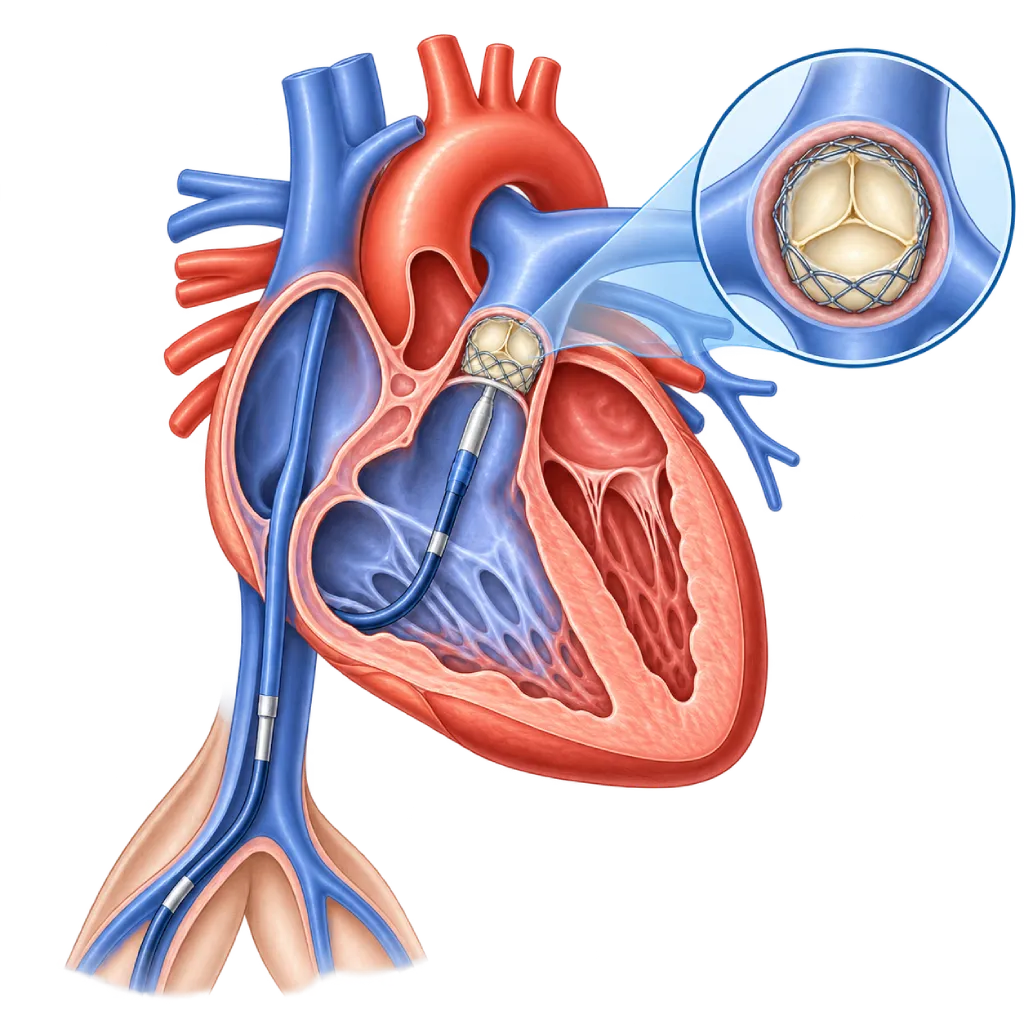
Transcatheter pulmonary valve replacement places an artificial valve in the outflow path from the right ventricle to the pulmonary artery through a catheter, without opening the chest. It is mainly for people with repaired congenital heart disease, such as tetralogy of Fallot, whose valve or artificial conduit is leaking or narrowed. It takes about 2 to 4 hours.
Sources01 / Understanding the procedure
About Transcatheter Pulmonary Valve Replacement
What Is Transcatheter Pulmonary Valve Replacement
The pulmonary valve controls blood flow from the right ventricle of the heart to the pulmonary artery, which carries blood to the lungs. In some congenital heart diseases, this valve or the right ventricular outflow path was repaired with surgery in childhood, for example with a patch or an artificial tube with a valve (conduit). Over time, the repaired valve or the valve inside the conduit can leak, narrow, or both.
Transcatheter pulmonary valve replacement is a way to place a new valve in that position through a catheter, without open-heart surgery.
Who Usually Needs It
The majority of patients are older children, teenagers, and adults who previously had surgery for tetralogy of Fallot, truncus arteriosus, a Ross operation, or other congenital heart surgery with an artificial conduit to the pulmonary artery. Severe, long-standing pulmonary valve leakage makes the right ventricle enlarge and weaken, while narrowing makes the right ventricle work too hard. Both can cause tiring easily, shortness of breath during activity, or heart rhythm disorders, sometimes without being noticed.
How It Works in Brief
An artificial valve made from animal tissue mounted on a metal frame is folded at the tip of a catheter. The catheter is guided from a vein in the groin or neck, through the right atrium and right ventricle, and the valve is then expanded inside the conduit, an old biological valve, or a right ventricular outflow path of suitable size. Often the old conduit is first lined with a stent so the new valve has a firm place to rest.
Tests Before the Procedure
Echocardiography assesses the function of the valve and the right ventricle. Cardiac MRI can measure the size and function of the right ventricle and the severity of the leak, while a cardiac CT scan assesses the size of the conduit and its distance from the coronary arteries. An exercise test is sometimes used to assess your ability to be active. The results help the team decide the timing and method of replacement, before the right ventricle becomes too weak.
Surgery or a Catheter Procedure
The pulmonary valve can also be replaced through surgery, covered on the Congenital Heart Surgery page. Catheter replacement is considered when the size and shape of the right ventricular outflow path are suitable. If the path is too wide or irregular, there is another abnormality that needs surgery, or the valve risks compressing a coronary artery, surgery may be more suitable. The decision is made by the congenital heart team together with the patient and family.
02 / Understanding the procedure
When Is This Procedure Considered?
A doctor may consider this procedure in the situations below. The decision is always made after a thorough assessment.
- Narrowed or Leaking Artificial ConduitA valved conduit from the right ventricle to the pulmonary artery whose function has declined after many years.
- Pulmonary Valve Leakage After Tetralogy of Fallot SurgerySevere leakage that enlarges the right ventricle or causes symptoms.
- Worn-Out Biological Pulmonary ValveAn artificial pulmonary valve from previous surgery that is now narrowed or leaking.
- Enlarged or Weakened Right VentricleSeen on cardiac MRI or echocardiography even before symptoms are clear.
- Symptoms During Activity or Rhythm ProblemsTiring easily, shortness of breath, or an irregular heart rhythm related to the pulmonary valve.
Situations to Consider First
- Outflow Path Too Wide or IrregularIf the valve cannot rest firmly in place, surgical replacement may be more suitable.
- Risk of Compressing a Coronary ArteryIf a balloon test shows that a coronary artery is compressed, the valve is not placed through a catheter.
- Active InfectionEndocarditis or other infections need to be treated first before a new valve is placed.
- Blocked VeinsBlockage of the veins in the groin and neck can make the catheter route impossible.
- Need for Other Heart SurgeryFor example, closing a residual hole or repairing the tricuspid valve, making a combined operation more suitable.
03 / Before the procedure
How Should You Prepare?
This procedure is usually planned and preceded by an evaluation by the congenital heart team.
Imaging. Echocardiography, cardiac MRI or a cardiac CT scan, and if needed an exercise test and heart rhythm recording.
Surgical history. Bring records of previous surgeries and procedures, including the type and size of any conduit or valve that was placed, if available.
Teeth and sources of infection. Have your teeth checked and treat any source of infection first, because infection of the artificial valve is one of the problems that must be prevented after this procedure.
Medications and fasting. Bring a list of your medications. Blood thinners may need to be adjusted according to the team's instructions. Follow the fasting instructions before general anesthesia.
Children and teenagers. A parent or guardian accompanies the child and signs the consent form. Explain to your child in simple words what will happen.
Possible pregnancy. Women of childbearing age should tell the doctor if they might be pregnant, because the procedure uses X-rays.
Unsure about the preparation instructions? Ask our team before the procedure.
04 / During the procedure
How Is the Procedure Done?
The procedure is performed by the congenital heart team in the cardiac catheterization lab, usually under general anesthesia, and takes about 2 to 4 hours.
Measuring Pressures and the Conduit
A catheter is inserted through a vein in the groin or neck. The doctor measures the pressures in the right side of the heart and images the conduit with contrast dye to determine the valve size.
Testing the Coronary Arteries
A balloon is inflated inside the conduit while the coronary arteries are imaged with contrast. This step makes sure the new valve will not compress a coronary artery.
Preparing the Conduit with a Stent
If needed, the conduit is widened with a balloon and then lined with a stent. The stent gives the valve a place to rest and reduces the risk of the valve frame breaking later on.
Placing the New Valve
The valve is expanded inside the conduit or stent. Its function is assessed with pressure measurements and angiography, then the catheter is removed and the puncture is closed.
05 / Benefits and risks
Benefits and Risks Should Be Discussed Together.
Every medical procedure has benefits and risks. Your doctor will explain both for your situation before the procedure.
Benefits You Can Expect
- Fewer Repeat OperationsHelps delay or reduce the number of open-heart operations needed over a lifetime.
- Short RecoveryUsually a one-night hospital stay, with a return to school or work within a few days.
- Less Strain on the Right VentricleTreating the leak or narrowing helps the right ventricle work with less effort.
- Exercise Capacity Can ImproveSome patients feel fitter after the narrowing of the conduit is treated.
The benefits of a procedure are not a promise of a cure. Ask how the results will affect your care plan.
Risks You Should Know
- Valve Infection (Endocarditis)It can occur years later; good dental hygiene and getting checked when you have a fever are very important.
- Compression of a Coronary ArteryRare but dangerous; this is why a balloon test is done before the valve is placed.
- Tear in the Artificial ConduitThe old conduit can tear when it is widened; this can often be sealed with a covered stent.
- Valve Frame FractureThe metal frame can crack over time, causing the valve to narrow again; a stent placed beforehand reduces this risk.
- Rhythm Problems and Puncture Site IssuesA temporary irregular rhythm, bleeding, or bruising in the groin or neck.
- Valve Wear and Repeat ProceduresThe valve can wear out over time and may eventually need further treatment, often through a catheter.
Risks can differ from person to person. A full explanation and consent are given after an individual assessment.
06 / After the procedure
What Is Recovery Like?
Patients usually stay in the hospital for one night, sometimes longer if needed.
Puncture site. Lie down as advised for the first few hours. A small bruise in the groin or neck is normal.
Activity. Light activities and returning to school or work usually happen within a few days. Avoid strenuous exercise and heavy lifting for about one week or as advised by your doctor.
Medications. An antiplatelet medication, such as aspirin, is usually given for several months or longer as advised by your doctor.
Protecting the valve from infection. Valve infection can occur many years after the procedure. Keep your teeth and skin clean, avoid tattoos and piercings, and tell your dentist that you have an artificial valve. See a doctor if a fever does not go away.
Lifelong follow-up. Echocardiography and regular checkups at a congenital heart clinic are still needed, because the valve can wear out and may eventually need further treatment.
Warning signs. Go to the emergency room right away if you have:
- Fever lasting more than a few days, chills, or feeling weak with no clear cause
- Shortness of breath or tiring easily that returns
- An irregular, pounding heartbeat, severe dizziness, or fainting
- Chest pain
- Bleeding or swelling that grows larger in the groin or neck
Know the Signs That Need Prompt Care.
Follow the team's guidance on warning signs. If severe or sudden symptoms appear after the procedure, go to the nearest emergency department right away or call Heart Attack Center 1500 258.
07 / Team and support
Doctors and Procedure Support
The doctors below are listed for this procedure. Listed by name, not by ranking.

dr. BRM Ario S. Kuncoro, Sp.JP(K)
Echocardiography & Cardiovascular Imaging
Cardiovascular Diagnostic Center +2 centers
Clinical focusAdvanced and 3D Echocardiography · Transesophageal Echocardiography
View Profile
dr. Radityo Prakoso, Sp.JP(K)
Pediatrics & Congenital Heart Disease
Congenital and Structural Heart Center
Clinical focusPediatric Cardiology Consultation · Congenital Heart Disease Care
View Profile
dr. Salomo Purba, Sp.BTKV, Subsp.JPK(K)
Pediatric and Congenital Heart Surgery
Congenital and Structural Heart Center +2 centers
Clinical focusPediatric Heart Surgery · Congenital Heart Defect Surgery
View Profile08 / Common questions
Before You Plan the Procedure.
Questions patients and parents often ask about transcatheter pulmonary valve replacement.
Why does my pulmonary valve need to be replaced when I feel healthy?
Pulmonary valve leakage often develops slowly, so the body adapts and symptoms are not felt. Meanwhile, the right ventricle can keep enlarging and weakening. If left too long, the right ventricle may not fully recover even after the valve is replaced. That is why the timing of replacement is based on cardiac MRI, echocardiography, and exercise test results, not on symptoms alone.
Is my child too small for this procedure?
This procedure is usually done in older children, teenagers, and adults, because the veins must be large enough for the catheter and the conduit must be wide enough for the valve. In smaller children, the team may recommend waiting or choosing surgery. Suitability is assessed for each patient based on the size of the blood vessels and the conduit, not on age alone.
How is this different from pulmonary valve replacement surgery?
In surgery, the chest is opened and the pulmonary valve is replaced directly with the help of a heart-lung machine, so the hospital stay and recovery are longer. In the catheter procedure, the valve is placed through a vein without opening the chest, and patients usually go home the next day. Surgery is still more suitable if the outflow path is too wide or there is another abnormality that needs repair, as covered on the Congenital Heart Surgery page.
How long can the new valve last?
In medium-term studies, the majority of valves were still working well after 5 to 7 years, but some patients needed a repeat procedure. The valve can wear out, narrow because of a cracked frame, or become infected. Many of these problems can be treated again through a catheter, for example by placing a new valve inside the old one. That is why regular follow-up is still needed.
Is this procedure painful?
The procedure is done under general anesthesia, so you or your child are asleep and do not feel anything. After waking up, the groin or neck may feel sore and slightly bruised for a few days. This pain is usually relieved with ordinary pain medication. Tell the nurse if the pain gets worse or the puncture site becomes swollen.
What do I need to take care of after the procedure?
The focus is on preventing valve infection. Brush your teeth twice a day, have regular dental checkups, and tell your dentist that you have an artificial valve, because certain dental procedures require preventive antibiotics. Avoid tattoos and piercings. If a fever does not go away within a few days, see a doctor and mention your valve history before taking antibiotics.
Sources and Medical Review
Translated from the Indonesian edition medically reviewed by dr. Radityo Prakoso, Sp.JP(K).
Sources as cited in the Indonesian edition.
- Baumgartner H, De Backer J, Babu-Narayan SV, et al. 2020 ESC Guidelines for the management of adult congenital heart disease. Eur Heart J. 2021;42(6):563-645.
- Stout KK, Daniels CJ, Aboulhosn JA, et al. 2018 AHA/ACC Guideline for the Management of Adults With Congenital Heart Disease: A Report of the American College of Cardiology/American Heart Association Task Force on Clinical Practice Guidelines. Circulation. 2019;139(14):e698-e800.
- Bonhoeffer P, Boudjemline Y, Saliba Z, et al. Percutaneous replacement of pulmonary valve in a right-ventricle to pulmonary-artery prosthetic conduit with valve dysfunction. Lancet. 2000;356(9239):1403-1405.
- Cheatham JP, Hellenbrand WE, Zahn EM, et al. Clinical and hemodynamic outcomes up to 7 years after transcatheter pulmonary valve replacement in the US Melody valve investigational device exemption trial. Circulation. 2015;131(22):1960-1970.
- McElhinney DB, Zhang Y, Levi DS, et al. Reintervention and survival after transcatheter pulmonary valve replacement. J Am Coll Cardiol. 2022;79(1):18-32.
- McElhinney DB, Sondergaard L, Armstrong AK, et al. Endocarditis after transcatheter pulmonary valve replacement. J Am Coll Cardiol. 2018;72(22):2717-2728.
- Morray BH, McElhinney DB, Cheatham JP, et al. Risk of coronary artery compression among patients referred for transcatheter pulmonary valve implantation: a multicenter experience. Circ Cardiovasc Interv. 2013;6(5):535-542.