
Quick answer
What Is Heart Valve Disease?
Heart valve disease is a problem with one or more of the heart's four valves that makes a valve narrowed or leaky. As a result, blood flow is blocked or goes backward, and the heart has to work harder.
SourcesSigns That Need Urgent Help
- Severe shortness of breath at rest or being unable to lie down
- A cough with pink, frothy sputum
- Fainting, especially during or shortly after activity
- Severe chest pain that does not ease with rest
- A very fast heartbeat with weakness, dizziness, or shortness of breath
- Sudden weakness on one side of the body, a drooping face, or slurred speech
If these signs appear, go to the nearest emergency department right away or call Heart Attack Center 1500 258.
01 / Understanding the condition
About Heart Valve Disease
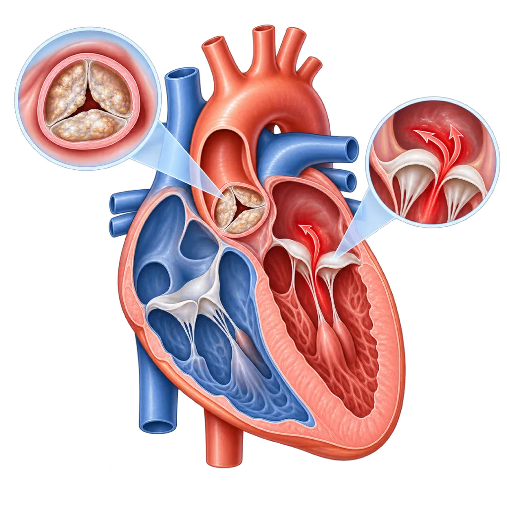
The heart has four valves. The mitral and tricuspid valves sit between the upper chambers (atria) and the lower chambers (ventricles), while the aortic and pulmonary valves sit at the exits of the ventricles. The valves work like one-way doors: they open to let blood flow forward, then close tightly so blood does not flow back.
Heart valve disease occurs when one or more valves do not work properly. There are two main forms. In stenosis, the valve leaflets thicken, stiffen, or stick together, so the valve does not open fully and blood flow is blocked. In regurgitation, or insufficiency, the valve does not close tightly, so some blood flows backward. One valve can have both problems at the same time, and more than one valve can be affected.
Types of Heart Valve Disease
The narrowing group includes aortic, mitral, pulmonary, and tricuspid stenosis, covered on the Heart Valve Stenosis page. The leaking group includes aortic, mitral, pulmonary, and tricuspid regurgitation, covered on the Valve Regurgitation / Insufficiency page. Some valve abnormalities are present from birth, covered on the Congenital Heart Disease page.
Why Valves Can Become Damaged
With aging, the valves can slowly calcify and thicken. In Indonesia, untreated rheumatic fever in childhood and adolescence is still an important cause of valve damage, especially to the mitral valve. Other causes include birth defects such as an aortic valve with only two leaflets (bicuspid), valve infection, enlargement of the heart's ventricles or atria, and widening of the aortic root.
How the Disease Progresses
Valve disease usually progresses slowly over many years. The heart adapts by thickening or enlarging, so many people have no symptoms in the early stages. The 2021 ESC/EACTS and 2020 ACC/AHA guidelines divide its course into several stages: at risk, mild to moderate, severe without symptoms, and severe with symptoms. These stages determine the monitoring schedule and the timing of treatment.
Valve disease can be monitored with painless tests. A severely damaged valve can be repaired or replaced, either through surgery or through a catheter. The decision is made together with a heart team made up of cardiologists, cardiac surgeons, and imaging specialists, taking into account your condition and your preferences.
02 / Understanding the condition
Types and Related Conditions
The following conditions belong to the Heart Valve Disease group. Open each one for a full explanation.
03 / Understanding the condition
Symptoms to Recognize.
Common Symptoms
- Shortness of breath during activity, when climbing stairs, or when lying flat
- Tiring easily and a reduced ability to be active
- Palpitations or a heartbeat that feels irregular
- Chest pain or pressure during activity
- Dizziness, lightheadedness, or nearly fainting during activity
- Swelling in the ankles or legs
- A heart murmur that a doctor hears during a routine checkup
Symptoms in Older Adults
In older adults, symptoms are often thought to be normal signs of aging, when they may actually come from a narrowed or leaky valve.
- Cutting back on activities without realizing it because of tiring quickly
- Getting out of breath when walking short distances
- Dizziness, unsteadiness, or nearly fainting during activity
- New palpitations with an irregular rhythm
Symptoms in Adult Women
Pregnancy increases blood volume and the heart's workload, so valve disease that caused no symptoms before may start to be felt.
- Shortness of breath that gets worse as the pregnancy progresses
- Palpitations that are new or get worse during pregnancy
- Waking up at night because of shortness of breath or coughing
- Tiring more quickly than the usual tiredness of pregnancy
Symptoms in Children
In children, valve disease can come from a birth defect or from rheumatic fever after a throat infection.
- Tiring quickly and often stopping to rest while playing
- Fast or labored breathing during light activity
- Difficulty gaining weight or growth lagging behind peers
- A history of fever with joint pain that moves from joint to joint after a throat infection
04 / Understanding the condition
What Causes This Condition?
- Calcification and thickening of the valves that develop with age
- Rheumatic heart disease, caused by rheumatic fever after a throat infection with streptococcal bacteria
- Birth defects, such as a two-leaflet (bicuspid) aortic valve or a pulmonary valve that is narrow from birth
- Infection of a heart valve (infective endocarditis)
- Enlargement of the ventricles or atria after a heart attack, in cardiomyopathy, or in atrial fibrillation
- Widening of the aortic root, including in Marfan syndrome and other connective tissue disorders
- Late effects of radiation therapy to the chest area, for example in cancer treatment
05 / Understanding the condition
Who Is at Higher Risk?
Age
The risk of valve calcification, especially of the aortic valve, rises after age 65. Mitral valve leakage due to changes in the valve tissue also becomes more common in older age.
Medical
A history of rheumatic fever, endocarditis, heart attack, heart failure, atrial fibrillation, high blood pressure, chronic kidney disease, or chest radiation therapy increases the chance of valve damage.
Genetic
A bicuspid aortic valve can be found in several members of a family. Marfan syndrome and other inherited connective tissue disorders make the valves and aortic root more likely to widen or leak.
Lifestyle
Smoking, high cholesterol, and untreated high blood pressure are linked to aortic valve calcification. Injecting drugs increases the risk of valve infection.
Other
Repeated untreated throat infections in childhood, crowded living conditions, and poor dental hygiene increase the risk of rheumatic heart disease and valve infection.
Groups at Higher Risk
- Older Adults: Calcification of the aortic valve and changes in the mitral valve tissue become more common after age 65, often without noticeable symptoms.
- Rheumatic Fever Survivors: People who had rheumatic fever in childhood or adolescence are at risk of valve damage years later.
- People with Congenital Heart Defects: This includes a bicuspid aortic valve and patients who have had surgery for a heart defect present from birth; they need lifelong heart checkups.
- People with Artificial Heart Valves: Artificial valves can narrow, leak, develop clots, or become infected, so they need regular checks.
- People with Heart Failure or Atrial Fibrillation: Enlarged ventricles or atria can pull on the valve ring, causing the mitral or tricuspid valve to leak.
06 / Understanding the condition
How Is It Diagnosed?
Testing usually begins with a heart murmur that a doctor hears through a stethoscope, or with symptoms of shortness of breath and tiring easily. The main next step is echocardiography, an ultrasound of the heart that shows the shape and movement of the valves, measures how narrow the valve is or how large the leak is, and assesses the size and strength of the ventricles.
If the images taken through the chest wall are not clear enough, your doctor may recommend echocardiography through the esophagus (transesophageal). In people who feel well even though their valve is severely damaged, an exercise test helps confirm whether they truly have no symptoms. Cardiac MRI and cardiac CT scans are used to measure the leak, the size of the ventricles, and the aortic root, or to plan a catheter procedure. Cardiac catheterization is done when other test results are not enough, or to check the coronary arteries before valve surgery.
Physical Examination
The doctor listens to your heart with a stethoscope for a murmur and checks your pulse, blood pressure, and signs of fluid buildup.
Echocardiography
The main test to see the shape and movement of the valves, measure how severe the narrowing or leak is, and assess the size and pumping strength of the ventricles.
Electrocardiogram (ECG)
Records the heart's electrical activity to look for atrial fibrillation, other rhythm problems, and signs of thickened heart muscle.
Chest X-Ray
Shows the size of the heart, enlargement of the atria or aorta, and fluid in the lungs.
Treadmill Stress Test
An exercise test to assess your ability to be active and confirm whether severe valve disease truly causes no symptoms, as your doctor sees fit.
Cardiac MRI
Measures the leak and the size of the ventricles in detail when echocardiography results are not clear enough.
Cardiac Catheterization
Measures pressures in the heart chambers when other test results do not agree, and checks the coronary arteries before valve surgery.
07 / Understanding the condition
Treatment Options.
Treatment is decided by a doctor after an assessment. The categories below can help you follow the conversation with your doctor.
Regular Monitoring
Mild to moderate valve disease usually only needs monitoring with regular checkups and echocardiography, from every few years to every 6 to 12 months depending on its severity.
Medicines to Relieve Symptoms
Diuretics, blood pressure medicines, rhythm-control medicines, and blood thinners help relieve symptoms and prevent complications, but they do not repair the valve. Your doctor decides which ones you need.
Valve Repair
The natural valve is kept and repaired so it closes tightly again, especially the mitral valve, through surgery or a clip placed through a catheter.
Surgical Valve Replacement
The damaged valve is replaced with a mechanical or biological artificial valve. A mechanical valve requires blood thinners for life.
Catheter-Based Valve Procedures
In selected patients, the valve is replaced or widened through a blood vessel without opening the chest, for example transcatheter aortic valve implantation or balloon widening of the mitral valve.
08 / Understanding the condition
Complications to Watch For.
Heart Failure
Years of excess workload make the ventricles enlarge or thicken and then weaken, causing shortness of breath, swelling, and tiring easily. Covered on the Heart Failure page.
Atrial Fibrillation
Enlarged atria, especially in mitral valve disease, are prone to an irregular rhythm. Covered on the Atrial Fibrillation page.
Stroke from a Blood Clot
Clots can form in an enlarged atrium or on an artificial valve, then travel through the bloodstream to the brain.
Pulmonary Hypertension
Raised pressure in the left atrium can pass back into the blood vessels of the lungs and strain the right ventricle. Covered on the Pulmonary Hypertension page.
Infective Endocarditis
Damaged valves or artificial valves are more easily infected by germs in the bloodstream. Covered on the Infective Endocarditis page.
09 / Understanding the condition
What Can You Do?
- Make sure a child's throat infection is fully treated as the doctor advises to prevent rheumatic fever
- If you have had rheumatic fever, take long-term preventive antibiotics on the schedule your doctor sets
- Keep your teeth and mouth clean and have regular dental checkups to lower the risk of valve infection
- Control your blood pressure, cholesterol, and blood sugar, and stop smoking
- Have an echocardiogram if your doctor finds a heart murmur, even if you have no symptoms yet
- If you have been diagnosed, go to your checkups on schedule and do not wait for severe symptoms
- Plan any pregnancy together with a cardiologist if you have valve disease
10 / Understanding the condition
When Should You See a Doctor?
See a cardiologist if a doctor finds a heart murmur, or if you start to tire easily and get short of breath during activities that used to feel light. In valve disease, symptoms often appear slowly, so they are easily put down to age. Go to the emergency room right away if you have severe shortness of breath at rest, fainting, or chest pain that does not ease.
- Shortness of breath, palpitations, or chest pain during activity
- Dizziness or nearly fainting during exercise or when climbing stairs
- Leg swelling or rapid weight gain over a few days
- A history of rheumatic fever, a congenital heart defect, or an artificial valve without regular checkups
- Fever lasting more than a few days for no clear reason in people with an artificial valve or a history of endocarditis
- Planning a pregnancy as a woman with valve disease
Don't Wait in an Emergency.
If symptoms are severe or come on suddenly, go to the nearest emergency department right away or call Heart Attack Center 1500 258, rather than waiting for an appointment.
11 / Common questions
Questions You May Have.
Frequently asked questions about heart valve disease.
Can heart valve disease be cured with medicine?
Medicine cannot open a narrowed valve or close a leaky one. Medicines are used to relieve symptoms, control blood pressure and heart rhythm, and prevent blood clots. When valve damage is severe, repairing or replacing the valve through surgery or a catheter becomes an option. For mild to moderate disease, regular monitoring is often enough. Your doctor chooses the option based on the severity of the disease and your condition.
What is the difference between a narrowed valve and a leaky valve?
A narrowed valve (stenosis) does not open fully, so the heart has to pump harder to push blood through it. A leaky valve (regurgitation or insufficiency) does not close tightly, so some blood flows backward and the heart pumps the same blood again. Both strain the heart in different ways, and one valve can have both. The details are covered on the Heart Valve Stenosis page and the Valve Regurgitation / Insufficiency page.
Can heart valve disease be inherited?
The majority of valve diseases are not passed down directly. However, some birth defects such as a bicuspid aortic valve can be found in more than one family member, and Marfan syndrome is inherited genetically. If a close relative has a bicuspid aortic valve or a widened aorta, your doctor may suggest echocardiography for other family members, even if they have no symptoms yet.
Can people with valve disease exercise?
In general, yes, and regular physical activity is still good for heart health. The type and intensity of exercise need to suit the affected valve and its severity. Mild valve disease usually does not limit activity, while people with severe aortic stenosis usually need to avoid strenuous and competitive sports. Discuss your exercise plans with your doctor, who can assess them with an exercise test.
How do I choose between a mechanical and a biological artificial valve?
A mechanical valve is durable but requires lifelong blood thinners of the vitamin K antagonist type, with regular blood tests. A biological valve made from animal tissue does not require long-term blood thinners in many patients, but it can wear out after many years and may need to be replaced again. The choice is made together with your doctor based on your age, pregnancy plans, bleeding risk, and preferences.
Do I need antibiotics before a tooth extraction?
Preventive antibiotics before certain dental procedures are only recommended for high-risk groups, such as people with an artificial valve, people who have had endocarditis, and some people with congenital heart disease. The majority of other valve diseases do not need them, but dental and oral hygiene remains important for everyone. Tell your dentist about your heart condition, and ask your cardiologist whether you belong to one of these groups.
Sources and Medical Review
Translated from the Indonesian edition medically reviewed by Dr. dr. Dafsah A. Juzar, Sp.JP(K).
Sources as cited in the Indonesian edition.
- Vahanian A, Beyersdorf F, Praz F, Milojevic M, Baldus S, Bauersachs J, et al. 2021 ESC/EACTS Guidelines for the management of valvular heart disease. Eur Heart J. 2022;43(7):561-632.
- Otto CM, Nishimura RA, Bonow RO, Carabello BA, Erwin JP 3rd, Gentile F, et al. 2020 ACC/AHA Guideline for the Management of Patients With Valvular Heart Disease: A Report of the American College of Cardiology/American Heart Association Joint Committee on Clinical Practice Guidelines. Circulation. 2021;143(5):e72-e227.
- Coffey S, Roberts-Thomson R, Brown A, Carapetis J, Chen M, Enriquez-Sarano M, et al. Global epidemiology of valvular heart disease. Nat Rev Cardiol. 2021;18(12):853-864.
- Iung B, Delgado V, Rosenhek R, Price S, Prendergast B, Wendler O, et al. Contemporary Presentation and Management of Valvular Heart Disease: The EURObservational Research Programme Valvular Heart Disease II Survey. Circulation. 2019;140(14):1156-1169.
- Kumar RK, Antunes MJ, Beaton A, Mirabel M, Nkomo VT, Okello E, et al. Contemporary Diagnosis and Management of Rheumatic Heart Disease: Implications for Closing the Gap: A Scientific Statement From the American Heart Association. Circulation. 2020;142(20):e337-e357.
- Delgado V, Ajmone Marsan N, de Waha S, Bonaros N, Brida M, Burri H, et al. 2023 ESC Guidelines for the management of endocarditis. Eur Heart J. 2023;44(39):3948-4042.
Doctors Who Treat This Condition

Dr. dr. Dudy A. Hanafy, Sp.BTKV, Subsp. JD(K), MARS
Adult Cardiac Surgery
Advanced Cardiovascular Surgical Care +1 center
Clinical focusMinimally Invasive Cardiac Surgery · Coronary Artery Bypass Surgery
View Profile
Dr. dr. Dafsah A. Juzar, Sp.JP(K)
Interventional Cardiology
Coronary Intervention Procedures +1 center
Clinical focusComplex Coronary Intervention · Cardiac Catheterization
View Profile
Dr. med. dr. Denio A. Ridjab, Sp.JP(K)
Interventional Cardiology
Coronary Intervention Procedures +2 centers
Clinical focusInterventional Cardiology · Coronary Artery Disease
View Profile