
Quick answer
What Is Pericardial Effusion?
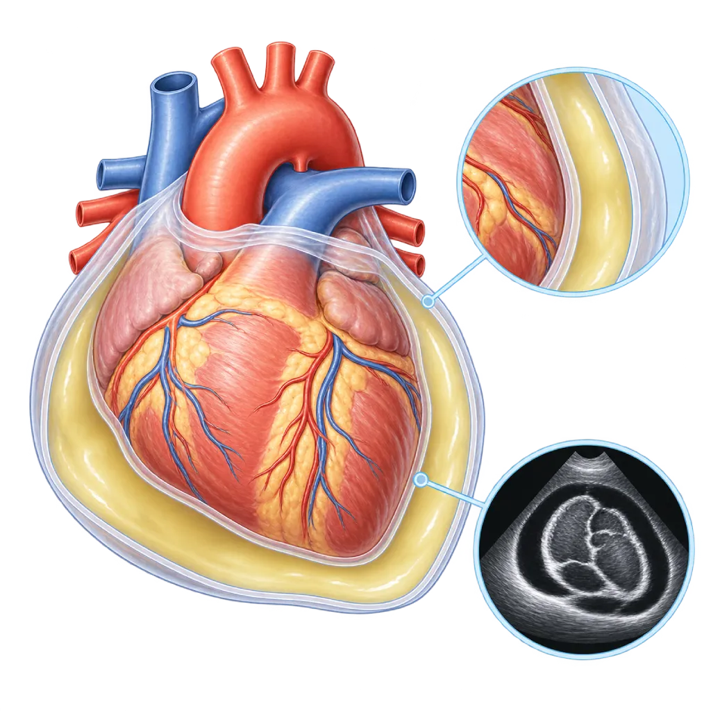
Pericardial effusion is excess fluid inside the sac that surrounds the heart (the pericardium). Most cases are small and cause no symptoms, but a large amount of fluid or fluid that builds up quickly can press on the heart and needs to be drained.
SourcesSigns That Need Urgent Help
- Severe shortness of breath or breathing that gets worse over a few hours
- Severe dizziness, nearly fainting, or fainting
- Pale, cold, and sweaty skin
- A very fast heartbeat with weakness
- Restlessness, confusion, or unusual drowsiness
- Veins in the neck that look very prominent
If these signs appear, go to the nearest emergency department right away or call Heart Attack Center 1500 258.
01 / Understanding the condition
About Pericardial Effusion
The heart is wrapped in the pericardium, a thin sac with two layers. Normally there is a small amount of fluid between the two layers, about 10 to 50 mL, which acts as a lubricant. Pericardial effusion means the fluid inside this sac increases beyond normal. Depending on the cause, the fluid may be clear fluid, blood, pus, or fluid containing cancer cells.
Part of the Pericardial Disease Family
An effusion often accompanies inflammation of the heart lining, covered on the Pericarditis page along with its subtypes. However, an effusion can also occur without inflammation, for example in heart failure, severe kidney disease, or an underactive thyroid gland. In countries with many TB cases, such as Indonesia, TB should always be considered as a possible cause, especially when the effusion comes with fever, night sweats, and weight loss. When the fluid presses on the heart and disrupts its pumping, the situation becomes an emergency, covered on the Cardiac Tamponade page.
How Quickly the Fluid Builds Up
The pericardial sac can stretch slowly. Fluid that builds up over weeks to months can reach a large volume before causing symptoms. In contrast, fluid that builds up over hours, for example from bleeding after a heart procedure, can press on the heart even when the amount is relatively small. For this reason, doctors assess the amount of fluid, how quickly it is increasing, and its effect on the filling of the heart.
Size and Duration
With echocardiography, doctors measure the thickness of the fluid layer. An effusion is called small if it is less than 10 mm, moderate if it is 10 to 20 mm, and large if it is more than 20 mm. An effusion that lasts more than three months is called chronic. A small effusion without symptoms is generally not dangerous, while a large effusion that persists needs monitoring because some can progress to tamponade.
Treatment Follows the Cause
The 2015 ESC guidelines for pericardial diseases recommend treatment based on the cause and on whether the heart is being compressed. If there are signs of inflammation, the effusion is treated like pericarditis with anti-inflammatory medication. If there is an underlying disease, that disease is treated. The fluid is drained with a needle (pericardiocentesis) if it is pressing on the heart, if a bacterial infection or cancer is suspected, or if it is large and persistent without a clear cause. The course of the condition depends more on its cause than on the amount of fluid.
02 / Understanding the condition
Symptoms to Recognize.
Common Symptoms
Small effusions often cause no symptoms and are found by chance during a checkup. Symptoms appear when there is a lot of fluid or it builds up quickly.
- Shortness of breath, especially during activity or when lying down
- A feeling of fullness, heaviness, or discomfort in the chest
- Sharp chest pain if there is also inflammation of the heart lining
- Tiring easily and feeling weak
- Cough, hoarseness, hiccups, or difficulty swallowing because the fluid presses on nearby organs
- Palpitations
- Fever, if the cause is an infection or inflammation
03 / Understanding the condition
What Causes This Condition?
- Inflammation of the heart lining caused by a viral infection or with no clear cause (idiopathic)
- Tuberculosis (TB), an important cause in countries with many TB cases
- Cancer that spreads to the heart lining, such as lung cancer, breast cancer, or lymphoma
- Autoimmune diseases such as lupus and rheumatoid arthritis
- Severe kidney failure (uremia), heart failure, or an underactive thyroid gland
- Bleeding after heart surgery, a catheter procedure, a chest injury, or a tear in the aorta
- Radiation therapy to the chest and certain medications
04 / Understanding the condition
Who Is at Higher Risk?
Medical
Autoimmune diseases, severe kidney failure, cancer, hypothyroidism, heart failure, and a history of pericarditis increase the likelihood of an effusion.
Age
Effusions caused by cancer and systemic diseases are more common in older age, while effusions caused by viral inflammation can occur at any age.
Other
A history of heart surgery, catheter procedures, or pacemaker implantation, radiation therapy to the chest, and close contact with people who have TB.
Groups at Higher Risk
- Cancer Patients: Lung cancer, breast cancer, and lymphoma can spread to the heart lining, and radiation therapy to the chest can trigger an effusion.
- Patients After Heart Surgery: Fluid or blood can collect in the heart sac within a few days to weeks after surgery or a catheter procedure.
- People with Kidney Failure: The buildup of metabolic waste and fluid in severe kidney failure can trigger an effusion, especially when dialysis is not yet adequate.
- People with Autoimmune Diseases: Lupus and other autoimmune diseases can attack the heart lining and cause fluid that keeps coming back.
- People with a History of TB Contact: Close contact with someone who has TB or a weakened immune system increases the chance of TB affecting the heart lining.
05 / Understanding the condition
How Is It Diagnosed?
Pericardial effusion is often found by chance, for example during a chest X-ray or echocardiography done for another reason. Echocardiography is the main test: the doctor can see the fluid, measure the thickness of the layer, and assess whether the fluid is starting to press on the heart chambers. When there is a lot of fluid, an ECG may show smaller electrical waves or waves that alternate in size. A chest X-ray may show an enlarged heart shadow.
The next step is to find the cause. Blood tests assess inflammation (CRP), thyroid function, kidney function, and troponin, depending on your symptoms. CT or cardiac MRI helps show trapped (loculated) fluid, a thickened lining, or a mass. If fluid is drained, a sample is examined in the laboratory to look for germs, TB, or cancer cells.
Echocardiography
The main test to see the fluid, measure the thickness of the layer, and assess whether the fluid is pressing on the heart.
Electrocardiogram (ECG)
Looks for signs of inflammation, smaller electrical waves, or an alternating pattern when there is a lot of fluid.
Chest X-Ray
Shows an enlarged heart shadow as well as lung abnormalities such as TB or a tumor.
C-Reactive Protein (CRP) Test
Assesses whether inflammation is present, which determines whether the effusion is treated like pericarditis.
Thyroid Function Test
Looks for an underactive thyroid gland, a cause of effusion that is easily missed.
Kidney Function Test
Assesses severe kidney failure as a cause, and is also checked before using medications or contrast agents.
Cardiac MRI
Shows trapped fluid, thickening or inflammation of the lining, and masses around the heart.
06 / Understanding the condition
Treatment Options.
Treatment is decided by a doctor after an assessment. The categories below can help you follow the conversation with your doctor.
Regular Monitoring
A small effusion without symptoms or inflammation can generally just be monitored with echocardiography on a schedule set by your doctor.
Anti-Inflammatory Medication and Colchicine
If the effusion comes with signs of inflammation, it is treated like pericarditis. Your doctor determines the type of medication, how long to take it, and how to stop it.
Treating the Cause
For example, anti-TB medication, antibiotics, cancer treatment, thyroid hormone, more adequate dialysis, or control of an autoimmune disease.
Pericardiocentesis
Drainage of the fluid with a needle and a small tube guided by echocardiography or X-ray, when the fluid presses on the heart or needs to be tested.
Pericardial Window or Surgery
An operation to create a channel so the fluid can keep draining out, considered for effusions that recur, contain pus, or result from bleeding.
07 / Understanding the condition
Complications to Watch For.
Cardiac Tamponade
The fluid presses on the heart so its chambers cannot fill completely. This emergency is covered on the Cardiac Tamponade page.
Recurrent Effusion
Fluid can form again after being drained, especially in cancer or autoimmune disease, so a pericardial window operation is sometimes needed.
Constrictive Pericarditis
Long-lasting inflammation, especially from TB or a bacterial infection, can make the lining thick and stiff. Covered on the Constrictive Pericarditis page.
Complications of Fluid Drainage
Pericardiocentesis is generally safe when guided by imaging, but it can rarely cause bleeding, rhythm disturbances, or puncture of nearby organs.
08 / Understanding the condition
What Can You Do?
- Take anti-inflammatory medication and colchicine for as long as recommended if the effusion comes with inflammation
- Complete your TB treatment in full if you are diagnosed with TB
- Keep autoimmune, kidney, and thyroid conditions under control with regular checkups
- Attend your follow-up echocardiography as scheduled, even if you have no symptoms
- After heart surgery, promptly report any new shortness of breath, weakness, or chest pain
- Do not stop or change your medication on your own
09 / Understanding the condition
When Should You See a Doctor?
See a doctor if you have shortness of breath that gets worse when lying down, a feeling of fullness in the chest, or tire easily for no clear reason, especially if you have cancer, an autoimmune disease, or kidney disease, or have recently had heart surgery. If an effusion was found by chance, follow-up is still needed to find the cause and monitor the amount of fluid. Go to the emergency room right away if you have:
- Severe shortness of breath or breathing that gets worse over a few hours
- Severe dizziness, nearly fainting, or fainting
- Pale, cold skin with a very fast heartbeat
- Unusual restlessness or confusion
Don't Wait in an Emergency.
If symptoms are severe or come on suddenly, go to the nearest emergency department right away or call Heart Attack Center 1500 258, rather than waiting for an appointment.
10 / Common questions
Questions You May Have.
Frequently asked questions about pericardial effusion.
Is pericardial effusion the same as an enlarged heart?
No. An enlarged heart means the heart chambers or the heart muscle itself have become larger, covered on the Cardiomegaly page. In pericardial effusion, what increases is the fluid inside the sac that surrounds the heart. On a chest X-ray, both can look like a large heart shadow, so doctors use echocardiography to tell them apart clearly.
Does every pericardial effusion need to be drained?
No. A small effusion without symptoms can generally just be monitored, and an effusion caused by inflammation often shrinks with anti-inflammatory medication. The fluid is drained if it presses on the heart, if a bacterial infection or cancer is suspected so the fluid needs to be tested, or if a large effusion persists without a clear cause. Your doctor weighs the benefits and risks of the procedure based on your condition.
Does pericardial effusion mean I have cancer?
Not necessarily. Many effusions are caused by viral inflammation, a cause that cannot be found, kidney failure, heart failure, an underactive thyroid, or TB. Cancer is one possible cause, especially in people already known to have cancer. For this reason, your doctor looks for the cause step by step through blood tests, imaging, and, if needed, testing of the fluid.
How is fluid around the heart drained?
The procedure is called pericardiocentesis. After local anesthesia, the doctor inserts a thin needle, usually from below the breastbone, guided by echocardiography or X-ray. A small tube is then placed to drain the fluid, generally for a few days until the fluid decreases. The fluid that comes out is tested in the laboratory to find the cause. If the fluid keeps coming back, your doctor may consider a pericardial window operation.
Can pericardial effusion come back?
Yes, especially if the cause has not been treated, such as cancer, autoimmune disease, kidney failure, or recurring inflammation. An effusion caused by viral inflammation that is treated well generally rarely comes back. Follow-up echocardiography on schedule helps detect increasing fluid before it causes serious symptoms. Take your medication as recommended and do not stop it on your own.
How often do I need checkups?
It depends on the size of the effusion, its cause, and your symptoms. A small effusion without symptoms often does not need close monitoring. A moderate effusion is usually rechecked with echocardiography every few months, and a large effusion more often. Your doctor will set the exact schedule. Come in sooner if shortness of breath, weakness, or a feeling of fullness in the chest gets worse.
Sources and Medical Review
Translated from the Indonesian edition medically reviewed by dr. BRM Ario S. Kuncoro, Sp.JP(K).
Sources as cited in the Indonesian edition.
- Adler Y, Charron P, Imazio M, Badano L, Barón-Esquivias G, Bogaert J, et al. 2015 ESC Guidelines for the diagnosis and management of pericardial diseases: The Task Force for the Diagnosis and Management of Pericardial Diseases of the European Society of Cardiology (ESC). Eur Heart J. 2015;36(42):2921-2964.
- Schulz-Menger J, Collini V, Gröschel J, Adler Y, Brucato A, Christian V, et al. 2025 ESC Guidelines for the management of myocarditis and pericarditis. Eur Heart J. 2025;46(40):3952-4041.
- Imazio M, Adler Y. Management of pericardial effusion. Eur Heart J. 2013;34(16):1186-1197.
- Klein AL, Abbara S, Agler DA, Appleton CP, Asher CR, Hoit B, et al. American Society of Echocardiography Clinical Recommendations for Multimodality Cardiovascular Imaging of Patients with Pericardial Disease: Endorsed by the Society for Cardiovascular Magnetic Resonance and Society of Cardiovascular Computed Tomography. J Am Soc Echocardiogr. 2013;26(9):965-1012.
- Vakamudi S, Ho N, Cremer PC. Pericardial Effusions: Causes, Diagnosis, and Management. Prog Cardiovasc Dis. 2017;59(4):380-388.
- Mayosi BM, Burgess LJ, Doubell AF. Tuberculous Pericarditis. Circulation. 2005;112(23):3608-3616.
Doctors Who Treat This Condition

dr. Sri Diniharini, Sp.JP(K)
Echocardiography & Cardiovascular Imaging
Cardiovascular Diagnostic Center +1 center
Clinical focusAdvanced Echocardiography · Transesophageal Echocardiography
View Profile
dr. BRM Ario S. Kuncoro, Sp.JP(K)
Echocardiography & Cardiovascular Imaging
Cardiovascular Diagnostic Center +2 centers
Clinical focusAdvanced and 3D Echocardiography · Transesophageal Echocardiography
View Profile
dr. Radityo Prakoso, Sp.JP(K)
Pediatrics & Congenital Heart Disease
Congenital and Structural Heart Center
Clinical focusPediatric Cardiology Consultation · Congenital Heart Disease Care
View Profile