
Quick answer
What Is Wolff-Parkinson-White Syndrome?
Wolff-Parkinson-White (WPW) syndrome is a condition present from birth in which an extra electrical pathway between the upper and lower chambers of the heart can trigger episodes of a fast heartbeat. Most people with it can be treated well, and catheter ablation stops the episodes in many patients.
SourcesSigns That Need Urgent Help
- Fainting, especially during exercise or while the heart is racing
- A very fast, irregular heartbeat with severe dizziness
- Chest pain or severe shortness of breath
- Cold sweats, paleness, and severe weakness during an episode
- An episode that does not stop after the vagal maneuvers your doctor taught you
- A baby who is very limp, pale gray, or refuses to feed
If these signs appear, go to the nearest emergency department right away or call Heart Attack Center 1500 258.
01 / Understanding the condition
About Wolff-Parkinson-White Syndrome
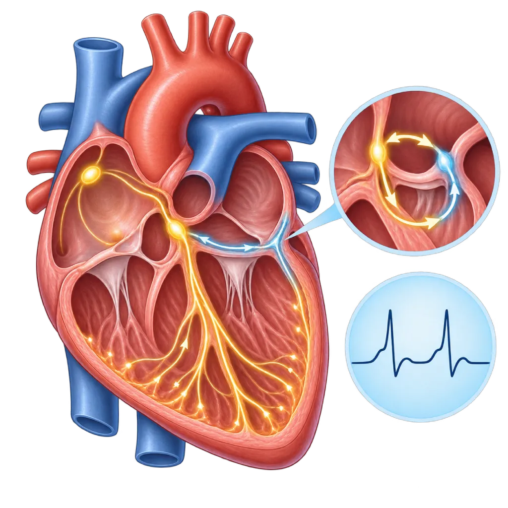
Wolff-Parkinson-White syndrome, or WPW, is named after the three doctors who described it in 1930. People with WPW are born with an extra electrical pathway, often called the bundle of Kent, that connects the upper chambers (atria) and lower chambers (ventricles) of the heart. Normally, every signal from the atria must pass through the AV node, a gate that holds the signal back briefly before passing it on to the ventricles. The extra pathway bypasses that gate, so part of the ventricles receives the signal early. WPW is a commonly seen type of pre-excitation, covered on the Pre-Excitation Syndrome page.
WPW Pattern and WPW Syndrome
On an ECG, the extra pathway shows up as a short PR interval, a delta wave (a slurred start of the ventricular wave), and a widened ventricular wave. If these signs are present without symptoms, doctors call it a WPW pattern. If they come with episodes of tachycardia, it is called WPW syndrome. The WPW pattern is found in about 1 to 3 of every 1,000 people, and some of them never have an episode.
How Episodes Happen
The common type of episode is called AVRT. The signal travels down through the AV node and then back up through the extra pathway, forming a loop that keeps circling. The heart then beats fast and regularly, often 150 to 250 times per minute, in episodes that start and stop suddenly. This type of tachycardia is also covered on the Supraventricular Tachycardia page.
A less common situation, but the main concern, is atrial fibrillation in people with WPW. The chaotic rhythm of the atria can be conducted very rapidly through the extra pathway because it is not filtered by the AV node. In a small number of cases, this can turn into ventricular fibrillation and cause cardiac arrest, sometimes without any earlier symptoms. That is why everyone with a WPW pattern or WPW syndrome should have their risk assessed by a cardiologist.
Treatment
The extra pathway can be disabled with catheter ablation. The 2019 ESC guidelines recommend ablation for people with WPW syndrome who have repeated episodes, and in many patients the episodes do not come back afterward. For people with a WPW pattern and no symptoms, the decision is made after assessing the risk of the extra pathway, taking into account age, occupation, and the sports they play.
02 / Understanding the condition
Symptoms to Recognize.
Common Symptoms
People with a WPW pattern often have no symptoms. Symptoms appear during an episode of tachycardia.
- A very fast, regular, pounding heartbeat that starts suddenly
- Episodes that stop as suddenly as they start
- A fast, irregular heartbeat, a possible sign of atrial fibrillation
- Dizziness or light-headedness
- Shortness of breath or chest discomfort
- Weakness, anxiety, or sweating during an episode
Symptoms in Infants
WPW is one of the causes of tachycardia in babies. Babies cannot describe their symptoms, so an episode is recognized from their behavior, skin color, and breathing.
- Constant fussiness or, on the other hand, looking very limp and sleepy
- Feeding poorly or refusing to feed
- Fast breathing or appearing to gasp for air
- Pale or grayish, sweaty skin
Symptoms in Children
Children often describe an episode as their heart suddenly racing or their chest feeling strange.
- Complaining that their heart is suddenly racing or pounding
- Suddenly stopping play and looking pale and weak
- Complaining of chest or stomach pain during an episode
- Dizziness or feeling faint, especially during exercise
03 / Understanding the condition
What Causes This Condition?
- An extra electrical pathway present from birth (the bundle of Kent) that connects the upper and lower chambers
- Leftover tissue connecting the upper and lower chambers that did not disappear as the heart developed before birth
- Congenital heart defects that often come with extra pathways, especially Ebstein anomaly
- Changes in certain genes, such as PRKAG2, in a small number of cases that run in families
- Episode triggers such as caffeine, alcohol, lack of sleep, stress, or strenuous physical activity
04 / Understanding the condition
Who Is at Higher Risk?
Genetic
Most cases of WPW occur without a family history. A small number are inherited, sometimes together with thickening of the heart muscle due to changes in the PRKAG2 gene.
Medical
Ebstein anomaly and some other congenital heart defects more often come with one or more extra pathways.
Age
WPW can cause episodes from infancy. Episodes also often appear for the first time in school-age children, teenagers, or young adults.
Lifestyle
Too much caffeine, energy drinks, alcohol, stimulants, and lack of sleep do not cause WPW, but they can trigger episodes in people who have it.
Groups at Higher Risk
- Children and Teenagers: PACES/HRS guidance recommends a risk assessment for children and teenagers with a WPW pattern, because in some children serious events can happen without earlier symptoms.
- Athletes and High-Risk Workers: Competitive athletes, drivers, pilots, and people who work at heights are advised to have a risk assessment of the extra pathway, because fainting during activity can endanger themselves and others.
- People with Ebstein Anomaly: This congenital abnormality of the tricuspid valve, covered on the Ebstein Anomaly page, often comes with one or more extra pathways.
- Families with Inherited WPW: In a small number of families, WPW is inherited together with thickening of the heart muscle due to changes in certain genes. Your doctor may suggest an ECG for family members.
05 / Understanding the condition
How Is It Diagnosed?
WPW is often first discovered on an ECG, either during an episode or at a routine checkup. Your doctor will ask about any history of a racing heartbeat, dizziness, or fainting, and about heart disease in your family. The ECG between episodes is distinctive enough to show an extra pathway, and the shape of the delta wave helps estimate where it is.
Next, your doctor assesses the risk of the extra pathway. A pattern that comes and goes on a Holter monitor, or that disappears suddenly during a treadmill test, usually means a pathway that cannot conduct signals very rapidly. Echocardiography looks for any accompanying structural abnormalities. If you have episodes, or if you are an athlete or a worker for whom fainting would be dangerous, your doctor may recommend an electrophysiology study. This catheter-based test precisely measures what the extra pathway can do and maps its location, so ablation can be done in the same session.
Electrocardiogram (ECG)
Shows a short PR interval, a delta wave, and a widened ventricular wave, and helps estimate where the extra pathway is.
Holter Monitoring
Records the heart rhythm for 24 to 48 hours to capture episodes and see whether the WPW pattern is constant or comes and goes.
Long-Term Heart Rhythm Monitoring
A recorder worn for longer, or one you activate yourself when your heart races, if episodes are infrequent.
Treadmill Stress Test
Shows whether the WPW pattern suddenly disappears as the heart rate rises, a clue that the extra pathway is low risk.
Echocardiography
Looks for structural abnormalities that can come with WPW, such as Ebstein anomaly or thickening of the heart muscle, and assesses pumping strength.
Electrophysiology Study
Measures the extra pathway's ability to conduct fast signals, looks for other pathways, and maps their location, usually followed by ablation.
06 / Understanding the condition
Treatment Options.
Treatment is decided by a doctor after an assessment. The categories below can help you follow the conversation with your doctor.
Vagal Maneuvers
Simple techniques that stimulate the vagus nerve and can stop a fast, regular AVRT episode. Your doctor will teach you a technique that is safe for you.
Treating Episodes in the Emergency Room
Episodes that do not stop are treated with injected medicine or a controlled electric shock. In atrial fibrillation with WPW, doctors avoid drugs that only slow the AV node and choose other drugs or electrical cardioversion.
Catheter Ablation
The extra pathway is disabled with a catheter inserted through a blood vessel in the groin. The 2019 ESC guidelines recommend ablation for WPW syndrome with repeated episodes and for people who have had atrial fibrillation with pre-excitation.
Risk Assessment for a WPW Pattern
For a WPW pattern without symptoms, your doctor assesses the risk of the extra pathway with noninvasive tests or an electrophysiology study. Ablation is recommended if the pathway is high risk and can be considered if it is low risk.
Antiarrhythmic Medicines
For people who have not yet had or cannot have ablation, certain antiarrhythmic medicines can reduce episodes. Your doctor decides on the type and dose, because not all rhythm medicines are suitable for people with WPW.
07 / Understanding the condition
Complications to Watch For.
Recurring AVRT Episodes
Repeated episodes of a racing heartbeat can disrupt school, work, sports, and your sense of safety, although they usually do not damage the heart.
Atrial Fibrillation with Pre-Excitation
The chaotic rhythm of the atria is conducted through the extra pathway without being filtered by the AV node, so the ventricles beat very fast and irregularly. This needs urgent treatment.
Sudden Cardiac Arrest
This is rare. Atrial fibrillation conducted very rapidly can turn into ventricular fibrillation, sometimes in people who previously had no symptoms. This risk can be reduced with ablation.
Weakened Heart Pumping
In a small number of children, uncoordinated contraction of the ventricles caused by the extra pathway, or long-lasting episodes, can weaken the heart's pumping. This usually improves after ablation.
Fainting and Injury
A very fast episode can lower blood pressure, causing you to faint and be injured in a fall, especially while driving or exercising.
08 / Understanding the condition
What Can You Do?
- Keep a copy of the ECG that shows your WPW pattern and bring it to every medical visit
- Tell every doctor and emergency room staff member that you have WPW before you are given heart medicine
- Learn your episode triggers and cut back on them, such as too much caffeine, energy drinks, or alcohol
- Have a risk assessment before taking up competitive sports or a job where fainting would be dangerous
- Learn vagal maneuvers from your doctor and practice them when you are not having an episode
- Take your medicines as advised and do not stop them on your own
09 / Understanding the condition
When Should You See a Doctor?
A WPW pattern on your ECG needs to be assessed by a cardiologist, even if you have never had an episode. This assessment determines whether your extra pathway only needs monitoring or should be treated with ablation. Make an appointment if any of the following applies to you, and go to the emergency room right away if warning signs appear.
- Your ECG report mentions a WPW pattern, pre-excitation, or a delta wave
- Episodes of a racing heartbeat that start and stop suddenly
- Your child has been diagnosed with WPW and is going to take part in competitive sports
- You work as a driver or pilot, or at heights
- Fainting, chest pain, or a very fast, irregular heartbeat: go to the emergency room right away
Don't Wait in an Emergency.
If symptoms are severe or come on suddenly, go to the nearest emergency department right away or call Heart Attack Center 1500 258, rather than waiting for an appointment.
10 / Common questions
Questions You May Have.
Frequently asked questions about Wolff-Parkinson-White syndrome.
Is WPW dangerous?
Most people with WPW live normal lives, and their episodes can be treated well. However, a small number of extra pathways can conduct signals very rapidly if atrial fibrillation occurs, and in rare cases this can cause cardiac arrest. Because this risk cannot be judged from symptoms alone, everyone with a WPW pattern or WPW syndrome should be checked by a cardiologist.
Can WPW be cured?
The extra pathway can be disabled with catheter ablation. After a successful ablation, the delta wave usually disappears from the ECG, and in many patients the episodes do not come back. A small number of pathways can start conducting signals again, so a repeat ablation is needed. Your doctor will explain the chances and risks based on where your extra pathway is located.
I have a WPW pattern with no symptoms. Does it need treatment?
Not always. The risk of a WPW pattern without symptoms is assessed first, for example with a Holter monitor, a treadmill test, or an electrophysiology study. If the pathway proves to be high risk, guidelines recommend ablation. If it is low risk, you can choose monitoring or ablation after discussing the benefits and risks. Athletes and people whose work would be dangerous if they fainted are generally advised to have an electrophysiology study.
Is WPW inherited?
Usually not. Most cases of WPW occur without a family history. In a small number of families, WPW is inherited, sometimes together with thickening of the heart muscle due to changes in certain genes such as PRKAG2. If more than one family member has WPW or there is thickening of the heart muscle, your doctor may suggest an ECG for immediate family members and further testing.
Can people with WPW exercise?
Many people with WPW can exercise after being assessed by a doctor. Guidelines recommend a risk assessment of the extra pathway for competitive athletes, usually with an electrophysiology study. If the pathway is high risk, ablation generally allows athletes to return to training after a recovery period. Stop exercising and get checked if your heart races very fast or you feel dizzy or nearly faint while training.
What are the risks of ablating an extra pathway?
Ablation is done through a blood vessel in the groin under local anesthesia and sedation. The risks are small, such as bruising or bleeding at the puncture site. For pathways located close to the AV node, there is a small chance of a conduction problem that requires a pacemaker. Your doctor will explain the risks based on where your pathway is before the procedure.
Sources and Medical Review
Translated from the Indonesian edition medically reviewed by Dr. dr. Dicky A. Hanafy, Sp.JP(K).
Sources as cited in the Indonesian edition.
- Brugada J, Katritsis DG, Arbelo E, et al. 2019 ESC Guidelines for the management of patients with supraventricular tachycardia. The Task Force for the management of patients with supraventricular tachycardia of the European Society of Cardiology (ESC). Eur Heart J. 2020;41(5):655-720.
- Page RL, Joglar JA, Caldwell MA, et al. 2015 ACC/AHA/HRS Guideline for the Management of Adult Patients With Supraventricular Tachycardia. Circulation. 2016;133(14):e506-e574.
- Cohen MI, Triedman JK, Cannon BC, et al. PACES/HRS Expert Consensus Statement on the Management of the Asymptomatic Young Patient with a Wolff-Parkinson-White (WPW, Ventricular Preexcitation) Electrocardiographic Pattern. Heart Rhythm. 2012;9(6):1006-1024.
- Etheridge SP, Escudero CA, Blaufox AD, et al. Life-threatening event risk in children with Wolff-Parkinson-White syndrome: a multicenter international study. JACC Clin Electrophysiol. 2018;4(4):433-444.
- Pappone C, Vicedomini G, Manguso F, et al. Wolff-Parkinson-White syndrome in the era of catheter ablation: insights from a registry study of 2169 patients. Circulation. 2014;130(10):811-819.
- Delise P, Sciarra L. Sudden cardiac death in patients with ventricular preexcitation. Card Electrophysiol Clin. 2020;12(4):519-525.
- Sharma S, Drezner JA, Baggish A, et al. International recommendations for electrocardiographic interpretation in athletes. J Am Coll Cardiol. 2017;69(8):1057-1075.
Doctors Who Treat This Condition

Dr. dr. Dicky A. Hanafy, Sp.JP(K)
Arrhythmia and Electrophysiology
Arrhythmia and Device Center +1 center
Clinical focusElectrophysiology and Arrhythmia Ablation · Pacemaker and ICD Implantation
View Profile
dr. Sunu B. Raharjo, Sp.JP(K), Ph.D.
Arrhythmia and Electrophysiology
Arrhythmia and Device Center
Clinical focusClinical and Interventional Electrophysiology · Cardiac Arrhythmia Ablation
View Profile
dr. Radityo Prakoso, Sp.JP(K)
Pediatrics & Congenital Heart Disease
Congenital and Structural Heart Center
Clinical focusPediatric Cardiology Consultation · Congenital Heart Disease Care
View Profile