
Quick answer
What Is Signal-Averaged Electrocardiogram?
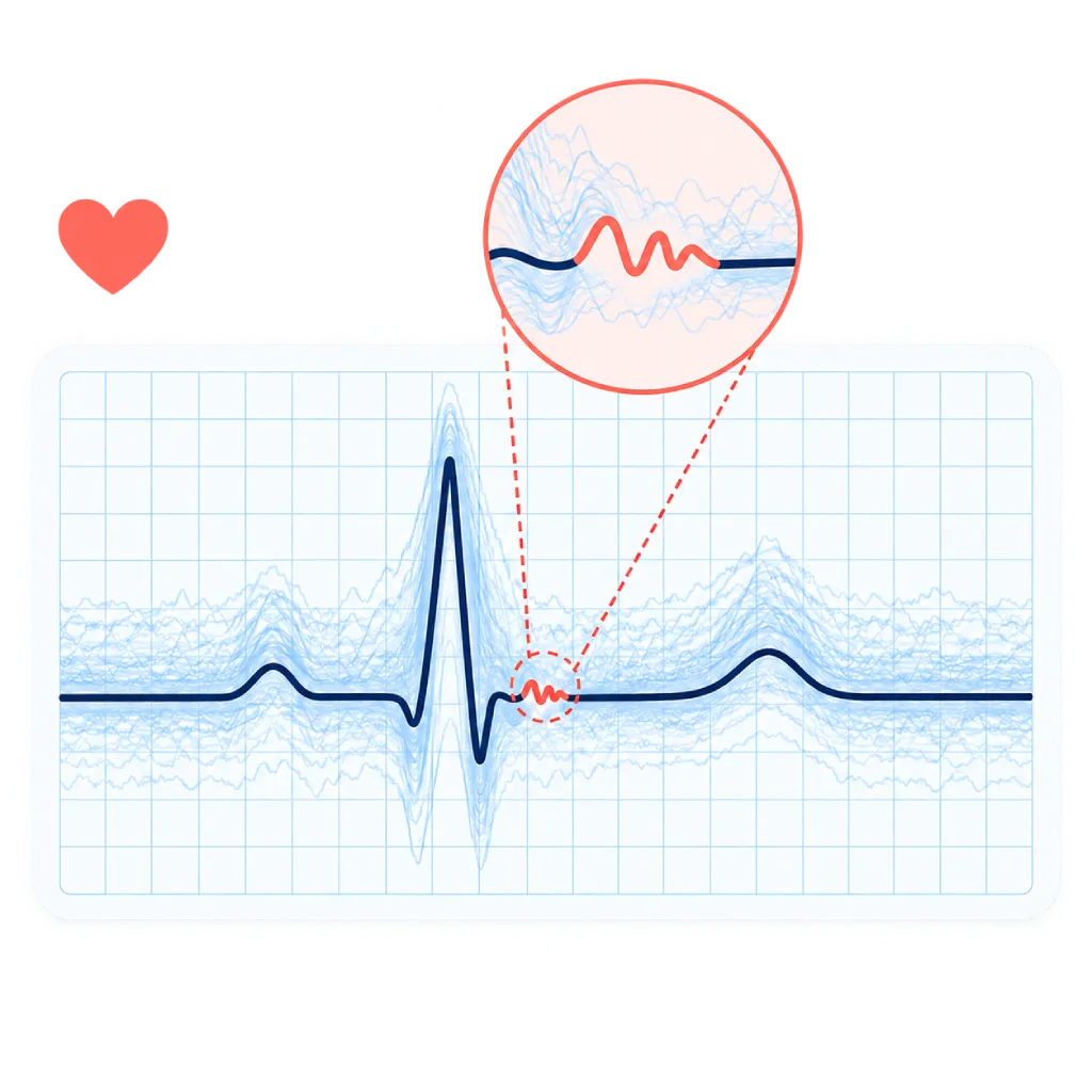
A signal-averaged electrocardiogram (SAECG) is a special ECG recording that combines hundreds of heartbeats to remove signal interference. The result can show late potentials, very small electrical signals that indicate slowed electrical flow in part of the heart muscle, which can trigger ventricular tachycardia. The test is needle-free and painless.
Sources01 / Understanding the procedure
About Signal-Averaged Electrocardiogram
What Is a Signal-Averaged Electrocardiogram
A standard ECG records the heart's electrical activity for a few seconds. An SAECG records for longer, and then a computer averages hundreds of beats that have the same shape. Signal interference from the body's muscles and the surroundings cancels out, while very small heart signals remain visible. The small signals being looked for are called late potentials.
How It Works at a Glance
Electrodes are placed on the chest, back, and sides of the body to record the heart's electrical activity from three directions. While you lie still, the device discards beats with a different shape, then stacks and averages the rest. At the end of the QRS wave, the part of the beat when the heart's ventricles are activated, a small tail of signal may appear, indicating slowed electrical flow in part of the heart muscle.
What Is Assessed
- Filtered QRS duration: the total activation time of the ventricles on the filtered recording.
- LAS40: the duration of the very low-voltage signal at the end of the QRS.
- RMS40: the voltage in the terminal 40 milliseconds of the QRS.
The commonly used cutoffs are a filtered QRS longer than 114 milliseconds, an LAS40 longer than 38 milliseconds, and an RMS40 below 20 microvolts. Late potentials are considered present when two or more of these three values are outside the cutoffs. In the assessment of arrhythmogenic right ventricular cardiomyopathy, a single value outside the cutoff already counts as one of the minor criteria.
What the Results Mean
Late potentials indicate that part of the heart muscle conducts electricity more slowly, for example scar tissue after a heart attack, or muscle replaced by fat and connective tissue in arrhythmogenic right ventricular cardiomyopathy. Areas like these can become a source of ventricular tachycardia. A normal result indicates a lower risk of dangerous rhythm disturbances, but an abnormal result alone is not enough to conclude that the risk is high.
The Role of This Test Today
SAECG is mainly used as one of the diagnostic criteria for arrhythmogenic right ventricular cardiomyopathy and as additional information when fainting or palpitations are suspected to come from the ventricles. The decision to implant an ICD is not based on this test, but on the heart's pumping function, echocardiography or cardiac MRI, and an electrophysiology study if needed.
02 / Understanding the procedure
When Is This Procedure Considered?
A doctor may consider this procedure in the situations below. The decision is always made after a thorough assessment.
- Suspected Arrhythmogenic Right Ventricular CardiomyopathyAs one of the diagnostic criteria, together with an ECG and heart imaging.
- Fainting or Palpitations Suspected to Come from the VentriclesAdditional information to decide whether further tests are needed.
- History of Heart Attack with Rhythm DisturbancesAssesses scar tissue that can trigger ventricular tachycardia.
- Frequent Extra Beats from the Right VentricleHelps distinguish harmless conditions from arrhythmogenic cardiomyopathy.
- Screening Family Members of Patients with Arrhythmogenic CardiomyopathyPart of the evaluation of family members at risk.
Situations to Consider First
- Bundle Branch Block or Wide QRSThe standard cutoffs do not apply, so the results are hard to interpret.
- Pacemaker-Paced RhythmThe pacemaker signal makes late potentials impossible to assess.
- Difficulty Lying StillMovement, shivering, or coughing adds signal interference.
- Very Frequent Extra BeatsBeats with a different shape are discarded, so the recording takes longer.
- Oily or Very Hairy SkinElectrode contact is poorer, so the area is cleaned or shaved.
03 / Before the procedure
How Should You Prepare?
An SAECG does not require fasting or special preparation.
Clothing. Wear a separate top and bottom so your chest and back can be uncovered easily.
Skin. Do not use lotion, oil, or powder on your chest and back on the day of the test.
Medicines. Take your medicines as usual unless your doctor tells you otherwise. Bring a list of your medicines, because heart rhythm medicines can affect the results.
Important information. Tell the staff if you have a pacemaker or an ICD.
Previous results. Bring the results of any ECG, Holter, echocardiography, or cardiac MRI you have had.
Unsure about the preparation instructions? Ask our team before the procedure.
04 / During the procedure
How Is the Procedure Done?
The whole test is usually finished within 20 to 30 minutes, including preparation.
Skin Preparation
The skin where the electrodes will go is cleaned, shaved if needed, and lightly rubbed so the electrodes stick well and signal interference is reduced.
Placing the Electrodes
Electrodes are placed in specific positions on the chest, back, and both sides of the body to record the heart's electrical activity from three directions.
Recording
You lie still on your back, breathe normally, and do not talk for about 10 to 20 minutes, until enough beats have been recorded.
Analysis and Reading
A computer averages the beats and calculates the three main values. The doctor reads the results together with your ECG, Holter, and heart imaging.
05 / Benefits and risks
Benefits and Risks Should Be Discussed Together.
Every medical procedure has benefits and risks. Your doctor will explain both for your situation before the procedure.
Benefits You Can Expect
- Needle-Free and PainlessIt only uses electrodes placed on the skin.
- Shows Signals Not Visible on a Standard ECGAveraging hundreds of beats reveals very small electrical signals.
- Helps Diagnose Arrhythmogenic CardiomyopathyIt is one of the diagnostic criteria for this disease.
- A Normal Result Is ReassuringA normal result indicates a lower risk of dangerous rhythm disturbances.
- Guides Further TestingHelps your doctor choose the right next test.
The benefits of a procedure are not a promise of a cure. Ask how the results will affect your care plan.
Risks You Should Know
- Mild Skin IrritationThe electrode adhesive can cause brief redness.
- Results Cannot Be AssessedWith a wide QRS, a paced rhythm, or too much signal interference.
- An Abnormal Result Is Not Necessarily DangerousSome abnormal results are not followed by rhythm disturbances, so other tests are needed.
- Does Not Replace Other TestsThis test complements an ECG, Holter monitoring, and heart imaging rather than replacing them.
Risks can differ from person to person. A full explanation and consent are given after an individual assessment.
06 / After the procedure
What Is Recovery Like?
There is no recovery period. You can return to your activities right away.
Skin. Mild redness from the electrode adhesive usually fades on its own within a few hours.
If the result is abnormal. Your doctor may recommend Holter monitoring, echocardiography, cardiac MRI, or an electrophysiology study to complete the assessment.
Warning signs. Go to the emergency room right away or contact the medical team if you have:
- Fainting, especially during activity
- A very fast heartbeat with dizziness, shortness of breath, or chest pain
- Chest pain that does not improve with rest
- Sudden shortness of breath
Know the Signs That Need Prompt Care.
Follow the team's guidance on warning signs. If severe or sudden symptoms appear after the procedure, go to the nearest emergency department right away or call Heart Attack Center 1500 258.
07 / Team and support
Doctors and Procedure Support
The doctors below are listed for this procedure. Listed by name, not by ranking.

Dr. med. dr. Denio A. Ridjab, Sp.JP(K)
Interventional Cardiology
Coronary Intervention Procedures +2 centers
Clinical focusInterventional Cardiology · Coronary Artery Disease
View Profile
Dr. dr. Faris Basalamah, Sp.JP(K)
Interventional Cardiology
Coronary Intervention Procedures +1 center
Clinical focusComplex Coronary Intervention · Cardiac Electrophysiology
View Profile
dr. Sunu B. Raharjo, Sp.JP(K), Ph.D.
Arrhythmia and Electrophysiology
Arrhythmia and Device Center
Clinical focusClinical and Interventional Electrophysiology · Cardiac Arrhythmia Ablation
View Profile08 / Common questions
Before You Plan the Procedure.
Frequently asked questions about the signal-averaged electrocardiogram. Your doctor will explain what the results mean for your condition.
What is the difference between an SAECG and a standard ECG?
A standard ECG records for a few seconds and is enough to see the heart rhythm and signs of a heart attack. An SAECG records for longer and then averages hundreds of beats, so signal interference is reduced and very small signals at the end of each beat can be seen. These small signals are not visible on a standard ECG.
What are late potentials?
Late potentials are very small electrical signals at the end of the QRS wave. They appear when electrical flow slows as it passes through a damaged area of heart muscle, such as scar tissue. Areas like these can become the starting point of ventricular tachycardia, a fast rhythm disturbance that comes from the heart's ventricles.
Is this test painful or dangerous?
No. An SAECG only records the heart's electrical activity through electrodes placed on the skin, with no needles, no medicines, and no radiation. All you may feel is slight itching or redness from the electrode adhesive, and a cool sensation when the skin is cleaned before the electrodes are placed.
Why do I have to lie still for so long?
The signals being looked for are very small, much smaller than the signals from your body's muscles when you move, talk, or shiver. When you lie still, signal interference is reduced and the device can collect enough clean beats to average. For this reason, the recording can take about 10 to 20 minutes.
Does an abnormal result mean I need an ICD?
Not automatically. An abnormal result indicates that part of the heart muscle conducts electricity more slowly, but many people with abnormal results never develop a dangerous rhythm disturbance. The decision to implant an ICD is based on the heart's pumping function, heart imaging, your history of symptoms, and an electrophysiology study if needed.
How is SAECG related to arrhythmogenic right ventricular cardiomyopathy?
Arrhythmogenic right ventricular cardiomyopathy is a heart muscle disease that often runs in families, in which the muscle of the right ventricle is replaced by fat and connective tissue. These areas conduct electricity more slowly and can produce late potentials. For this reason, the SAECG result is one of the minor criteria used to diagnose this disease.
Sources and Medical Review
Translated from the Indonesian edition medically reviewed by Dr. dr. Faris Basalamah, Sp.JP(K).
Sources as cited in the Indonesian edition.
- Breithardt G, Cain ME, El-Sherif N, et al. Standards for analysis of ventricular late potentials using high-resolution or signal-averaged electrocardiography: a statement by a Task Force Committee of the European Society of Cardiology, the American Heart Association, and the American College of Cardiology. Circulation. 1991;83(4):1481-1488.
- Marcus FI, McKenna WJ, Sherrill D, et al. Diagnosis of arrhythmogenic right ventricular cardiomyopathy/dysplasia: proposed modification of the Task Force Criteria. Circulation. 2010;121(13):1533-1541.
- Gatzoulis KA, Arsenos P, Trachanas K, et al. Signal-averaged electrocardiography: past, present, and future. J Arrhythm. 2018;34(3):222-229.
- Bailey JJ, Berson AS, Handelsman H, Hodges M. Utility of current risk stratification tests for predicting major arrhythmic events after myocardial infarction. J Am Coll Cardiol. 2001;38(7):1902-1911.
- Zeppenfeld K, Tfelt-Hansen J, de Riva M, et al. 2022 ESC Guidelines for the management of patients with ventricular arrhythmias and the prevention of sudden cardiac death. Eur Heart J. 2022;43(40):3997-4126.