
Quick answer
What Is Ischemic Stroke?
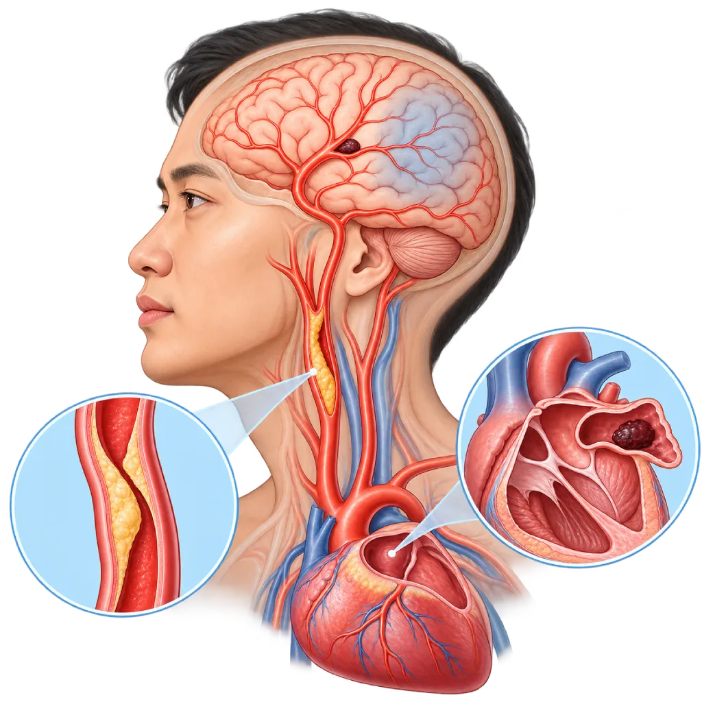
Ischemic stroke happens when a blood vessel in the brain is blocked by a clot or plaque, so part of the brain does not get enough blood. It is an emergency: medicine or a procedure to open the blockage brings major benefit when given within the first few hours.
Sources01 / Understanding the condition
About Ischemic Stroke
Ischemic stroke is a type of stroke caused by a blocked blood vessel in the brain, and the majority of strokes are this type. The part of the brain beyond the blockage does not get enough oxygen. At its center, brain cells die quickly, while the surrounding area can still be saved if blood flow is restored promptly. This area is the target of emergency treatment. An overview of all types of stroke is covered on the Stroke page.
Where the Blockage Comes From
Doctors classify ischemic stroke by its source. First, cholesterol plaque in large arteries, such as the carotid artery in the neck, which narrows blood flow or releases clots to the brain. Second, clots from the heart, called cardioembolism. Third, blockage of small blood vessels deep in the brain (lacunar stroke) caused by long-standing high blood pressure and diabetes. The rest have less common causes, such as a tear in the wall of a neck artery or a blood clotting disorder. In some patients, the cause is not found even after a complete workup, which is called cryptogenic stroke.
The Role of the Heart
Strokes caused by clots from the heart tend to be more extensive because the clots are often large enough to block a major artery of the brain. The main cause is atrial fibrillation, an irregular rhythm that lets blood pool and clot in the pouch of the left atrium. Atrial fibrillation often comes and goes without symptoms, so it may only be found when the heart rhythm is recorded for several days to several months. This condition is covered on the Atrial Fibrillation page. Other sources include clots in the left ventricle after a heart attack, mechanical artificial heart valves, mitral stenosis, valve infection, and patent foramen ovale in strokes at a young age.
Emergency Treatment
The AHA/ASA 2019 and ESO guidelines recommend two ways to open the blockage. Clot-dissolving medicine given through an IV (thrombolysis) is given within 4.5 hours of symptom onset in eligible patients. If the blockage is in a large artery, the clot can be removed with a catheter (thrombectomy), usually within 6 hours, and up to 24 hours in selected patients based on imaging results. Both can be done one after the other. After the emergency phase, attention shifts to finding the source of the clot and preventing another stroke.
02 / Understanding the condition
Symptoms to Recognize.
Common Symptoms
Symptoms come on suddenly, usually on one side of the body, and depend on which blood vessel is blocked.
- A drooping face or an uneven smile
- Weakness or paralysis of an arm or leg on one side
- Numbness or tingling on one side of the face or body
- Slurred speech, trouble finding words, or trouble understanding speech
- Darkened vision in one eye, or loss of part of the field of vision
- Spinning dizziness, unsteady walking, or uncoordinated hand movements
- Trouble swallowing or choking when drinking
03 / Understanding the condition
What Causes This Condition?
- Cholesterol plaque in the carotid artery or brain arteries that narrows blood flow or releases clots (atherosclerosis)
- Clots from the heart, especially in atrial fibrillation
- Clots in the left ventricle after a heart attack, a mechanical artificial heart valve, or a heart valve infection
- Blockage of small blood vessels deep in the brain due to long-standing high blood pressure and diabetes
- Clots from a vein that cross through an opening in the wall between the atria, covered on the Patent Foramen Ovale page
- A tear in the wall of a neck artery (dissection), more common at a young age
- Blood clotting disorders, such as antiphospholipid syndrome, or inflammation of the blood vessels
04 / Understanding the condition
Who Is at Higher Risk?
Medical
High blood pressure, atrial fibrillation, diabetes, high LDL cholesterol, coronary artery disease, heart failure, chronic kidney disease, sleep apnea, and a history of TIA or previous stroke.
Lifestyle
Smoking, physical inactivity, a diet high in salt and saturated fat, obesity, and excessive alcohol. These factors can be changed, and controlling them lowers the risk of stroke.
Age
Risk increases with age, especially after 55. Under age 50, less common causes such as neck artery tears and patent foramen ovale are found more often.
Genetic
A history of stroke or heart disease in close family members at a young age, inherited high cholesterol, and inherited blood clotting disorders.
Groups at Higher Risk
- People with Atrial Fibrillation: The risk of stroke is about five times higher than in people with a normal rhythm, especially without suitable blood thinners.
- People with Hypertension and Diabetes: Both damage large and small blood vessels in the brain and are among the main causes of lacunar stroke.
- People with Carotid Artery Narrowing: Plaque in a neck artery can release clots to the brain, especially if it has already caused symptoms.
- Heart Attack Survivors: A clot can form in a left ventricle whose wall moves weakly and then travel to the brain.
- People Who Have Had a TIA: A temporary stroke attack is a warning sign that a more severe ischemic stroke may follow within days.
05 / Understanding the condition
How Is It Diagnosed?
As soon as the patient arrives, the emergency team asks when the symptoms started or when the patient was last seen normal, checks blood sugar, blood pressure, and level of consciousness, then rates the severity of the symptoms with the NIHSS scale. A head CT scan is done as soon as possible. If there is no bleeding and time still allows, clot-dissolving medicine can be given right away. CT angiography or MRI shows whether there is a blockage in a large artery that can be removed with a catheter.
After the emergency phase, tests focus on finding the source of the clot: the blood vessels of the neck and brain, the heart rhythm, the structure of the heart, and blood tests for sugar, HbA1c, and cholesterol. If the cause has not been found, long-term rhythm recording and transesophageal echocardiography may be added. In young people, the doctor also looks for a neck artery tear, patent foramen ovale, and blood clotting disorders.
A non-contrast head CT scan as soon as possible in the emergency room to rule out bleeding before clot-dissolving medicine is given.
CT angiography and CT perfusion or MRI to find a blockage in a large artery and assess brain tissue that can still be saved.
Blood Sugar Test
Blood sugar is checked before treatment because low blood sugar can cause symptoms that look like a stroke.
Vascular Ultrasound
Ultrasound of the carotid and vertebral arteries in the neck to look for plaque and narrowing.
Long-Term Heart Rhythm Monitoring
An ECG followed by rhythm recording for several days to several weeks to look for atrial fibrillation that comes and goes.
Implantable Loop Recorder (ILR)
A small rhythm recorder under the skin of the chest, considered after a stroke with no clear cause to look for atrial fibrillation over many months.
Echocardiography
Echocardiography through the chest or through the esophagus (transesophageal) to look for clots, valve abnormalities, or an opening in the wall of the heart.
06 / Understanding the condition
Treatment Options.
Treatment is decided by a doctor after an assessment. The categories below can help you follow the conversation with your doctor.
Intravenous Thrombolysis
Clot-dissolving medicine through an IV within 4.5 hours of symptom onset or of when the patient was last seen normal, in eligible patients. The doctor weighs the benefit against the risk of bleeding.
Mechanical Thrombectomy
A clot in a large artery is removed with a catheter inserted from the groin or arm. Usually within 6 hours, and up to 24 hours in selected patients.
Stroke Unit Care
Monitoring of blood pressure, blood sugar, temperature, and swallowing by a dedicated team, along with gradual mobilization and early rehabilitation.
Antiplatelet Medicines
For strokes caused by plaque or blockage of small vessels. In a minor stroke or high-risk TIA, two antiplatelet medicines may be given for a short period. The type and duration are decided by the doctor.
Anticoagulant Medicines
For strokes caused by atrial fibrillation or clots from the heart. When to start after a stroke is decided by the doctor based on the size of the stroke and the risk of bleeding.
Procedures on the Source of the Clot
Carotid endarterectomy or stenting for a narrowing that has caused symptoms, closure of a patent foramen ovale in selected patients, or LAA closure when anticoagulants cannot be used long term.
Statins and Risk Factor Control
Statins, blood pressure medicines, blood sugar control, and quitting smoking to lower the risk of another stroke and heart attack.
07 / Understanding the condition
Complications to Watch For.
Brain Swelling
A large stroke can make the brain swell in the first few days. In certain situations, a neurosurgeon opens part of the skull to relieve the pressure.
Bleeding in the Stroke Area
Damaged brain tissue can bleed, either on its own or after clot-dissolving medicine, so the patient is closely monitored.
Pneumonia from Swallowing Problems
Food or fluid can enter the airways. For this reason, swallowing ability is checked before the patient is given food or drink.
Paralysis and Speech Problems
Weakness on one side of the body and speech problems can partly persist and improve gradually with rehabilitation.
Blood Clots in the Leg Veins
Lack of movement during a hospital stay makes it easier for clots to form in the leg veins. Covered on the Deep Vein Thrombosis page.
Depression and Memory Problems
These often appear after a stroke, affect recovery, and can be treated if recognized early.
Recurrent Stroke
The risk of another stroke is higher in the first weeks, especially if the source of the clot has not been found or treated.
08 / Understanding the condition
What Can You Do?
- Control your blood pressure to the target set by your doctor and measure it regularly at home
- If you have atrial fibrillation, take your blood thinner as recommended and do not stop it on your own
- Take statins and other medicines as prescribed to keep your LDL cholesterol on target
- Quit smoking and limit alcohol
- Be physically active for at least 150 minutes per week
- Control your blood sugar if you have diabetes
- Learn the stroke warning signs (SeGeRa Ke RS) and teach them to your family
09 / Understanding the condition
When Should You See a Doctor?
Ischemic stroke is an emergency. If stroke warning signs (SeGeRa Ke RS) appear, call 119 or go to the nearest emergency room right away, even if the symptoms last only a few minutes and then go away. Note the time the symptoms started, do not give food, drink, or medicine by mouth, and do not wait until the next day.
Make a consultation appointment after you go home if:
- You need follow-up on the cause of your stroke and prevention medicines
- Your doctor recommends heart rhythm recording or echocardiography to look for the source of the clot
- You often have palpitations with an irregular rhythm
- Your blood pressure, blood sugar, or cholesterol has not yet reached target
- You have side effects from blood thinners, such as unusual nosebleeds or bruising
10 / Common questions
Questions You May Have.
Frequently asked questions about ischemic stroke and how to prevent it.
What do the 4.5-hour and 24-hour limits mean in ischemic stroke?
They are the time limits for treatment to open the blockage. Clot-dissolving medicine through an IV is given within 4.5 hours of symptom onset or of when the patient was last seen normal. Clot removal with a catheter is usually done within 6 hours, and in selected patients up to 24 hours based on CT or MRI results. The sooner it is done, the greater the benefit, so do not wait until close to the time limit.
What if the symptoms are already there when I wake up?
The start time is counted from the last time you were seen normal, for example before going to sleep. Even so, go to the emergency room right away. In some patients, MRI or CT perfusion can show that the stroke is still recent and that brain tissue can still be saved, so the doctor may still consider treatment to open the blockage.
Why does a cardiologist also examine patients after an ischemic stroke?
Some ischemic strokes come from clots that form in the heart, especially in atrial fibrillation. This rhythm often comes and goes without symptoms, so it needs to be recorded for several days to several months. Echocardiography looks for clots, valve abnormalities, or a patent foramen ovale. The results guide the choice of medicine: anticoagulants if the source is the heart, antiplatelets if the source is plaque.
What is cryptogenic stroke?
Cryptogenic stroke means the cause of an ischemic stroke has not been found even though standard tests have been done. In this situation, the doctor may suggest long-term heart rhythm recording, including a recorder placed under the skin, because atrial fibrillation is sometimes only recorded after months. In young people, patent foramen ovale and blood clotting disorders are also looked for.
When are blood thinners started after a stroke caused by atrial fibrillation?
The timing depends on the size of the stroke and the risk of bleeding in the brain. For a small stroke, the medicine can usually be started within a few days, while for a large stroke it is usually delayed longer. The decision is made together with the neurologist and the cardiologist based on CT or MRI results. Do not start, stop, or change your medicine on your own.
Can an ischemic stroke happen again?
Yes, especially in the first weeks and months. The risk drops when the source of the clot is found and treated, for example with anticoagulants for atrial fibrillation or surgery for carotid narrowing, and when blood pressure, cholesterol, and blood sugar are under control. Taking your medicines every day, attending regular checkups, quitting smoking, and staying active are steps you can take yourself.
Sources and Medical Review
Translated from the Indonesian edition medically reviewed by dr. Emanoel Oepangat, Sp.JP(K).
Sources as cited in the Indonesian edition.
- Powers WJ, Rabinstein AA, Ackerson T, et al. Guidelines for the Early Management of Patients With Acute Ischemic Stroke: 2019 Update to the 2018 Guidelines for the Early Management of Acute Ischemic Stroke: A Guideline for Healthcare Professionals From the American Heart Association/American Stroke Association. Stroke. 2019;50(12):e344-e418.
- Berge E, Whiteley W, Audebert H, et al. European Stroke Organisation (ESO) guidelines on intravenous thrombolysis for acute ischaemic stroke. Eur Stroke J. 2021;6(1):I-LXII.
- Turc G, Bhogal P, Fischer U, et al. European Stroke Organisation (ESO) - European Society for Minimally Invasive Neurological Therapy (ESMINT) Guidelines on Mechanical Thrombectomy in Acute Ischaemic Stroke. Eur Stroke J. 2019;4(1):6-12.
- Kleindorfer DO, Towfighi A, Chaturvedi S, et al. 2021 Guideline for the Prevention of Stroke in Patients With Stroke and Transient Ischemic Attack: A Guideline From the American Heart Association/American Stroke Association. Stroke. 2021;52(7):e364-e467.
- Dawson J, Béjot Y, Christensen LM, et al. European Stroke Organisation (ESO) guideline on pharmacological interventions for long-term secondary prevention after ischaemic stroke or transient ischaemic attack. Eur Stroke J. 2022;7(3):I-XLI.
- Sanna T, Diener HC, Passman RS, et al. Cryptogenic Stroke and Underlying Atrial Fibrillation. N Engl J Med. 2014;370(26):2478-86.
- Naylor R, Rantner B, Ancetti S, et al. European Society for Vascular Surgery (ESVS) 2023 Clinical Practice Guidelines on the Management of Atherosclerotic Carotid and Vertebral Artery Disease. Eur J Vasc Endovasc Surg. 2023;65(1):7-111.
Doctors Who Treat This Condition

dr. Emanoel Oepangat, Sp.JP(K)
Interventional Cardiology
Vascular Diagnostic and Therapeutic Center +1 center
Clinical focusCoronary Intervention · Peripheral Vascular Intervention
View Profile
dr. Suko Adiarto, Sp.JP(K), Ph.D.
Interventional Cardiology
Vascular Diagnostic and Therapeutic Center +1 center
Clinical focusCoronary Intervention · Peripheral Vascular Intervention
View Profile
dr. Siska S. Danny, Sp.JP(K)
Interventional Cardiology
Coronary Intervention Procedures
Clinical focusPCI for Acute Coronary Syndrome · Primary Coronary Intervention
View Profile