
Quick answer
What Is Stroke?
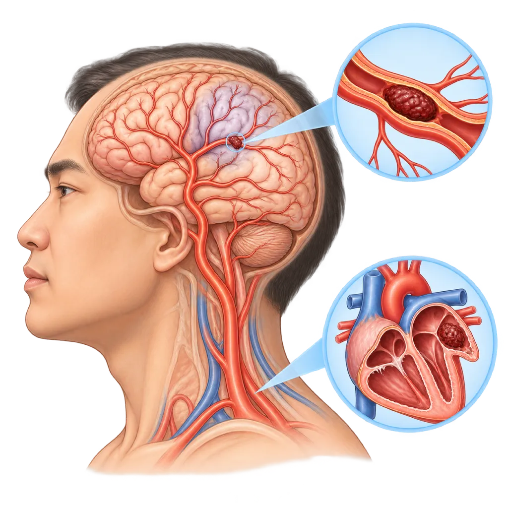
A stroke happens when blood flow to part of the brain is blocked or a blood vessel in the brain bursts, so brain cells are damaged within minutes. A stroke is an emergency: go to the emergency room right away if signs of a stroke appear.
Sources01 / Understanding the condition
About Stroke
A stroke is a sudden disturbance of brain function caused by a disruption of blood flow to part of the brain. The brain needs a constant supply of oxygen and sugar from the blood. When that supply is cut off, brain cells in the affected area begin to be damaged within minutes, so the functions they control, such as movement, speech, or vision, are also affected.
Types of Stroke
Strokes are divided into two main groups. Ischemic stroke happens when a blood vessel in the brain is blocked by a clot or plaque, and it is the more common type. Hemorrhagic stroke happens when a blood vessel in or around the brain bursts. There are also transient ischemic attacks (TIA) and strokes with mild symptoms, covered on the Minor Stroke and TIA page, as well as blockage of a retinal artery, known as an eye stroke. A list of all the types appears on this page.
Time Is Critical
Neurologists use the phrase "time is brain." In ischemic stroke, clot-dissolving medicine can be given by infusion if the patient arrives within 4.5 hours of symptom onset, and a blockage in a large vessel can be removed with a catheter in selected patients up to 24 hours. Before that, a CT or MRI of the head must be done to make sure there is no bleeding, because the two types of stroke are treated differently. That is why it helps to recognize the signs with the SeGeRa Ke RS reminder and to note the time symptoms started or the time the person was last seen normal.
How Stroke Is Linked to the Heart
Many strokes start in the heart or in the large blood vessels of the neck. Atrial fibrillation, an irregular heart rhythm, makes blood clot easily in the left atrium, and these clots can break off and travel to the brain. This condition is covered on the Atrial Fibrillation page. Cholesterol plaque in the carotid arteries of the neck can narrow blood flow or release fragments to the brain, covered on the Atherosclerosis page. In some strokes in young people, a clot from a vein crosses through a small opening in the wall between the heart's upper chambers, covered on the Patent Foramen Ovale page. Heart attacks and heart valve disease can also be sources of clots.
That is why, after an ischemic stroke or TIA, heart tests such as an ECG, rhythm recording over several days, and echocardiography are part of the search for the cause. The risk factors are also the same as for heart disease: high blood pressure, diabetes, high cholesterol, smoking, and lack of exercise. Controlling them protects both the brain and the heart.
02 / Understanding the condition
Types and Related Conditions
The following conditions belong to the Stroke group. Open each one for a full explanation.
03 / Understanding the condition
Symptoms to Recognize.
Common Symptoms
Stroke symptoms appear suddenly, usually on one side of the body, and can occur during activity or on waking up.
- An uneven face or a drooping mouth when smiling
- Weakness, heaviness, or difficulty moving an arm or leg on one side
- Numbness or tingling in the face, arm, or leg on one side
- Slurred speech, trouble finding words, or speech that does not make sense
- Sudden blurred, darkened, or double vision
- Spinning dizziness, staggering, or trouble keeping your balance
- Difficulty swallowing or choking when drinking
- A severe headache unlike any you have had before
04 / Understanding the condition
What Causes This Condition?
- A clot that forms in a brain or neck blood vessel narrowed by cholesterol plaque (atherosclerosis)
- A clot from the heart that travels to the brain, for example in atrial fibrillation or after a heart attack
- Blockage of small blood vessels deep inside the brain due to long-standing high blood pressure and diabetes
- Rupture of a small blood vessel inside the brain, mainly due to uncontrolled high blood pressure
- Rupture of an aneurysm, a bulge in the wall of a brain artery, covered on the Cerebral Aneurysm page
- Less common causes: a tear in the wall of a neck artery, blood clotting disorders, inflammation of blood vessels, or congenital blood vessel abnormalities
05 / Understanding the condition
Who Is at Higher Risk?
Medical
High blood pressure is the main risk factor. Other factors are diabetes, high LDL cholesterol, atrial fibrillation and other heart diseases, chronic kidney disease, and a previous TIA or stroke.
Lifestyle
Smoking, a diet high in salt and low in vegetables and fruit, physical inactivity, excess body weight, and heavy alcohol use. The INTERSTROKE study showed that about 90 percent of stroke risk is linked to factors that can be changed.
Age
Risk increases with age, especially after 55, although stroke can also happen at a young age.
Genetic
A history of stroke in a parent or sibling, inherited high cholesterol, and rare inherited disorders of blood clotting or blood vessels.
Other
Breathing problems during sleep (sleep apnea), long-term stress and depression, pregnancy and the postpartum period, and estrogen-containing birth control pills in women who smoke or have migraine with aura.
Groups at Higher Risk
- People with Hypertension: High blood pressure damages the walls of the brain's blood vessels and is the main risk factor for both ischemic and hemorrhagic stroke.
- People with Atrial Fibrillation: An irregular rhythm makes clots more likely to form in the heart. Blood thinners lower the risk of stroke in the majority of people with this condition.
- People with Diabetes: Diabetes speeds up damage to large and small blood vessels, including those in the brain.
- Older Adults: Stroke risk increases with age, and many older adults have more than one risk factor at the same time.
- TIA or Stroke Survivors: People who have had a TIA or stroke have a higher risk of another stroke, especially in the first months.
06 / Understanding the condition
How Is It Diagnosed?
When a stroke is suspected, testing happens at the same time as treatment. The emergency team asks when the symptoms started or when the person was last seen normal, checks blood pressure, blood sugar, and level of consciousness, and then rates the severity of symptoms using the NIHSS stroke scale. A head CT is done as quickly as possible to rule out bleeding. If a blockage in a large vessel is suspected, CT angiography or MRI is used to determine whether the blockage can be removed with a catheter.
Once the condition is stable, doctors look for the cause so the stroke does not happen again. Tests include imaging of the blood vessels in the neck and brain, an ECG and heart rhythm recording over several days, echocardiography, and blood tests for sugar, cholesterol, and blood clotting. For strokes in young people without a clear cause, doctors may add transesophageal echocardiography and special blood clotting tests.
A head CT without contrast right after arriving at the emergency room to distinguish a stroke caused by a blockage from one caused by bleeding, because they are treated differently.
Brain MRI to see damaged areas of the brain in more detail, including small strokes that do not show on CT.
CT angiography or MR angiography to see the blood vessels of the brain and neck and look for blockages, narrowing, or aneurysms.
Blood Sugar Test
Blood sugar is checked right away because blood sugar that is too low or too high can cause symptoms that look like a stroke.
Vascular Ultrasound
Ultrasound of the carotid arteries in the neck to look for plaque and narrowing that can release clots to the brain.
Long-Term Heart Rhythm Monitoring
An ECG followed by rhythm recording for several days to several weeks to look for atrial fibrillation that comes and goes.
Echocardiography
A heart ultrasound to look for clots, valve abnormalities, or an opening in the heart's dividing wall as a source of clots.
07 / Understanding the condition
Treatment Options.
Treatment is decided by a doctor after an assessment. The categories below can help you follow the conversation with your doctor.
Clot-Dissolving Medicine (Thrombolysis)
For ischemic stroke, medicine is given by infusion if the patient arrives within 4.5 hours of symptom onset and there is no bleeding or other contraindication. Your doctor determines whether it is suitable.
Clot Removal with a Catheter (Thrombectomy)
For blockages in large vessels, the clot is removed with a catheter inserted from the groin up to the brain. This can be done up to 24 hours in selected patients.
Hemorrhagic Stroke Treatment
Blood pressure is lowered in a controlled way, the effect of blood thinners is reversed, and in certain situations a neurosurgeon removes the clot or secures the ruptured vessel.
Stroke Unit Care
Care by a specialized team to monitor blood pressure, blood sugar, temperature, and swallowing ability, and to prevent pneumonia and vein clots.
Preventing Another Stroke
Antiplatelet or anticoagulant medicines based on the cause, statins, blood pressure medicines, and procedures to address the source of clots, such as carotid surgery or LAA closure. Your doctor determines the type of medicine.
Rehabilitation After Stroke
Physical therapy, speech therapy, and occupational therapy start as early as possible once the condition is stable to restore everyday abilities.
08 / Understanding the condition
Complications to Watch For.
Paralysis and Movement Problems
Weakness on one side of the body, stiff muscles, or balance problems that need rehabilitation exercises and can improve gradually.
Speech and Swallowing Problems
Difficulty speaking, understanding words, or swallowing. Swallowing problems can let food enter the airway and cause pneumonia.
Memory and Mood Problems
Problems with thinking and memory, depression, and anxiety often appear after a stroke and need to be recognized so they can be treated.
Blood Clots in the Leg Veins
Lying down for long periods makes clots more likely to form in the leg veins, and these can break off and travel to the lungs. Covered on the Deep Vein Thrombosis page.
Seizures
Some stroke survivors, especially after a hemorrhagic stroke or a large stroke, develop seizures later on.
Recurrent Stroke
The risk of another stroke is higher in the first months, so medicines and risk factor control need to be followed regularly.
09 / Understanding the condition
What Can You Do?
- Check your blood pressure regularly and keep it within the target agreed with your doctor
- Quit smoking and avoid secondhand smoke
- Control your blood sugar and cholesterol, and take your medicines as prescribed
- If you have atrial fibrillation, discuss blood thinners with your cardiologist
- Stay active for at least 150 minutes per week, for example by brisk walking
- Limit salt, eat more vegetables and fruit, and avoid heavy drinking
- Learn the SeGeRa Ke RS signs and teach them to your family
10 / Understanding the condition
When Should You See a Doctor?
A stroke always needs emergency help. If you or someone near you shows SeGeRa Ke RS signs, call 119 or go to a nearby emergency room right away, even if the symptoms improve on their own. Note the time they started, do not give food, drinks, or medicine by mouth, and do not drive yourself.
Make an appointment if:
- You had brief stroke symptoms some time ago that have not been checked
- You have just come home after a stroke and need follow-up on the cause and preventive medicines
- You have atrial fibrillation, often feel an irregular heartbeat, or have heart valve disease
- You have several risk factors, such as hypertension, diabetes, high cholesterol, or smoking
11 / Common questions
Questions You May Have.
Frequently asked questions about stroke, its emergency signs, and its link to the heart.
What are SeGeRa Ke RS and BE FAST?
Both are reminders of stroke signs. SeGeRa Ke RS, from the Indonesian Ministry of Health, stands for Indonesian words meaning an uneven smile, weakness on one side of the body, slurred speech, numbness on one side, sudden blurred vision, and a severe headache or unsteadiness. The English acronym BE FAST stands for balance, eyes, face, arm, speech, and time to get help right away. Even one sign is enough reason to go to the emergency room immediately.
What should family members do if they suspect a stroke?
Call 119 or take the person to a nearby emergency room as quickly as possible. Note the time the symptoms started or the time the person was last seen normal, because this information determines treatment options. If the person is vomiting or drowsy, lay them on their side. Do not give food, drinks, or medicine by mouth, including aspirin, and do not waste time on unproven methods, such as pricking the fingertips.
How is stroke related to heart disease?
The heart can be a source of clots that block blood vessels in the brain, especially in atrial fibrillation, after a heart attack, or in valve disease. In some strokes in young people, a clot from a vein crosses through a patent foramen ovale. Plaque in the carotid arteries of the neck forms through the same process as plaque in the coronary arteries. That is why heart tests are part of the search for the cause of a stroke.
Can people recover from a stroke?
Many stroke survivors improve, especially in the first three to six months, when the brain is actively forming new nerve pathways. How much recovery happens depends on the size and location of the damage, how quickly treatment was given, and how consistently rehabilitation is followed. Physical therapy, speech therapy, and family support help survivors become independent again. Some symptoms may remain, so rehabilitation goals are tailored to each person's condition.
Can stroke happen at a young age?
Yes. Although it is more common in older adults, stroke also occurs in people under 50. The causes are more varied, such as a tear in the wall of a neck artery, a patent foramen ovale, blood clotting disorders, heart disease, or stimulant drug use. Risk factors such as hypertension, smoking, and obesity are also increasingly found in young people, so the search for the cause is usually more extensive.
How can I prevent another stroke?
Prevention is tailored to the cause of the stroke. It generally includes antiplatelet or anticoagulant medicines, statins, and blood pressure medicines, along with quitting smoking, staying active, and eating a low-salt diet. If the cause is narrowing of a carotid artery or a patent foramen ovale, your doctor may recommend a procedure. Take your medicines regularly and do not stop them on your own, because the risk of another stroke is higher in the first months.
Sources and Medical Review
Translated from the Indonesian edition medically reviewed by dr. Emanoel Oepangat, Sp.JP(K).
Sources as cited in the Indonesian edition.
- Powers WJ, Rabinstein AA, Ackerson T, et al. Guidelines for the Early Management of Patients With Acute Ischemic Stroke: 2019 Update to the 2018 Guidelines for the Early Management of Acute Ischemic Stroke: A Guideline for Healthcare Professionals From the American Heart Association/American Stroke Association. Stroke. 2019;50(12):e344-e418.
- Kleindorfer DO, Towfighi A, Chaturvedi S, et al. 2021 Guideline for the Prevention of Stroke in Patients With Stroke and Transient Ischemic Attack: A Guideline From the American Heart Association/American Stroke Association. Stroke. 2021;52(7):e364-e467.
- Greenberg SM, Ziai WC, Cordonnier C, et al. 2022 Guideline for the Management of Patients With Spontaneous Intracerebral Hemorrhage: A Guideline From the American Heart Association/American Stroke Association. Stroke. 2022;53(7):e282-e361.
- Berge E, Whiteley W, Audebert H, et al. European Stroke Organisation (ESO) guidelines on intravenous thrombolysis for acute ischaemic stroke. Eur Stroke J. 2021;6(1):I-LXII.
- Aroor S, Singh R, Goldstein LB. BE-FAST (Balance, Eyes, Face, Arm, Speech, Time): Reducing the Proportion of Strokes Missed Using the FAST Mnemonic. Stroke. 2017;48(2):479-81.
- O'Donnell MJ, Chin SL, Rangarajan S, et al. Global and regional effects of potentially modifiable risk factors associated with acute stroke in 32 countries (INTERSTROKE): a case-control study. Lancet. 2016;388(10046):761-75.
- Van Gelder IC, Rienstra M, Bunting KV, et al. 2024 ESC Guidelines for the management of atrial fibrillation developed in collaboration with the European Association for Cardio-Thoracic Surgery (EACTS). Eur Heart J. 2024;45(36):3314-414.
Doctors Who Treat This Condition

dr. Sunu B. Raharjo, Sp.JP(K), Ph.D.
Arrhythmia and Electrophysiology
Arrhythmia and Device Center
Clinical focusClinical and Interventional Electrophysiology · Cardiac Arrhythmia Ablation
View Profile
dr. Debby C. Soemitha, Sp.PD
Internal Medicine
Clinical focusDiabetes and Diabetes Complications · Obesity and Metabolic Disease
View Profile
dr. Emanoel Oepangat, Sp.JP(K)
Interventional Cardiology
Vascular Diagnostic and Therapeutic Center +1 center
Clinical focusCoronary Intervention · Peripheral Vascular Intervention
View Profile