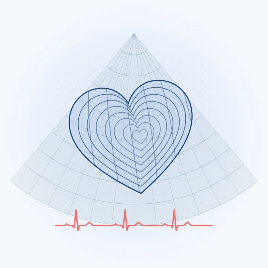
Understanding the technology
About This Technology.
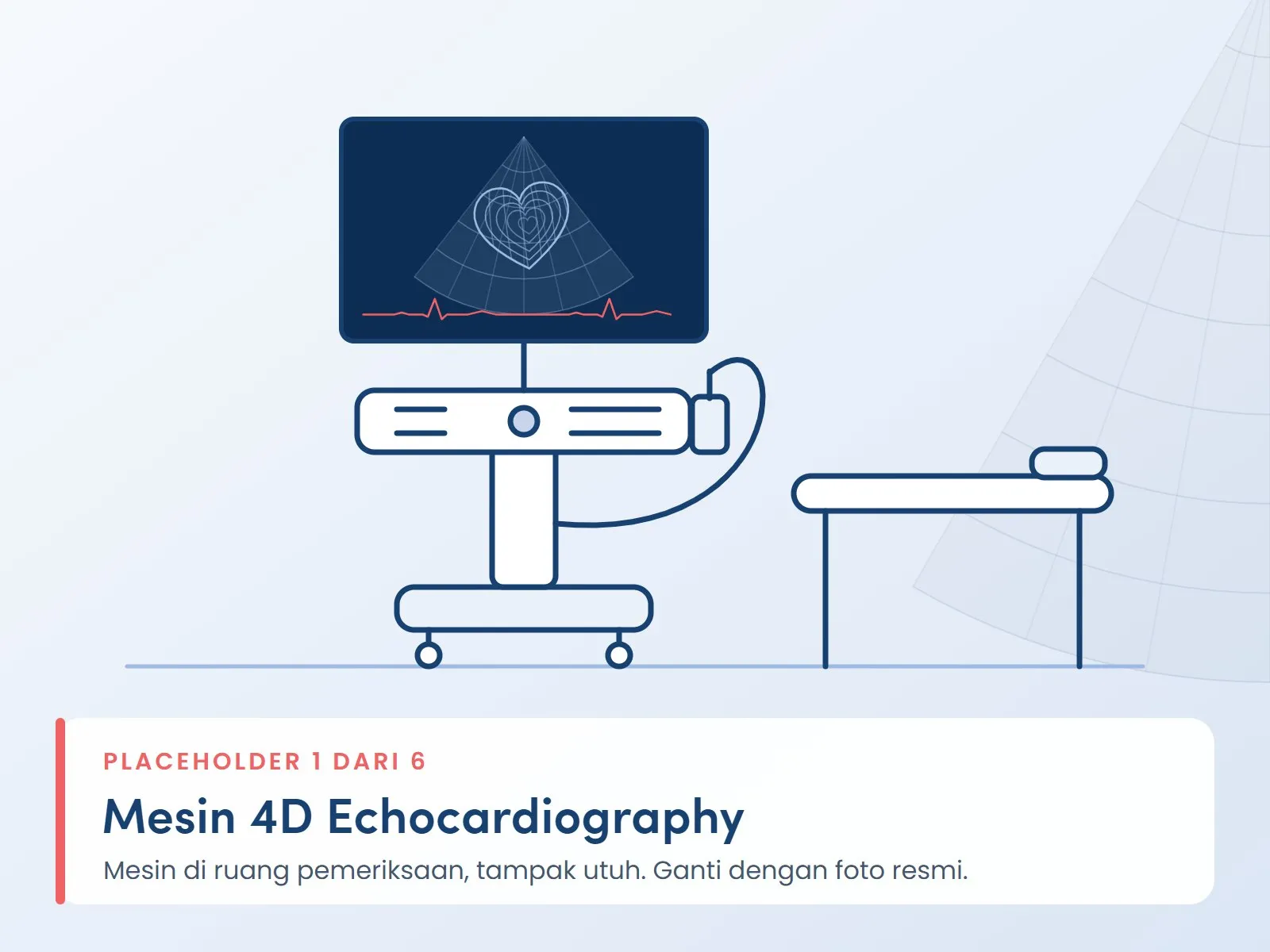
4D echocardiography is an ultrasound scan that records three-dimensional images of the beating heart in real time. Doctors use it to measure chamber volumes and pumping strength, to see the shape and movement of the valves, and to assess structural problems such as a hole in the wall dividing the heart. It uses sound waves, with no radiation.
What Is 4D Echocardiography?
Echocardiography, or echo, is a heart scan that uses ultrasound. A conventional echo shows the heart as two-dimensional slices, so the doctor has to combine several slices to picture the actual shape of the heart.
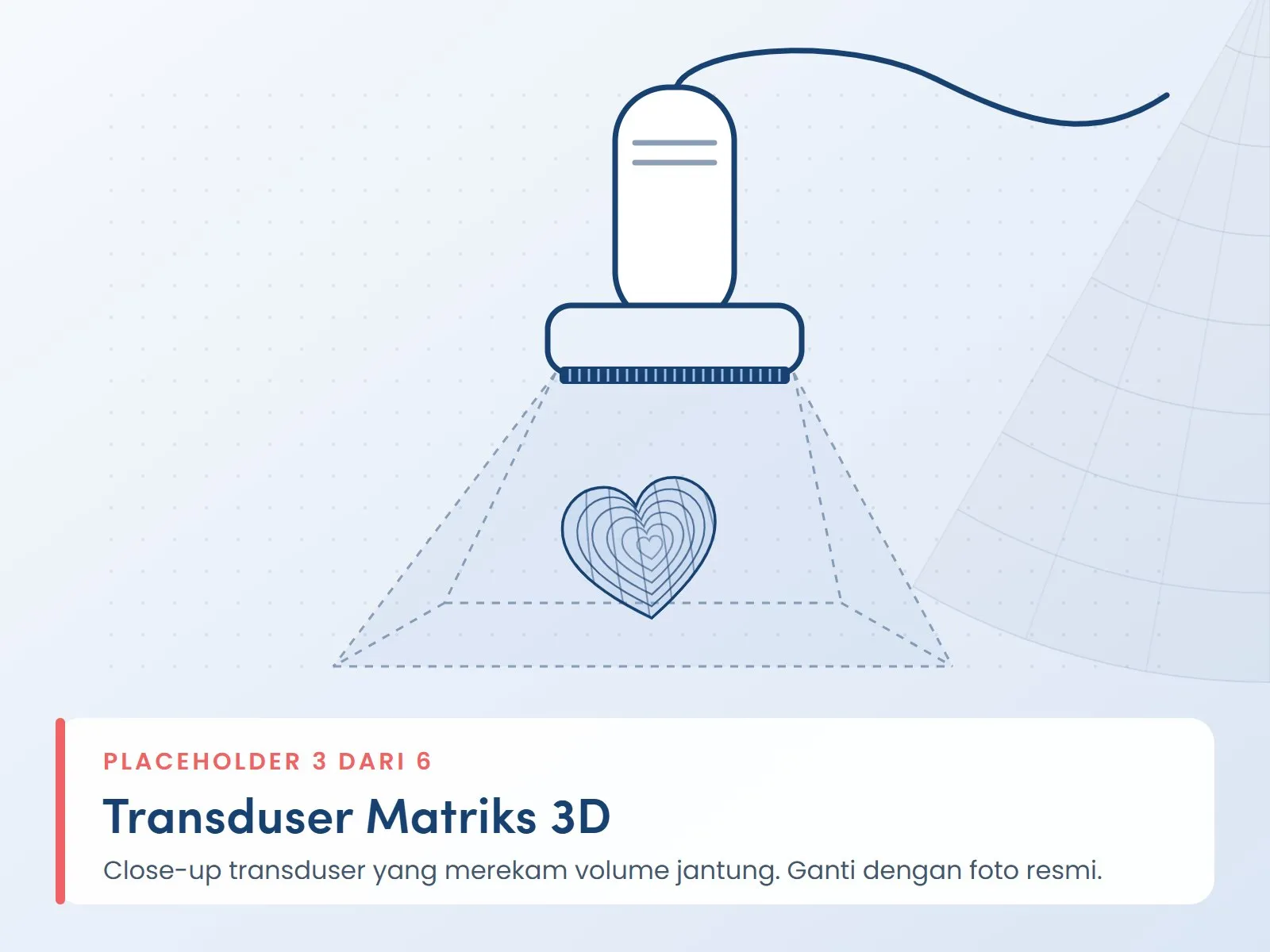
4D echocardiography uses a special transducer that records a whole volume of the heart at once. The result is a three-dimensional image that moves with each heartbeat; this element of time is what is called the fourth dimension. From a single recording, the doctor can rotate and crop the image from many angles without scanning again.
How This Technology Helps Your Doctor
- Measuring pumping strength. Left ventricular volumes and ejection fraction are calculated directly from the three-dimensional image, rather than estimated from formulas that assume a shape for the heart.
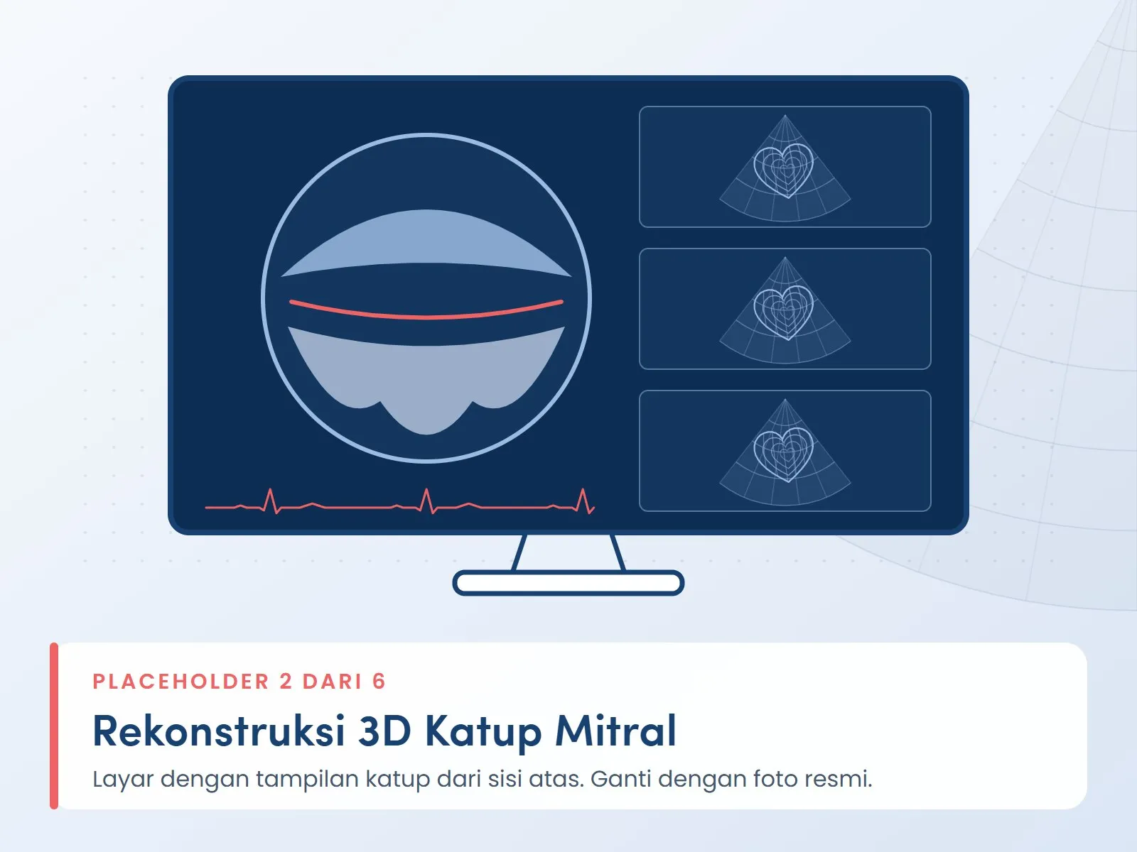
- Seeing the whole valve. The mitral valve can be displayed from above, much as a surgeon sees it, which makes the location and extent of a problem easier to understand.
- Assessing structural problems. The size and shape of a hole in the wall dividing the heart can be assessed from several sides.
- Planning procedures. Three-dimensional images help the heart team plan, and when needed guide, procedures on the valves and other heart structures.
Two Ways to Perform the Scan
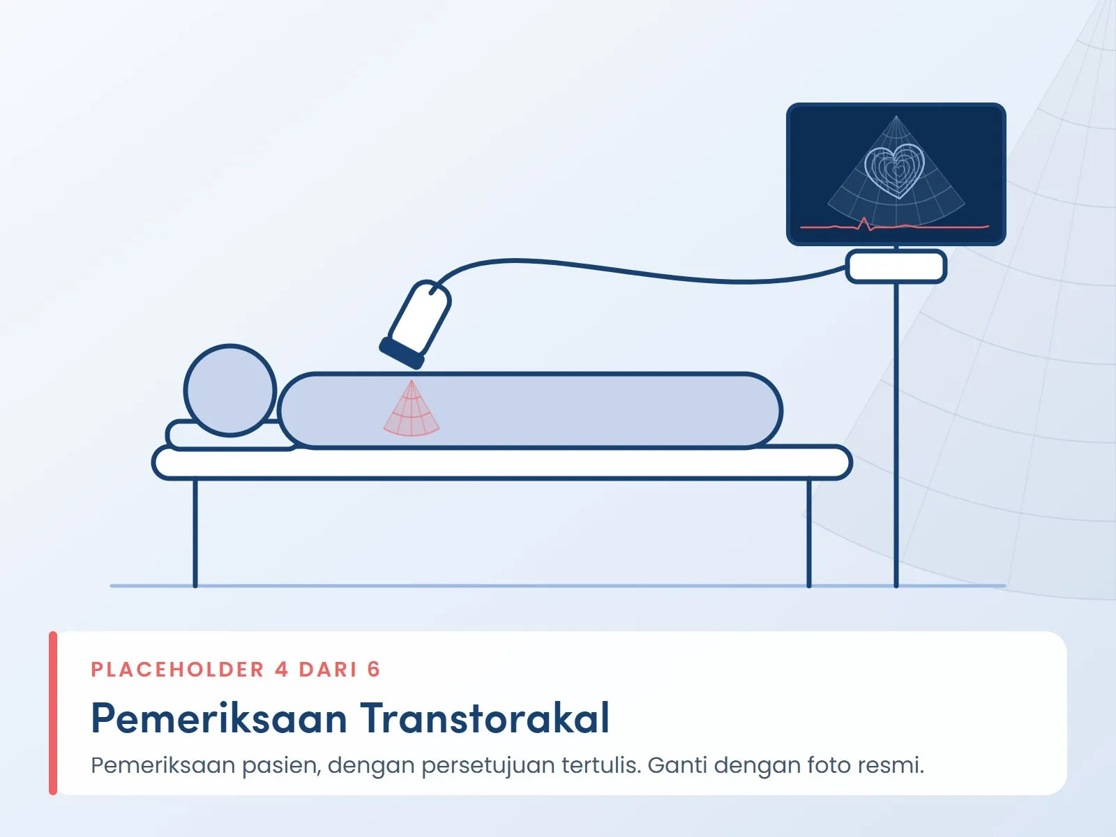
Transthoracic (TTE). The transducer is placed on your chest with the help of gel. This is the approach used most often, and it does not require sedation.
Transesophageal (TEE). A small probe is passed through the mouth into the esophagus, which lies directly behind the heart, under light sedation. This approach is used when your doctor needs clearer images of the valves or the wall dividing the heart.
A Complement, Not a Replacement
Three-dimensional images are used together with two-dimensional echo and Doppler in a single examination. Image quality can be affected by chest shape, body weight, lung disease, or an irregular heart rhythm. For this reason, your doctor decides when three-dimensional images are needed, and when another test such as a cardiac CT scan is more suitable.
After the Scan
The recording is analyzed and interpreted by a cardiologist. The results are discussed with you during your consultation, including whether further tests or regular follow-up are needed.
Clinical use
Uses and Benefits.
Whether this technology suits your condition is decided by a doctor after an evaluation.
- Measuring heart chamber volumes and left ventricular ejection fraction (pumping strength)
- Assessing the size and function of the right ventricle
- Viewing the shape and movement of the mitral, aortic, and tricuspid valves, including leaks or narrowing
- Measuring holes in the wall dividing the heart, such as an atrial septal defect (ASD)
- Evaluating congenital heart disease and structural heart abnormalities
- Planning and monitoring procedures on the valves and heart structures
- Regular monitoring of heart pumping function, for example in heart failure
Benefits for the Doctor's Assessment
A Complete View of the Heart from One Recording
A single volume recording can be rotated and cropped from many angles during analysis, so your doctor does not rely on two-dimensional slices alone.
Volume Measurement Without Shape Assumptions
Left ventricular volumes and ejection fraction are calculated directly from the three-dimensional image. ASE and EACVI guidance recommends this method when it is available and image quality is adequate.
A Surgeon's View of the Valve
The mitral valve can be displayed from above, helping the heart team understand the location and extent of a problem before planning valve repair.
No Radiation
The scan uses ultrasound waves, so it can be repeated for regular follow-up. The transthoracic approach is performed from the surface of the chest with no incision.
Brief Technical Information
- Type of examination
- Transthoracic (TTE) and transesophageal (TEE)
- Imaging modes
- 2D, Doppler, and real-time 3D (4D)
- Image source
- Ultrasound waves, no ionizing radiation
- Service location
- Cardiovascular Diagnostic Center
Documentation