
Quick answer
What Is Stress Echocardiography?
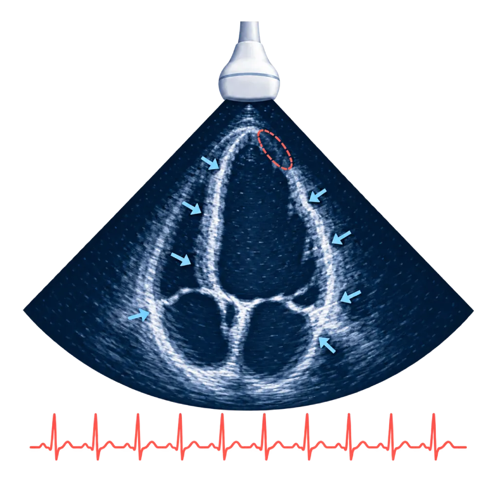
Stress echocardiography is a heart ultrasound done at rest and while the heart is working hard, using a treadmill, a stationary bike, or medication. If part of the heart wall does not contract strongly under stress, blood flow to that area may be reduced. This test does not use radiation.
Sources01 / Understanding the procedure
About Stress Echocardiography
What Is Stress Echocardiography
Stress echocardiography combines a heart ultrasound (echocardiography) with a stress test. The doctor records images of the heart at rest, then records them again while the heart is working hard. By comparing the two, the doctor can see how the heart muscle and valves respond to stress in ways that are not visible at rest.
How It Works in Brief
Under stress, healthy heart muscle contracts more strongly and its walls thicken. If one of the coronary arteries is narrowed, the part of the muscle it supplies does not get enough blood, so its movement weakens or lags behind. Sound waves from the ultrasound probe show these changes in motion directly on the screen, without radiation. If the images are unclear, the doctor may use a special ultrasound contrast agent made of very small bubbles, which is different from iodinated contrast.
What It Is Used For
- Coronary artery disease: looks for areas of heart muscle that do not get enough blood when working hard, and assesses how extensive they are.
- Heart muscle that is still viable: with a low dose of medication, the doctor assesses whether weakened muscle can still recover if its blood flow is restored.
- Valve disease: assesses changes in pressure and flow in narrowed or leaking valves, including when symptoms do not match the results of routine tests.
- Hypertrophic cardiomyopathy: checks whether an obstruction to blood flow out of the heart appears under stress.
What the Results Mean
A normal result means the whole heart wall contracts more strongly under stress, and the risk of cardiac events in the near future is generally low. If part of the wall lags behind, its location and extent help the doctor estimate which artery is narrowed and decide whether cardiac catheterization is needed. In valve disease, the results help determine the right time for a procedure.
02 / Understanding the procedure
When Is This Procedure Considered?
A doctor may consider this procedure in the situations below. The decision is always made after a thorough assessment.
- Chest Pain with Suspected Coronary Artery DiseaseLooks for heart wall motion that weakens under stress.
- Assessing Known Coronary NarrowingHelps decide whether a stent or bypass surgery is needed.
- Weak Heart Muscle After a Heart AttackAssesses whether the muscle can still recover, using a low dose of medication.
- Low-Flow Aortic StenosisDistinguishes truly severe valve narrowing from narrowing that only appears severe.
- Severe Valve Disease Without SymptomsBrings out symptoms or pressure changes that are hidden at rest.
- Hypertrophic Cardiomyopathy with SymptomsLooks for an obstruction to blood flow out of the heart that appears under stress.
Situations to Consider First
- Recent Heart Attack or Unstable AnginaThe stress test is postponed until your condition is stable.
- Uncontrolled Dangerous Rhythm DisordersThese need to be treated before the heart is put under stress.
- Very High Blood PressureBlood pressure needs to be controlled before the test.
- Currently Worsening Heart FailureThe test is done after your condition improves.
- Other Acute IllnessesFor example, pulmonary embolism, inflammation of the heart muscle, or a tear in the aorta.
03 / Before the procedure
How Should You Prepare?
Follow the instructions given when your test is scheduled.
Eating and drinking. Avoid heavy meals for about 3 hours beforehand. Avoid coffee, tea, and smoking on the day of the test.
Medications. Your doctor may ask you to temporarily stop medications that slow the heart rate, such as beta blockers, 1 to 2 days beforehand. Do not stop any medication on your own.
Clothing. Wear clothing and shoes that are comfortable for brisk walking.
Bring your records. Bring a list of your medications and any previous echocardiography or ECG results, and let the staff know if you have asthma or allergies, or are pregnant.
Unsure about the preparation instructions? Ask our team before the procedure.
04 / During the procedure
How Is the Procedure Done?
The whole test usually takes about 1 hour. ECG electrodes are attached to your chest, and your blood pressure is checked regularly.
Echocardiography at Rest
You lie on your left side. The doctor applies gel to your chest and places the ultrasound probe to record images of the heart from several angles.
Stress Test
You walk on a treadmill or pedal a stationary bike with increasing effort. If you cannot exercise, dobutamine is given through an IV at a dose increased step by step until your heart rate reaches the target.
Echocardiography Under Stress
Images are recorded again at peak stress. On the treadmill, you lie down again right away so the images can be taken within one to two minutes of stopping, before the heart slows down.
Recovery and Reading
You are monitored until your heart rate and blood pressure return to normal. The doctor compares the rest and stress images side by side.
05 / Benefits and risks
Benefits and Risks Should Be Discussed Together.
Every medical procedure has benefits and risks. Your doctor will explain both for your situation before the procedure.
Benefits You Can Expect
- No RadiationUses sound waves, so it can be repeated if needed.
- Seeing Wall Motion DirectlyShows the parts of the heart muscle that do not get enough blood when working hard.
- Assesses Valves and Pressures at the Same TimeUseful in valve disease and hypertrophic cardiomyopathy.
- Can Be Done with MedicationAn option for patients who cannot walk on a treadmill.
- A Normal Result Is ReassuringThe risk of cardiac events in the near future is generally low.
The benefits of a procedure are not a promise of a cure. Ask how the results will affect your care plan.
Risks You Should Know
- Chest Pain or Breathlessness Under StressThe test is stopped if symptoms appear, and the team is ready to manage them.
- Heart Rhythm DisturbancesUsually mild and brief; serious ones are rare.
- Blood Pressure ChangesBlood pressure can rise or fall during stress.
- Unclear ImagesIn some patients, images are hard to obtain, so an ultrasound contrast agent or another test is needed.
Risks can differ from person to person. A full explanation and consent are given after an individual assessment.
06 / After the procedure
What Is Recovery Like?
You can go home and resume your usual activities right away once your heart rate is back to normal. Feeling tired after the treadmill is normal and goes away with rest.
After stress medication. Palpitations, a warm feeling, or slight trembling usually go away within a few minutes after the infusion is stopped.
Warning signs to watch for. Go to the emergency room right away or contact the medical team if you experience:
- Chest pain that does not go away with rest
- Shortness of breath that gets worse
- A very fast or irregular heartbeat
- Severe dizziness or fainting
Know the Signs That Need Prompt Care.
Follow the team's guidance on warning signs. If severe or sudden symptoms appear after the procedure, go to the nearest emergency department right away or call Heart Attack Center 1500 258.
07 / Team and support
Doctors and Procedure Support
The doctors below are listed for this procedure. Listed by name, not by ranking.

dr. BRM Ario S. Kuncoro, Sp.JP(K)
Echocardiography & Cardiovascular Imaging
Cardiovascular Diagnostic Center +2 centers
Clinical focusAdvanced and 3D Echocardiography · Transesophageal Echocardiography
View Profile
Dr. med. dr. Denio A. Ridjab, Sp.JP(K)
Interventional Cardiology
Coronary Intervention Procedures +2 centers
Clinical focusInterventional Cardiology · Coronary Artery Disease
View Profile
dr. Sri Diniharini, Sp.JP(K)
Echocardiography & Cardiovascular Imaging
Cardiovascular Diagnostic Center +1 center
Clinical focusAdvanced Echocardiography · Transesophageal Echocardiography
View Profile4D Echocardiography
An ultrasound scan of the heart that records three-dimensional images in real time to assess pumping function, the valves, and…
The availability of technology and test support is confirmed with the team. Technology is chosen according to clinical need.
08 / Common questions
Before You Plan the Procedure.
Frequently asked questions about stress echocardiography. Your doctor will explain what the results mean for your condition.
How is it different from a standard treadmill test?
A standard treadmill test only assesses the ECG recording while the heart is working hard. Stress echocardiography adds ultrasound images of the heart before and after stress, so the doctor can see directly which parts of the wall are not moving normally. The results are more accurate, especially if your ECG is difficult to read from the start.
How is it different from myocardial perfusion imaging?
Both look for areas of heart muscle that do not get enough blood under stress. Stress echocardiography assesses wall motion with sound waves, without radiation, and can assess the valves at the same time. Myocardial perfusion imaging assesses blood flow with a radioactive tracer and is more suitable when ultrasound images are hard to obtain. Your doctor chooses the one that suits you.
What if I cannot manage walking on the treadmill?
The doctor can give dobutamine through an IV, which makes the heart beat faster and more strongly, as it does during exercise. The dose is increased step by step while the ECG, blood pressure, and ultrasound images are continuously monitored. Any palpitations or warm feeling usually go away within a few minutes after the infusion is stopped.
Is this test safe?
Stress echocardiography is generally safe because it is done under a doctor's supervision with continuous ECG and blood pressure monitoring. The test is stopped if symptoms or worrying changes appear. Serious complications, such as severe rhythm disturbances, are rare. This test also does not use radiation.
Why is stress echocardiography used in valve disease?
In some valve diseases, symptoms and pressure in the heart only become apparent under stress. Stress echocardiography shows changes in pressure and flow across a narrowed or leaking valve, as well as pressure in the lungs. The results help the doctor decide whether the valve needs to be repaired or replaced now, or can simply be monitored.
Do I need to stop taking my medications before the test?
Sometimes. Medications that slow the heart rate, such as beta blockers, can make it hard for the heart to reach the target level of stress, making the results less accurate. Your doctor will decide which medications to pause and for how long, depending on the purpose of the test. Do not stop any medication without your doctor's instructions.
Sources and Medical Review
Translated from the Indonesian edition medically reviewed by dr. BRM Ario S. Kuncoro, Sp.JP(K).
Sources as cited in the Indonesian edition.
- Vrints C, Andreotti F, Koskinas KC, et al. 2024 ESC Guidelines for the management of chronic coronary syndromes. Eur Heart J. 2024;45(36):3415-3537.
- Pellikka PA, Arruda-Olson A, Chaudhry FA, et al. Guidelines for performance, interpretation, and application of stress echocardiography in ischemic heart disease: from the American Society of Echocardiography. J Am Soc Echocardiogr. 2020;33(1):1-41.e8.
- Lancellotti P, Pellikka PA, Budts W, et al. The clinical use of stress echocardiography in non-ischaemic heart disease: recommendations from the European Association of Cardiovascular Imaging and the American Society of Echocardiography. Eur Heart J Cardiovasc Imaging. 2016;17(11):1191-1229.
- Praz F, Borger MA, Lanz J, et al. 2025 ESC/EACTS Guidelines for the management of valvular heart disease. Eur Heart J. 2025;46(44):4635-4736.