
Quick answer
What Is Microvascular Angina?
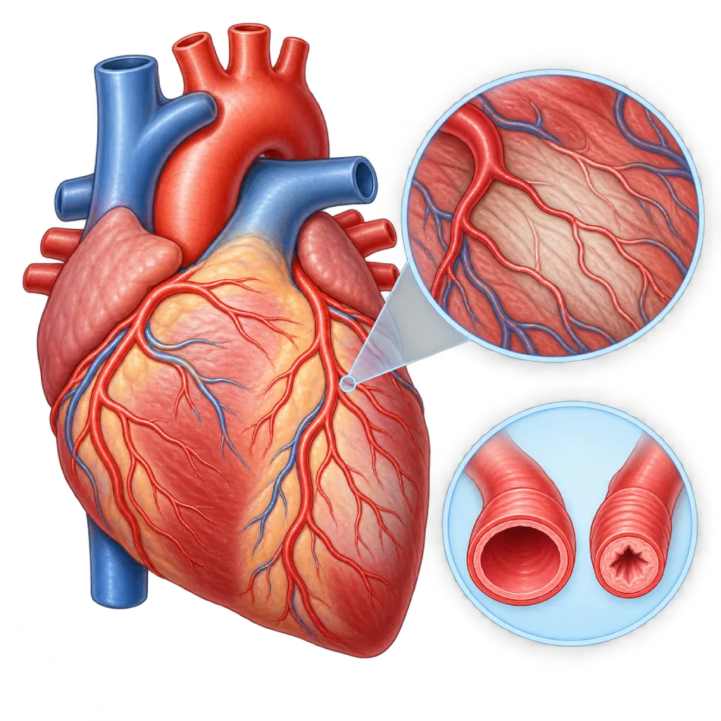
Microvascular angina is chest pain caused by a problem in the very small blood vessels of the heart muscle, so blood flow does not increase when needed. The large coronary arteries often look normal or only slightly narrowed on testing.
SourcesSigns That Need Urgent Help
- Chest pain that is more severe or lasts longer than your usual pattern
- Chest pain with cold sweats, nausea, or vomiting
- Severe shortness of breath or being unable to lie flat
- Fainting or nearly fainting
- A very fast or irregular heartbeat
If these signs appear, go to the nearest emergency department right away or call Heart Attack Center 1500 258.
01 / Understanding the condition
About Microvascular Angina
Microvascular angina is angina pectoris that arises from the coronary microcirculation, the network of very small blood vessels inside the heart muscle. These vessels are much smaller than the coronary arteries seen on angiography. It is these small vessels that regulate how much blood flows to the heart muscle. When they do not work properly, the heart muscle can run short of oxygen even though the large coronary arteries are not blocked.
Part of ANOCA and INOCA
Doctors call angina without significant blockage in the large coronary arteries ANOCA (angina with non-obstructive coronary arteries). When reduced blood flow is proven, the condition is called INOCA (ischemia with non-obstructive coronary arteries). Microvascular angina is one of its main causes, alongside spasm of the large coronary arteries, which is covered on the Vasospastic Angina page. Both mechanisms can occur together in the same person.
Two Main Mechanisms
First, the small vessels are less able to widen when the heart is working hard, so coronary flow reserve falls. Second, the small vessels tend to constrict excessively, which is called microvascular spasm. These problems are linked to damage to the inner lining of the blood vessels (endothelium) caused by hypertension, diabetes, high cholesterol, smoking, and chronic inflammation, as well as to thickening of the heart muscle.
Why It Is Often Recognized Late
Coronary angiography shows only the large arteries. As a result, many people are told that their heart is normal, even though their symptoms are real and keep coming back. The symptoms resemble typical angina, but they often last longer, can appear at rest or after activity has ended, and respond less well to short-acting nitrates. This pattern sometimes leads to the symptoms being mistaken for a stomach problem or anxiety alone. Microvascular angina is found more often in women, especially in middle age and after menopause, although men can have it too. People with diabetes, hypertension, and autoimmune diseases are also affected more often.
Treatment Matched to the Mechanism
With coronary function testing, doctors can find out which mechanism is dominant and choose suitable medicines. The CorMicA trial showed that treatment tailored to the results of coronary function testing improved angina symptoms and quality of life compared with usual care. Risk factor control and lifestyle changes remain the foundation of treatment.
02 / Understanding the condition
Symptoms to Recognize.
Common Symptoms
- Pain, pressure, or heaviness in the chest during activity or emotional stress
- Pain that lasts longer, sometimes more than 10 minutes after activity stops
- Chest pain that can also occur at rest
- Shortness of breath and tiredness during activity
- Short-acting nitrate medicine relieves the pain less well in some people
- Discomfort spreading to the back, neck, jaw, or arm
Symptoms in Adult Women
Microvascular angina is found more often in women, and the following symptoms often come with it.
- Unusual and prolonged tiredness
- Shortness of breath without obvious chest pain
- Sleep disturbed by chest discomfort
- Chest pain triggered by emotional stress
- Symptoms that start around the time of menopause
03 / Understanding the condition
What Causes This Condition?
- Small blood vessels of the heart that are less able to widen when oxygen demand rises
- Spasm of the small blood vessels that narrows blood flow excessively
- Damage to the inner lining of the vessels (endothelial dysfunction) due to hypertension, diabetes, high cholesterol, or smoking
- Thickening of the heart muscle, for example from long-standing hypertension, hypertrophic cardiomyopathy, or aortic stenosis
- Chronic inflammation, for example in autoimmune diseases such as lupus or rheumatoid arthritis
- Increased sensitivity of the heart's nerves to pain in some people
04 / Understanding the condition
Who Is at Higher Risk?
Lifestyle
Smoking, too little physical activity, excess body weight, and long-term stress are linked to poor function of the small blood vessels.
Medical
Hypertension, diabetes, high cholesterol, chronic kidney disease, and autoimmune diseases such as lupus or rheumatoid arthritis raise the risk.
Age
It is found more often in middle age, especially in women after menopause, when the protection from the hormone estrogen decreases.
Other
A history of high blood pressure in pregnancy or preeclampsia, as well as thickening of the heart muscle from any cause, is linked to coronary microcirculation problems.
Groups at Higher Risk
- Middle-Aged and Postmenopausal Women: Angina without blockage, including microvascular angina, is found more often in women than in men.
- People with Diabetes and Hypertension: Long-term high blood sugar and high blood pressure damage the inner lining of the heart's small blood vessels.
- People with Autoimmune Diseases: Chronic inflammation in lupus, rheumatoid arthritis, or scleroderma can disrupt the function of the heart's small blood vessels.
- People with Chest Pain and a Normal Angiogram: If angina symptoms persist even though angiography found no blockage, microvascular angina should be considered.
05 / Understanding the condition
How Is It Diagnosed?
The first step is to make sure there is no significant narrowing in the large coronary arteries, usually with CT coronary angiography or coronary angiography. If the large arteries are not blocked but the angina symptoms are real, the doctor assesses how the small blood vessels are working.
The 2024 ESC guidelines and the EAPCI consensus recommend invasive coronary function testing in this situation. During catheterization, a sensor-tipped wire measures coronary flow reserve (CFR) and microvascular resistance (IMR) after a medicine that widens the vessels is given. An acetylcholine test then assesses the tendency toward spasm in both the large and small vessels. Methods without a catheter can also be used, such as a cardiac PET scan, cardiac MRI with a stress medicine, or Doppler echocardiography, to estimate blood flow to the heart muscle.
An ECG, blood tests, and echocardiography help rule out other causes, such as thickening of the heart muscle or valve disease. According to the COVADIS criteria, the diagnosis is made when typical symptoms of ischemia occur together with evidence of small vessel dysfunction and no significant blockage in the large arteries.
CT Coronary Angiography
Rules out significant narrowing of the large coronary arteries without a catheter.
Coronary Angiography
Shows the large coronary arteries and, in the same procedure, can be followed by invasive coronary function testing.
Fractional Flow Reserve (FFR)
The sensor-tipped wire used for FFR can also measure coronary flow reserve (CFR) and microvascular resistance (IMR).
Cardiac PET Scan
Measures blood flow to the heart muscle at rest and under medicine-induced stress, so flow reserve can be calculated without a catheter.
Cardiac MRI
MRI with a stress medicine can show reduced blood flow in the inner layer of the heart muscle and rule out cardiomyopathy.
Echocardiography
Assesses heart muscle thickening and the valves; Doppler of the left coronary artery can estimate coronary flow reserve.
An acetylcholine test during catheterization to see whether the small or large vessels are prone to spasm.
06 / Understanding the condition
Treatment Options.
Treatment is decided by a doctor after an assessment. The categories below can help you follow the conversation with your doctor.
Risk Factor Control
Blood pressure, cholesterol, and blood sugar are brought under control. Statins and ACE inhibitors or ARBs are often used because they can improve blood vessel function.
Antianginal Medicines Matched to the Mechanism
If the small vessels are less able to widen, beta blockers are often the first choice. If there is microvascular spasm, calcium channel blockers are more suitable. Your doctor decides the type and combination.
Additional Medicines
Ranolazine, nicorandil, or trimetazidine may be considered if symptoms persist. Short-acting nitrates can still help some people during an episode.
Lifestyle Changes
Quitting smoking, regular exercise, losing excess weight, and a heart-healthy diet help improve the function of the inner lining of the blood vessels.
Cardiac Rehabilitation
Supervised, structured exercise training can increase exercise capacity and reduce symptoms in angina without blockage.
Stress and Pain Management
Relaxation techniques, counseling, and enough sleep help, because stress and pain sensitivity can make symptoms worse.
07 / Understanding the condition
Complications to Watch For.
Recurrent Angina
Pain that keeps coming back can limit work and activities and lead people to visit the emergency room or undergo tests again and again.
Heart Attack
Small vessel dysfunction is linked to a higher risk of heart attack, including heart attacks that occur without blockage of the large arteries, covered on the Myocardial Infarction with Non-Obstructive Coronary Arteries (MINOCA) page.
Heart Failure with Preserved Ejection Fraction
Microcirculation problems are linked to a stiff heart muscle, covered on the Heart Failure with Preserved Ejection Fraction page.
Anxiety and Reduced Quality of Life
Unexplained symptoms often cause worry and a feeling of not being believed. A clear diagnosis helps you and your family understand the condition.
08 / Understanding the condition
What Can You Do?
- Quit smoking and avoid other people's cigarette smoke
- Keep your blood pressure, blood sugar, and cholesterol at target
- Stay active regularly, for example brisk walking for 30 minutes on the majority of days
- Manage stress in a way that suits you, such as breathing exercises, relaxation, or counseling
- Get enough sleep, and get checked if you snore loudly or stop breathing during sleep
- Take your medicines as prescribed and note your symptom pattern to discuss at follow-up visits
09 / Understanding the condition
When Should You See a Doctor?
See a cardiologist if you often feel pain or pressure in your chest, especially if earlier tests were called normal but the symptoms continue. Your symptoms are real and can be investigated further. Bring your previous test results, including angiography or coronary CT results, so the doctor can decide the next step. Chest pain at rest that you are having for the first time, or pain that is more severe and lasts longer than usual, needs to be checked in the emergency room right away.
- Recurrent chest pain with angiography or coronary CT results that show no significant blockage.
- Tiredness and shortness of breath that limit your daily activities.
- Symptoms that do not improve with the angina medicines you are taking.
- You have hypertension, diabetes, or an autoimmune disease and are starting to notice symptoms in your chest.
Don't Wait in an Emergency.
If symptoms are severe or come on suddenly, go to the nearest emergency department right away or call Heart Attack Center 1500 258, rather than waiting for an appointment.
10 / Common questions
Questions You May Have.
Frequently asked questions about microvascular angina.
My catheterization results were normal, so why do I still have chest pain?
Coronary angiography shows only the large arteries. Problems in the very small blood vessels of the heart muscle cannot be seen this way. If angina symptoms continue, your doctor can assess small vessel function with coronary function testing, PET, or cardiac MRI. A normal result on angiography does not mean your symptoms are not real.
Is microvascular angina dangerous?
Microvascular angina is usually not an emergency, but research shows a higher risk of heart events compared with people without this condition. The symptoms can also interfere with work and activities. With a clear diagnosis, risk factor control, and medicines matched to the mechanism, both the symptoms and the risk can be reduced. Regular follow-up helps monitor how it develops.
Why is microvascular angina more common in women?
The exact cause is not yet fully understood. Hormonal changes after menopause, the role of estrogen in blood vessel function, autoimmune diseases that are more common in women, and a history of high blood pressure in pregnancy are thought to play a part. Men can also have it, so angina symptoms with normal large arteries should be investigated in anyone.
Can microvascular angina be treated with a stent?
No. A stent is placed to open a narrowing in a large coronary artery, while the problem in microvascular angina lies in very small blood vessels spread throughout the heart muscle. Treatment consists of medicines chosen according to the mechanism, risk factor control, lifestyle changes, and structured exercise training.
What is a coronary function test?
A coronary function test is done during catheterization. A very thin sensor-tipped wire is passed into the coronary artery to measure flow reserve and small vessel resistance after a medicine that widens the vessels is given. An acetylcholine test can be added to check for a tendency toward spasm. The results help your doctor choose medicines that match the mechanism of your angina.
Can microvascular angina get better?
Many people find that their symptoms improve after their risk factors are controlled, they quit smoking, exercise regularly, and receive suitable treatment. The process is gradual and sometimes needs several medicine adjustments. Keep a record of your symptom pattern, such as triggers, timing, and duration, so your doctor can better assess how well the treatment is working.
Sources and Medical Review
Translated from the Indonesian edition medically reviewed by Dr. dr. Jajang Sinardja, Sp.JP(K).
Sources as cited in the Indonesian edition.
- Kunadian V, Chieffo A, Camici PG, Berry C, Escaned J, Maas AHEM, et al. An EAPCI Expert Consensus Document on Ischaemia with Non-Obstructive Coronary Arteries in Collaboration with European Society of Cardiology Working Group on Coronary Pathophysiology & Microcirculation Endorsed by Coronary Vasomotor Disorders International Study Group. Eur Heart J. 2020;41(37):3504-20.
- Ong P, Camici PG, Beltrame JF, Crea F, Shimokawa H, Sechtem U, et al. International standardization of diagnostic criteria for microvascular angina. Int J Cardiol. 2018;250:16-20.
- Vrints C, Andreotti F, Koskinas KC, Rossello X, Adamo M, Ainslie J, et al. 2024 ESC Guidelines for the management of chronic coronary syndromes. Eur Heart J. 2024;45(36):3415-537.
- Samuels BA, Shah SM, Widmer RJ, Kobayashi Y, Miner SES, Taqueti VR, et al. Comprehensive Management of ANOCA, Part 1: Definition, Patient Population, and Diagnosis: JACC State-of-the-Art Review. J Am Coll Cardiol. 2023;82(12):1245-63.
- Ford TJ, Stanley B, Good R, Rocchiccioli P, McEntegart M, Watkins S, et al. Stratified Medical Therapy Using Invasive Coronary Function Testing in Angina: The CorMicA Trial. J Am Coll Cardiol. 2018;72(23 Pt A):2841-55.
- Taqueti VR, Di Carli MF. Coronary Microvascular Disease Pathogenic Mechanisms and Therapeutic Options: JACC State-of-the-Art Review. J Am Coll Cardiol. 2018;72(21):2625-41.
Doctors Who Treat This Condition

Dr. med. dr. Denio A. Ridjab, Sp.JP(K)
Interventional Cardiology
Coronary Intervention Procedures +2 centers
Clinical focusInterventional Cardiology · Coronary Artery Disease
View Profile
Dr. dr. Dafsah A. Juzar, Sp.JP(K)
Interventional Cardiology
Coronary Intervention Procedures +1 center
Clinical focusComplex Coronary Intervention · Cardiac Catheterization
View Profile
Dr. dr. Jajang Sinardja, Sp.JP(K)
Interventional Cardiology
Coronary Intervention Procedures
Clinical focusAcute Coronary Syndrome · Primary Coronary Intervention
View Profile