
Quick answer
What Is Cardiac PET Scan?
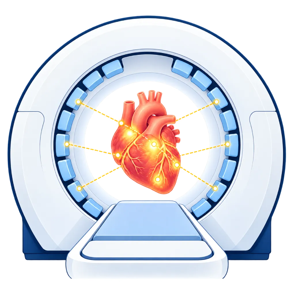
A cardiac PET scan is a nuclear medicine test that uses a low-dose radioactive tracer to assess blood flow to the heart muscle, heart muscle that is still alive, and inflammation or infection in the heart. The results help the doctor diagnose coronary artery disease and plan procedures such as stent placement or bypass surgery.
Sources01 / Understanding the procedure
About Cardiac PET Scan
What Is a Cardiac PET Scan
PET (positron emission tomography) is a nuclear medicine test. A low-dose radioactive tracer is injected through an IV line, and the PET scanner then captures the signals it emits and builds them into a three-dimensional image of the heart. The PET scanner is usually combined with CT (PET/CT) so that the location of each part of the heart is mapped precisely.
How It Works in Brief
The tracer follows the bloodstream and enters the heart muscle. Areas of muscle that receive enough blood flow appear bright, while areas that lack blood flow appear dim. In a blood flow test, scans are taken at rest and while the heart is stimulated by a medication that mimics the effect of exercise, and the two sets of images are then compared.
Types of Tests
- Myocardial perfusion: assesses blood flow to the heart muscle at rest and under stress. PET can also measure blood flow in numbers, which helps identify narrowing in several vessels at once or problems in the small blood vessels of the heart.
- Viability: uses FDG, a labeled form of sugar, to see whether weakened heart muscle is still alive and has a chance of improving if its blood flow is restored.
- Inflammation and infection: FDG is also used to look for cardiac sarcoidosis, as well as infection of artificial valves or pacemaker leads.
Reading the Results
If blood flow is even at rest and under stress, a significant narrowing is unlikely. An area that is dim under stress but normal at rest indicates that the heart muscle is not getting enough blood flow. An area that is dim in both may mean scar tissue. In a viability test, low blood flow with continued FDG uptake indicates that the heart muscle is still alive.
Compared with Other Tests
Compared with SPECT perfusion imaging, PET generally provides sharper images, shorter scan times, and a lower radiation dose, and it can measure blood flow. PET is available only at certain nuclear medicine facilities, so the doctor chooses the test that fits the clinical question and your condition.
02 / Understanding the procedure
When Is This Procedure Considered?
A doctor may consider this procedure in the situations below. The decision is always made after a thorough assessment.
- Chest Pain or Shortness of Breath Suspected to Come from the HeartTo look for signs of reduced blood flow caused by coronary artery disease.
- Unclear Results from Other TestsFor example, an inconclusive treadmill test or SPECT, or images affected by body size.
- Assessing the Benefit of a Stent or BypassA viability test shows heart muscle that can still improve.
- Suspected Small Vessel Disease of the HeartBlood flow measurement helps when symptoms are present but the large vessels look normal.
- Suspected Cardiac SarcoidosisTo look for inflammation and monitor the response to treatment.
- Suspected Infection of an Artificial Valve or PacemakerHelps find infections that are hard to see on other tests.
Situations to Consider First
- PregnancyUsually avoided. Tell the staff if you are pregnant or might be pregnant.
- Severe Asthma or Breathlessness from Narrowed AirwaysCertain stress medications can trigger breathlessness, so the doctor will choose a different medication.
- Severe Heart Conduction Disorder Without a PacemakerStress medications can slow the heart rhythm.
- Very Low Blood PressureStress medications can lower blood pressure further.
- Still Consuming CaffeineCaffeine blocks the action of stress medications, so the test may be rescheduled.
- Very High Blood SugarThis can reduce image quality in tests using FDG.
03 / Before the procedure
How Should You Prepare?
Preparation differs by type of test, so follow the written instructions from the staff.
Caffeine. Avoid coffee, tea, chocolate, soft drinks, energy drinks, and medications containing caffeine for at least 12 to 24 hours beforehand.
Eating and drinking. For a blood flow test, you usually fast for about 4 to 6 hours. For an inflammation test, follow a high-fat diet with almost no carbohydrates the day before, then fast as instructed. You may still drink plain water.
Medications. Some heart or asthma medications may need to be stopped temporarily. Do not stop any medication without your doctor's direction, and bring a list of your medications.
Diabetes. Tell the staff early on, because the dose of your diabetes medication or insulin may need to be adjusted.
Important information. Tell the staff if you are pregnant or breastfeeding, have asthma, or find it hard to lie still for a long time with your arms above your head.
Clothing. Wear comfortable clothing without metal, and bring the results of previous heart tests.
Unsure about the preparation instructions? Ask our team before the procedure.
04 / During the procedure
How Is the Procedure Done?
A blood flow test usually takes about 1 hour, while a test with FDG can take 2 to 3 hours because of the waiting time for the tracer to be absorbed.
Preparation in the Examination Room
The staff place an IV line in your arm and ECG electrodes on your chest, then check your blood pressure. You lie on the scanner table with your arms above your head.
Scan at Rest
The tracer is injected through the IV line, and the scanner records images of your heart. A low-dose CT is also done to map the position of the heart.
Scan Under Stress
A stress medication is given through the IV line to mimic the effect of exercise. You may feel warm, slightly short of breath, or a brief heaviness in your chest. These sensations usually go away within a few minutes. The tracer is injected again and the heart is scanned again.
Test with FDG
For viability, inflammation, or infection, FDG is injected and you then rest quietly for about 1 hour before the scan.
05 / Benefits and risks
Benefits and Risks Should Be Discussed Together.
Every medical procedure has benefits and risks. Your doctor will explain both for your situation before the procedure.
Benefits You Can Expect
- Sharp and Accurate ImagesHelps detect reduced blood flow with high accuracy.
- Measures Blood Flow in NumbersHelps identify narrowing in several vessels or problems in the small blood vessels.
- Relatively Low Radiation DoseUsually lower than SPECT perfusion imaging.
- Helps Decide on a Stent or BypassShows heart muscle that is still alive and has a chance of improving.
- Assesses Heart Inflammation and InfectionHelps diagnose and monitor sarcoidosis and infections of devices in the heart.
- No IncisionOnly an IV line is needed, with no instruments inserted into the heart.
The benefits of a procedure are not a promise of a cure. Ask how the results will affect your care plan.
Risks You Should Know
- Radiation ExposureThe dose is low and kept as low as possible. The doctor weighs the benefits first.
- Effects of the Stress MedicationA brief feeling of warmth, breathlessness, chest pain, or headache. The team monitors you and can give an antidote.
- Brief Rhythm DisturbancesRarely caused by the stress medication and usually resolve quickly.
- Unclear ResultsMovement, caffeine, blood sugar, or not following the dietary preparation can affect the results.
- Allergic ReactionVery rare, to either the tracer or the stress medication.
Risks can differ from person to person. A full explanation and consent are given after an individual assessment.
06 / After the procedure
What Is Recovery Like?
There is no recovery period. You can usually go home and return to your activities right away.
Drink water. Drink plenty of water and urinate often to help your body clear the remaining tracer.
Close contact. After a test with FDG, the staff may advise you to limit close contact with pregnant women and babies for a few hours.
Food and medications. You can eat as usual and resume your medications as directed by your doctor.
Results. The images are read by a nuclear medicine specialist and then discussed with your cardiologist.
Warning signs to watch for. Go to the emergency room right away or contact the medical team if you experience:
- Chest pain that does not go away
- Severe shortness of breath
- Severe dizziness or fainting
- Itching, a rash, or swelling of the face
Know the Signs That Need Prompt Care.
Follow the team's guidance on warning signs. If severe or sudden symptoms appear after the procedure, go to the nearest emergency department right away or call Heart Attack Center 1500 258.
07 / Team and support
Doctors and Procedure Support
The doctors below are listed for this procedure. Listed by name, not by ranking.

Dr. med. dr. Denio A. Ridjab, Sp.JP(K)
Interventional Cardiology
Coronary Intervention Procedures +2 centers
Clinical focusInterventional Cardiology · Coronary Artery Disease
View Profile
dr. Puti S. Saus, Sp.JP(K)
Interventional Cardiology
Coronary Intervention Procedures
Clinical focusAcute Coronary Syndrome · Primary Coronary Intervention
View Profile
dr. Siska S. Danny, Sp.JP(K)
Interventional Cardiology
Coronary Intervention Procedures
Clinical focusPCI for Acute Coronary Syndrome · Primary Coronary Intervention
View Profile08 / Common questions
Before You Plan the Procedure.
Frequently asked questions about cardiac PET scans. The right test for your condition is discussed with your doctor.
What is the difference between a cardiac PET scan and a cardiac CT scan?
A cardiac CT scan mainly shows the structure of the coronary arteries, for example plaque and narrowing. A cardiac PET scan shows function, that is, whether the heart muscle is getting enough blood flow, is still alive, or is inflamed. The two can complement each other, and with PET/CT both tests are done with one machine.
Is a cardiac PET scan safe?
It is generally safe. The tracer used is radioactive, but the dose is low, and the majority of it decays within minutes to hours. The stress medication can cause brief symptoms such as a feeling of warmth or breathlessness, which are monitored directly by the team. Pregnant women generally do not undergo this test.
Why can't I drink coffee before a cardiac PET scan?
Caffeine in coffee, tea, chocolate, and soft drinks blocks the action of the stress medication used to widen the heart's blood vessels. As a result, areas with reduced blood flow may not show up. That is why you should avoid caffeine for at least 12 to 24 hours beforehand, or as instructed by the staff.
What is the difference between PET and SPECT perfusion imaging?
Both assess blood flow to the heart muscle using a radioactive tracer. PET generally provides sharper images, a shorter scan time, and a lower radiation dose, and it can measure blood flow in numbers. SPECT is more widely available. The doctor chooses the test based on your needs and availability.
Do I have to exercise during the test?
Usually not. In a cardiac PET scan, the heart is generally stimulated with a medication given through an IV line that mimics the effect of exercise, so you stay lying on the scanner table. This approach also suits people who have difficulty walking on a treadmill, for example because of knee pain or shortness of breath.
When will the cardiac PET scan results be ready?
The images are read by a nuclear medicine specialist, usually within a few days depending on the service schedule at the testing facility. Your cardiologist then discusses the results with you, including whether you need medication, further tests such as catheterization, or procedures such as stent placement or bypass surgery.
Sources and Medical Review
Translated from the Indonesian edition medically reviewed by dr. Puti S. Saus, Sp.JP(K).
Sources as cited in the Indonesian edition.
- Dilsizian V, Bacharach SL, Beanlands RS, et al. ASNC imaging guidelines/SNMMI procedure standard for positron emission tomography (PET) nuclear cardiology procedures. J Nucl Cardiol. 2016;23(5):1187-1226.
- Knuuti J, Wijns W, Saraste A, et al. 2019 ESC Guidelines for the diagnosis and management of chronic coronary syndromes. Eur Heart J. 2020;41(3):407-477.
- Gulati M, Levy PD, Mukherjee D, et al. 2021 AHA/ACC/ASE/CHEST/SAEM/SCCT/SCMR Guideline for the Evaluation and Diagnosis of Chest Pain. Circulation. 2021;144(22):e368-e454.
- Chareonthaitawee P, Beanlands RS, Chen W, et al. Joint SNMMI-ASNC expert consensus document on the role of 18F-FDG PET/CT in cardiac sarcoid detection and therapy monitoring. J Nucl Med. 2017;58(8):1341-1353.
- Delgado V, Ajmone Marsan N, de Waha S, et al. 2023 ESC Guidelines for the management of endocarditis. Eur Heart J. 2023;44(39):3948-4042.