
Quick answer
What Is Coarctation of the Aorta?
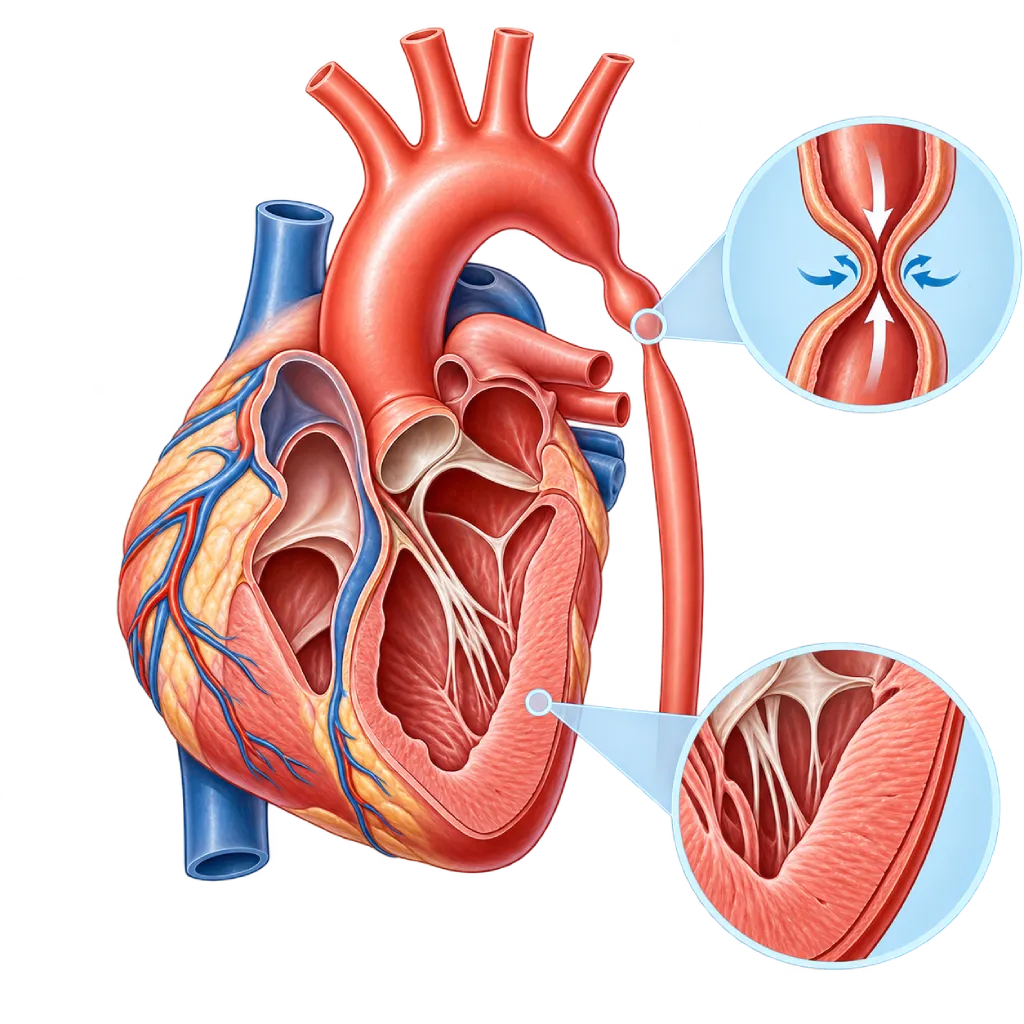
Coarctation of the aorta is a congenital narrowing of the aorta, the body's main artery, usually near its arch. As a result, blood pressure in the arms is high, blood flow to the lower body is reduced, and the left side of the heart works harder.
SourcesSigns That Need Urgent Help
- A baby who is very limp, hard to wake, or refuses to feed
- A baby breathing very fast with pale, grayish, cold skin
- A baby's diaper barely wet all day
- Severe chest or back pain that comes on suddenly
- A sudden, very severe headache, especially with vomiting or weakness on one side of the body
- Severe shortness of breath at rest, or fainting
If these signs appear, go to the nearest emergency department right away or call Heart Attack Center 1500 258.
01 / Understanding the condition
About Coarctation of the Aorta
The aorta is the main artery that carries oxygen-rich blood from the left ventricle of the heart to the rest of the body. In coarctation of the aorta, part of the aorta is narrowed from birth. The narrowing is usually in the descending aorta, just past the branch to the left arm, near the site of the ductus arteriosus. The ductus arteriosus is a channel in the fetus that normally closes a few days after birth. Coarctation is found in about 5 to 8 of every 100 cases of congenital heart disease and is more common in males. An overview of other congenital defects is covered on the Congenital Heart Disease page.
What Happens to Blood Flow
The narrowing works like a pinched hose. Before the narrow segment, blood pressure rises, so the arms and head receive high pressure. After the narrow segment, blood flow to the abdomen, kidneys, and legs is reduced, so the pulse in the groin feels weak and delayed. The left ventricle has to pump harder, and over time its muscle thickens. The body can also form bypass vessels (collaterals) in the chest wall to carry blood around the narrow segment.
Different in Babies and Adults
With severe coarctation, a baby may look healthy at birth and then get worse within a few days, when the ductus arteriosus closes. The baby becomes short of breath, has trouble feeding, looks pale, and may go into shock. With a mild narrowing, there are often no symptoms. The condition is found only when a child, teenager, or adult is examined because of high blood pressure at a young age or a heart murmur.
Commonly Associated Defects
Coarctation often occurs together with a bicuspid aortic valve, an aortic valve that has only two leaflets. Other defects that may come with it include ventricular septal defect, patent ductus arteriosus, and a small aortic arch. Coarctation is also more common in girls with Turner syndrome. Some people with coarctation have widened blood vessels in the brain, covered on the Cerebral Aneurysm page.
Needs Lifelong Monitoring
The narrowing can be repaired with surgery or a catheter procedure. However, repair is not the end of care. The ESC 2020 and AHA/ACC 2018 guidelines recommend lifelong follow-up because high blood pressure, renarrowing, and widening of the aorta can still appear years later, even if you feel well.
02 / Understanding the condition
Symptoms to Recognize.
Common Symptoms
- Blood pressure in the arms higher than in the legs
- Weak or delayed pulse in the groin or feet
- Recurring headaches
- Recurring nosebleeds with no clear cause
- Legs that feel cold or tire quickly when walking
- Calf cramps or pain during exercise
- A heart murmur found during a doctor's examination
Symptoms in Newborns
In severe coarctation, symptoms usually appear within the first few days to two weeks, when the ductus arteriosus closes.
- Fast or labored breathing
- Trouble feeding, tiring quickly, or sweating during feeds
- Pale or grayish skin, with cold hands and feet
- Fussiness or looking very sleepy
- Pulse in the groin that is hard to feel
- Fewer wet diapers
Symptoms in Children
Many children have no complaints at all; the condition is often found because of high blood pressure or a heart murmur during a routine checkup.
- High blood pressure at a routine checkup
- Legs that tire or ache quickly when playing and running
- Recurring headaches or nosebleeds
- A newly found heart murmur
- Feet that feel colder than the hands
Symptoms in Adults
Applies to adults who have not been diagnosed as well as those who had a repair in childhood.
- High blood pressure at a young age or that is hard to control with medicine
- Frequent headaches
- Calf pain when walking long distances
- Shortness of breath or tiring quickly during activity
- Blood pressure that rises excessively during exercise
03 / Understanding the condition
What Causes This Condition?
- Abnormal development of the aorta as the fetus grows, usually in the early weeks of pregnancy
- Ductus arteriosus tissue that extends into the aortic wall and shrinks along with the duct when it closes after birth
- Reduced blood flow through the left side of the fetal heart, for example when there is a valve or left ventricle abnormality
- Genetic factors, for example Turner syndrome in girls
- In many children, the exact cause is unknown
04 / Understanding the condition
Who Is at Higher Risk?
Genetic
Turner syndrome, some other genetic syndromes, and a history of congenital heart disease in a parent or sibling increase the likelihood of coarctation.
Medical
A bicuspid aortic valve and other defects of the left side of the heart are often found together with coarctation. Uncontrolled diabetes in the mother during pregnancy increases the risk of congenital heart disease in general.
Other
Coarctation is more common in males. After repair, residual narrowing and repair at an older age increase the likelihood of persistent high blood pressure.
Lifestyle
Smoking, excess weight, and high salt intake do not cause coarctation, but they worsen high blood pressure and the strain on the blood vessels in people who have it.
Groups at Higher Risk
- Girls with Turner Syndrome: About one in ten children with Turner syndrome has coarctation, so heart and aortic checks are recommended as soon as the diagnosis is made.
- People with a Bicuspid Aortic Valve: A two-leaflet aortic valve often occurs together with coarctation, so both should be checked when one of them is found.
- Children and Young Adults with High Blood Pressure: The cause of high blood pressure at a young age needs to be investigated, including by comparing blood pressure and pulses in the arms and legs.
- People with Repaired Coarctation: High blood pressure, renarrowing, and widening of the aorta can appear years after surgery or a catheter procedure.
- Women Planning a Pregnancy: Pregnancy adds strain on the circulation, so women with coarctation need an assessment of the aorta and blood pressure before becoming pregnant.
05 / Understanding the condition
How Is It Diagnosed?
Diagnosis starts with a simple examination: the doctor measures blood pressure in the arms and legs, then feels the pulses at the wrist and groin at the same time. Much higher pressure in the arms and a weak or delayed pulse in the legs point to coarctation. In newborns, oxygen saturation screening on the right hand and a foot helps find critical congenital heart disease, although coarctation is not always picked up this way.
Echocardiography is the main test to see where the narrowing is, measure how much it obstructs blood flow, assess the left ventricular muscle, and look for associated defects such as a bicuspid aortic valve. In teenagers and adults, cardiac MRI or cardiac CT scan gives a complete picture of the entire aorta, including collateral vessels and widening. Cardiac catheterization measures the pressure difference directly and is often combined with a widening procedure. Monitoring blood pressure over 24 hours and an exercise test help find high blood pressure that does not show up during a clinic visit.
Physical Examination
Comparing blood pressure in the arms and legs and feeling the groin pulse; a pressure difference and weak foot pulses are early clues.
Pulse oximetry screening in newborns: sensors on the right hand and a foot look for critical congenital heart disease before the baby goes home.
Echocardiography
Shows the location and severity of the narrowing, blood flow speed, the thickness and strength of the left ventricle, and a bicuspid aortic valve.
Cardiac MRI
Maps the whole aorta without X-rays, including collaterals, widening, and renarrowing; often used for regular monitoring.
Cardiac CT Scan
Detailed images of the aorta in a short time, useful before a procedure or when a stent is already in place and MRI is not suitable.
Cardiac Catheterization
Measures the pressure difference directly across the narrow area and can go straight on to balloon widening or stent placement.
Ambulatory Blood Pressure Monitoring
Records blood pressure over 24 hours to find hidden high blood pressure, including after coarctation has been repaired.
06 / Understanding the condition
Treatment Options.
Treatment is decided by a doctor after an assessment. The categories below can help you follow the conversation with your doctor.
Infant Stabilization with Prostaglandin
In babies with severe coarctation, a prostaglandin-type medicine given by IV keeps the ductus arteriosus open until surgery, together with intensive care.
Surgical Repair of the Aorta
The narrow segment is removed and the ends of the aorta are joined back together, or the segment is widened with a patch. This approach is commonly used in babies and young children. Covered on the Congenital Heart Surgery page.
Balloon Angioplasty
A balloon at the tip of a catheter is inflated to widen the narrow segment, especially in children and for renarrowing. Covered on the Balloon Angioplasty for Aortic Coarctation page.
Aortic Stent Placement
In teenagers and adults whose aortic size allows it, the ESC 2020 guidelines favor a catheter procedure with a stent when it is technically feasible.
Blood Pressure Medicines
The doctor chooses medicine classes such as beta blockers, ACE inhibitors, or ARBs to control high blood pressure before and after repair. The type and dose are decided by the doctor.
Lifelong Follow-Up
Regular checks of arm and leg blood pressure, echocardiography, and aortic MRI or CT to find high blood pressure, renarrowing, or widening early.
07 / Understanding the condition
Complications to Watch For.
Persistent Hypertension
High blood pressure can persist or come back even after the narrowing has been repaired, especially when the repair is done at an older age.
Left Ventricular Thickening and Heart Failure
A left ventricle that keeps pumping against resistance can thicken and then weaken. In babies with severe coarctation, heart failure can develop in the first weeks of life.
Renarrowing (Recoarctation)
The aorta can narrow again at the repair site, more often when the repair was done in infancy. Covered on the Aortic Recoarctation Repair page.
Aortic Aneurysm and Dissection
The aortic wall at the repair site or at the aortic root can widen and, although rarely, tear. Covered on the Aortic Dissection page.
Brain Aneurysm
Widened blood vessels in the brain are more common in people with coarctation and can rupture, especially when high blood pressure is not controlled.
Early Coronary Artery Disease
Long-term high blood pressure speeds up narrowing of the coronary arteries, so heart risk factors need to be controlled from a young age.
Bicuspid Aortic Valve Problems
A two-leaflet valve can narrow or leak with age and sometimes needs repair, so assessing the valve is part of routine checkups.
08 / Understanding the condition
What Can You Do?
- Coarctation cannot be reliably prevented, but good care during pregnancy helps lower the risk of congenital heart defects in general
- Control diabetes before and during pregnancy, take folic acid as recommended, and avoid smoking and alcohol
- Make sure your baby has oxygen saturation screening before leaving the place of birth
- Ask for blood pressure measurements in the arms and legs if a child or teenager is found to have high blood pressure
- Attend follow-up visits as scheduled after repair, including regular aortic imaging, even if you feel well
- Measure your blood pressure at home and manage your weight, salt intake, and smoking habits
- Discuss the type and intensity of exercise with your doctor, especially if you have high blood pressure or a widened aorta
- Talk with a cardiologist about your pregnancy plans so your aorta and blood pressure can be assessed first
09 / Understanding the condition
When Should You See a Doctor?
Take a baby to the emergency room right away if the baby looks very limp, has trouble feeding, breathes fast, or has pale, grayish, cold skin. In children and adults, sudden severe chest or back pain and a very severe headache also need urgent care. For symptoms that do not come on suddenly, make an appointment with a cardiologist if:
- A child, teenager, or young adult is found to have high blood pressure with no clear cause.
- Blood pressure in the arms is much higher than in the legs, or the pulse in the feet feels weak.
- Headaches, nosebleeds, or leg cramps during activity keep coming back.
- A doctor finds a heart murmur or a bicuspid aortic valve.
- You had coarctation repaired but have not had a checkup in a long time.
- You have coarctation and are planning a pregnancy.
Don't Wait in an Emergency.
If symptoms are severe or come on suddenly, go to the nearest emergency department right away or call Heart Attack Center 1500 258, rather than waiting for an appointment.
10 / Common questions
Questions You May Have.
Frequently asked questions about coarctation of the aorta.
Is coarctation of the aorta cured after it is repaired?
Repair makes blood flow much more smoothly and lowers the strain on the heart, but coarctation is considered a lifelong condition. Some people still have high blood pressure, the aorta can narrow again, and widening can appear at the repair site. For this reason, guidelines recommend regular checkups with a doctor who understands congenital heart disease, including arm and leg blood pressure checks and regular aortic imaging, even if you feel well.
Why can a baby look healthy at birth and then suddenly become ill?
While the ductus arteriosus is still open, blood can get past the narrow part of the aorta through this shortcut. The duct usually closes within a few days after birth, and its tissue can squeeze the aorta as well. At that point, blood flow to the lower body drops sharply, so the baby becomes short of breath, pale, and limp. This is why a baby who looks ill in the first week needs to be taken to the emergency room right away.
Is surgery or a catheter procedure better?
The choice depends on age, the shape and length of the narrowing, the size of the aorta, and any previous procedures. Babies and young children usually have surgery. In older children, a balloon may be used, especially for renarrowing. In teenagers and adults, the ESC 2020 guidelines favor stent placement when it is technically feasible. The congenital heart team will weigh the benefits and risks of each option with you.
Can people with coarctation exercise?
Most people with repaired coarctation, with no residual narrowing and well-controlled blood pressure, can exercise regularly. The doctor usually first assesses blood pressure at rest and during an exercise test, the size of the aorta, and the aortic valve. If there is still high blood pressure, narrowing, or widening of the aorta, exercise that involves heavy straining or lifting heavy weights may need to be limited. Ask about safe limits for you or your child.
Can women with coarctation get pregnant?
Many women with repaired coarctation can have a safe pregnancy. Before pregnancy, the doctor assesses blood pressure, any residual narrowing, the size of the aorta, and the aortic valve, then adjusts medicines if needed. During pregnancy, blood pressure is monitored more closely, and fetal echocardiography is recommended because the risk of congenital heart disease in the baby is slightly higher. Plan your pregnancy together with a cardiologist and an obstetrician.
Is coarctation passed on to children?
Most cases of coarctation occur without a family history. However, children of parents with congenital heart disease have a higher chance of a heart defect than the general population, although the chance is still small. If there is a genetic syndrome such as Turner syndrome, the inheritance pattern is different and should be discussed in genetic counseling. Fetal echocardiography during pregnancy helps find defects earlier.
Sources and Medical Review
Translated from the Indonesian edition medically reviewed by dr. Salomo Purba, Sp.BTKV, Subsp.JPK(K).
Sources as cited in the Indonesian edition.
- Baumgartner H, De Backer J, Babu-Narayan SV, et al. 2020 ESC Guidelines for the management of adult congenital heart disease. Eur Heart J. 2021;42(6):563-645.
- Stout KK, Daniels CJ, Aboulhosn JA, et al. 2018 AHA/ACC Guideline for the Management of Adults With Congenital Heart Disease: A Report of the American College of Cardiology/American Heart Association Task Force on Clinical Practice Guidelines. Circulation. 2019;139(14):e698-e800.
- Brown ML, Burkhart HM, Connolly HM, et al. Coarctation of the aorta: lifelong surveillance is mandatory following surgical repair. J Am Coll Cardiol. 2013;62(11):1020-1025.
- Forbes TJ, Kim DW, Du W, et al. Comparison of surgical, stent, and balloon angioplasty treatment of native coarctation of the aorta: an observational study by the CCISC (Congenital Cardiovascular Interventional Study Consortium). J Am Coll Cardiol. 2011;58(25):2664-2674.
- Connolly HM, Huston J, Brown RD, et al. Intracranial aneurysms in patients with coarctation of the aorta: a prospective magnetic resonance angiographic study of 100 patients. Mayo Clin Proc. 2003;78(12):1491-1499.
- Martin GR, Ewer AK, Gaviglio A, et al. Updated Strategies for Pulse Oximetry Screening for Critical Congenital Heart Disease. Pediatrics. 2020;146(1):e20191650.
Doctors Who Treat This Condition

dr. Radityo Prakoso, Sp.JP(K)
Pediatrics & Congenital Heart Disease
Congenital and Structural Heart Center
Clinical focusPediatric Cardiology Consultation · Congenital Heart Disease Care
View Profile
dr. Salomo Purba, Sp.BTKV, Subsp.JPK(K)
Pediatric and Congenital Heart Surgery
Congenital and Structural Heart Center +2 centers
Clinical focusPediatric Heart Surgery · Congenital Heart Defect Surgery
View Profile
dr. Suko Adiarto, Sp.JP(K), Ph.D.
Interventional Cardiology
Vascular Diagnostic and Therapeutic Center +1 center
Clinical focusCoronary Intervention · Peripheral Vascular Intervention
View Profile