
Quick answer
What Is Marfan Syndrome?
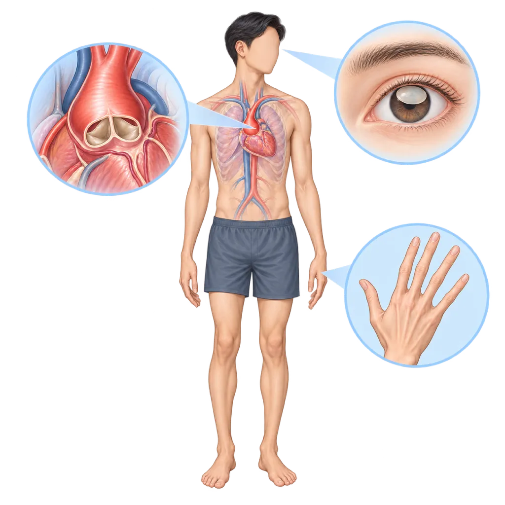
Marfan syndrome is an inherited connective tissue disorder caused by a change in the FBN1 gene. It can weaken and widen the aorta, shift the lens of the eye, and make the body tall with long arms and fingers.
SourcesSigns That Need Urgent Help
- Sudden, severe chest, back, or abdominal pain that feels like tearing
- Fainting or sudden weakness with chest pain
- Sudden shortness of breath with chest pain on one side
- A drooping face, slurred speech, or weakness on one side of the body
- Vision that suddenly goes dark or looks as if a curtain is covering it
- Severe shortness of breath at rest or being unable to lie flat
If these signs appear, go to the nearest emergency department right away or call Heart Attack Center 1500 258.
01 / Understanding the condition
About Marfan Syndrome
Marfan syndrome is an inherited disorder of the connective tissue, the tissue that strengthens and supports many parts of the body. It is caused by a change in the FBN1 gene, which makes the protein fibrillin-1. Because connective tissue is found throughout the body, this syndrome can affect the heart and blood vessels, eyes, bones, joints, and lungs. Marfan syndrome is found in about 1 in 5,000 people, both men and women.
Effects on the Aorta and Heart Valves
The part that needs special attention is the aorta, the large artery that leaves the heart. In Marfan syndrome, the wall of the aorta is weaker, so its root, just above the aortic valve, slowly widens. This widening is called an aortic aneurysm, covered on the Aortic Aneurysm page. If it keeps widening without treatment, the inner layer of the aortic wall can tear. This emergency is called aortic dissection, covered on the Aortic Dissection page. The mitral valve also often bulges back toward the left atrium (prolapse) and can leak.
Features in the Eyes and Bones
About half of people with the syndrome have a shifted lens of the eye (ectopia lentis), often with severe nearsightedness. Bone features include a tall body with long arms, legs, and fingers, a sunken or protruding chest, a curved spine, and flexible joints. Some people also have a sudden collapsed lung or widening of the membrane that surrounds the spinal nerves (dural ectasia), which can cause lower back pain. These features can increase with growth and differ from person to person, even within one family. This is why some people are only diagnosed in adulthood or after another family member is diagnosed.
Lifelong Monitoring
Marfan syndrome cannot yet be cured, but its effects can be controlled. The 2024 European Society of Cardiology (ESC) and 2022 American College of Cardiology/American Heart Association (ACC/AHA) guidelines recommend regular aortic checks, medicines that reduce the strain on the aortic wall, and planned surgery before the aorta reaches a size at which it is prone to tearing. With regular monitoring, many people with Marfan syndrome can now go to school, work, have a family, and live into old age. Care involves cardiologists, cardiac surgeons, eye doctors, orthopedic doctors, and genetic counselors.
02 / Understanding the condition
Symptoms to Recognize.
Common Symptoms
Widening of the aorta usually causes no symptoms, so body and eye features are often the first clue.
- A tall, slender body with long arms, legs, and fingers
- Severe nearsightedness or a lens of the eye that has shifted out of place
- A sunken or protruding chest
- A sideways curve of the spine (scoliosis)
- Very flexible joints and flat feet
- Stretch marks on the skin without significant weight change
- A high roof of the mouth with crowded teeth
03 / Understanding the condition
What Causes This Condition?
- A change (variant) in the FBN1 gene, which makes fibrillin-1, a protein that forms connective tissue
- Inherited in an autosomal dominant pattern: one changed copy of the gene is enough, with a 50% chance for each child
- About one in four cases arises from a new gene change with no family history
- Faulty fibrillin weakens the aortic wall, the supports of the eye lens, and other connective tissue
04 / Understanding the condition
Who Is at Higher Risk?
Genetic
Having a parent with Marfan syndrome gives a 50% chance of inheriting the FBN1 gene change. A family history of aortic dissection or sudden death indicates a higher aortic risk.
Medical
Uncontrolled high blood pressure, a severely leaking aortic valve, rapid widening of the aorta, and pregnancy and the period after giving birth add strain to an already weak aortic wall.
Lifestyle
Contact sports, high-intensity competitive sports, heavy weightlifting while holding your breath, smoking, and the use of stimulants such as cocaine can trigger surges of pressure in the aorta.
Groups at Higher Risk
- Children of People with Marfan Syndrome: Each child has a 50% chance of inheriting the gene change, so early heart and eye examinations and genetic testing are recommended.
- Women Planning a Pregnancy: Pregnancy and the period after giving birth increase the risk of aortic dissection, so counseling and aortic checks before pregnancy are recommended.
- Families with a History of Aortic Dissection: Aortic dissection or sudden death at a young age in the family may indicate an inherited aortic disease, including Marfan syndrome.
- Very Tall Teenagers and Young Adults: A very tall build with long arms, severe nearsightedness, or a sunken chest should be checked, especially before taking part in competitive sports.
05 / Understanding the condition
How Is It Diagnosed?
Marfan syndrome is diagnosed using the Ghent criteria, revised in 2010. The doctor combines several clues: the size of the aortic root compared with body size (Z-score), a shifted lens of the eye, the result of an FBN1 gene test, and a score of body features, for example chest shape, arm span, and signs at the wrist and thumb. Family history is also taken into account.
Echocardiography measures the aortic root and assesses the valves. This test is usually repeated 6 months after diagnosis to see how quickly the aorta is widening, then at least once a year if the size is stable. CT or MRI maps the whole aorta from the start and is repeated periodically. An eye doctor examines the lens with a slit lamp. Genetic testing helps confirm the diagnosis and distinguish it from similar conditions, such as Loeys-Dietz syndrome and vascular Ehlers-Danlos syndrome, which need different monitoring. Once the gene change is found, close family members can be tested in a more targeted way.
Physical Examination
The doctor assesses body features to calculate a systemic score, for example signs at the wrist and thumb, arm span, chest shape, and the spine.
Echocardiography
Measures the diameter of the aortic root and compares it with body size (Z-score), and assesses the aortic and mitral valves.
Cardiac MRI
MRI maps the whole aorta without X-rays, making it suitable for repeated monitoring at a young age.
Cardiac CT Scan
CT with contrast maps the aorta from the chest to the abdomen, especially before surgery or when certain parts of the aorta are not clearly visible.
An eye examination with a slit lamp by an eye doctor after the pupils are dilated, to look for a shifted lens.
An FBN1 gene test from a blood sample to confirm the diagnosis, distinguish it from similar conditions, and test family members.
06 / Understanding the condition
Treatment Options.
Treatment is decided by a doctor after an assessment. The categories below can help you follow the conversation with your doctor.
Beta Blockers or ARBs
Both classes of medicine reduce the pressure and strain on the aortic wall so that its widening slows down. Your doctor decides the type, dose, and whether to combine them.
Regular Aortic Monitoring
Echocardiography, CT, or MRI is scheduled to follow the size of the aorta, so surgery can be planned before the aorta is at risk of tearing.
Aortic Root Replacement Surgery
Usually recommended when the aortic root reaches about 50 mm, or about 45 mm if there are additional risk factors. If the valve is still in good condition, the native valve can be kept; if not, a Bentall procedure is performed.
Mitral Valve Repair or Replacement
Considered when mitral valve leakage is severe and causes symptoms or affects the heart's pumping function.
Treatment of the Eyes, Bones, and Lungs
Eye doctors, orthopedic doctors, and lung doctors treat lens displacement, scoliosis, chest shape, and collapsed lung as needed.
Pregnancy Planning
Counseling before pregnancy helps assess the risk. If the aortic root is already larger than about 45 mm, surgery before pregnancy is usually advised.
Genetic Counseling
Helps families understand the inheritance pattern and decide who needs to be tested, including options when planning a pregnancy.
07 / Understanding the condition
Complications to Watch For.
Widening of the Aortic Root
The weak aortic wall slowly widens, usually without symptoms. This widening is monitored with imaging and covered on the Aortic Aneurysm page.
Aortic Dissection
A tear in the inner layer of the aortic wall that causes sudden severe pain and needs emergency care. Covered on the Aortic Dissection page.
Aortic and Mitral Valve Leakage
A widened aortic root pulls on the aortic valve so that it leaks, while the mitral valve can prolapse. Severe leakage causes shortness of breath and needs to be repaired.
Heart Rhythm Disorders
Some people with the syndrome develop rhythm disorders, especially if a valve leaks severely or pumping function declines, so the doctor may recommend periodic rhythm recordings.
Shifted Lens and Retinal Detachment
A shifted lens blurs vision. People with the syndrome are also at higher risk of retinal detachment, glaucoma, and cataracts at a younger age.
Collapsed Lung (Pneumothorax)
Small air sacs at the top of the lung can burst so that the lung collapses, marked by chest pain on one side and sudden shortness of breath.
08 / Understanding the condition
What Can You Do?
- Have echocardiography and aortic imaging on schedule, even if you have no symptoms
- Take your aorta-protecting medicines regularly and do not stop them without your doctor's guidance
- Keep your blood pressure at the target set by your doctor
- Choose light to moderate exercise, such as walking or leisurely swimming, and avoid contact sports and heavy weightlifting
- Discuss pregnancy plans with a cardiologist before becoming pregnant
- Carry a record of your diagnosis and tell every doctor or emergency room staff member that you have Marfan syndrome
- Do not smoke, and avoid stimulants such as cocaine or amphetamines
- Encourage close family members to have heart checks and genetic counseling
09 / Understanding the condition
When Should You See a Doctor?
See a cardiologist if you or your child have the features below, or if a family member has Marfan syndrome. If you have already been diagnosed, sudden and severe chest or back pain is a reason to go to the emergency room right away.
- A very tall, slender body with long arms and fingers, along with severe nearsightedness
- A shifted lens of the eye found during an eye examination
- A widened aortic root or mitral valve prolapse on echocardiography results
- A parent, sibling, or child with Marfan syndrome
- A family history of aortic dissection or sudden death at a young age
- Plans for pregnancy in women with Marfan syndrome
Don't Wait in an Emergency.
If symptoms are severe or come on suddenly, go to the nearest emergency department right away or call Heart Attack Center 1500 258, rather than waiting for an appointment.
10 / Common questions
Questions You May Have.
Frequently asked questions about Marfan syndrome.
Can Marfan syndrome be cured?
Marfan syndrome cannot yet be cured because it comes from a gene change. However, its effects can be controlled. Regular aortic checks, medicines that reduce the strain on the aortic wall, and planned surgery before the aorta reaches a size at which it is prone to tearing can prevent aortic dissection. With care like this, many people with Marfan syndrome can go to school, work, have a family, and live into old age.
Can people with Marfan syndrome exercise?
Yes, and physical activity is still recommended. Choose light to moderate aerobic exercise, such as walking, leisurely cycling, or leisurely swimming. Avoid contact sports, high-intensity competitive sports, and heavy weightlifting that makes you hold your breath, because these can suddenly raise the pressure in the aorta. Safe limits differ from person to person, so discuss the type of exercise with your cardiologist.
Can women with Marfan syndrome become pregnant?
Many women with Marfan syndrome go through pregnancy well, but pregnancy needs planning. Pregnancy and the period after giving birth add strain to the aorta. Guidelines recommend an aortic check before pregnancy. If the aortic root is larger than about 45 mm, surgery before pregnancy is usually advised. During pregnancy, the size of the aorta is monitored regularly by a team of cardiologists and obstetricians.
How often does the aorta need to be checked?
After diagnosis, echocardiography is usually repeated 6 months later to see how quickly the aorta is widening. If the size is stable, the test is done at least once a year. If the size is approaching the threshold for surgery or is widening quickly, the test is done more often. CT or MRI of the whole aorta is also repeated periodically to monitor parts of the aorta that cannot be seen with echocardiography.
Will my child definitely inherit Marfan syndrome?
Not necessarily. Marfan syndrome is inherited in an autosomal dominant pattern, which means each child of an affected person has a 50% chance of inheriting the gene change. A child who does not inherit it will not pass the syndrome on. About one in four people with the syndrome have no family history because their gene change is new. Genetic counseling helps you understand the options for testing your child and planning a pregnancy.
What happens if the aortic root needs surgery?
The surgery is planned in advance to replace the widened aortic root with an artificial tube. If the aortic valve is still in good condition, the surgeon may keep the native valve. If the valve cannot be kept, the aortic root and valve are replaced together, which is covered on the Bentall Procedure page. Planned surgery carries a much lower risk than emergency surgery once the aorta has already torn.
Sources and Medical Review
Translated from the Indonesian edition medically reviewed by dr. Radityo Prakoso, Sp.JP(K).
Sources as cited in the Indonesian edition.
- Mazzolai L, Teixido-Tura G, Lanzi S, Boc V, Bossone E, Brodmann M, et al. 2024 ESC Guidelines for the management of peripheral arterial and aortic diseases. Eur Heart J. 2024;45(36):3538-700.
- Isselbacher EM, Preventza O, Hamilton Black J 3rd, Augoustides JG, Beck AW, Bolen MA, et al. 2022 ACC/AHA Guideline for the Diagnosis and Management of Aortic Disease: A Report of the American Heart Association/American College of Cardiology Joint Committee on Clinical Practice Guidelines. Circulation. 2022;146(24):e334-482.
- Loeys BL, Dietz HC, Braverman AC, Callewaert BL, De Backer J, Devereux RB, et al. The revised Ghent nosology for the Marfan syndrome. J Med Genet. 2010;47(7):476-85.
- Milewicz DM, Braverman AC, De Backer J, Morris SA, Boileau C, Maumenee IH, et al. Marfan syndrome. Nat Rev Dis Primers. 2021;7(1):64.
- Pitcher A, Spata E, Emberson J, Davies K, Halls H, Holland L, et al. Angiotensin receptor blockers and beta blockers in Marfan syndrome: an individual patient data meta-analysis of randomised trials. Lancet. 2022;400(10355):822-31.
- Regitz-Zagrosek V, Roos-Hesselink JW, Bauersachs J, Blomstrom-Lundqvist C, Cifkova R, De Bonis M, et al. 2018 ESC Guidelines for the management of cardiovascular diseases during pregnancy. Eur Heart J. 2018;39(34):3165-241.
Doctors Who Treat This Condition

dr. BRM Ario S. Kuncoro, Sp.JP(K)
Echocardiography & Cardiovascular Imaging
Cardiovascular Diagnostic Center +2 centers
Clinical focusAdvanced and 3D Echocardiography · Transesophageal Echocardiography
View Profile
dr. Suko Adiarto, Sp.JP(K), Ph.D.
Interventional Cardiology
Vascular Diagnostic and Therapeutic Center +1 center
Clinical focusCoronary Intervention · Peripheral Vascular Intervention
View Profile
dr. Radityo Prakoso, Sp.JP(K)
Pediatrics & Congenital Heart Disease
Congenital and Structural Heart Center
Clinical focusPediatric Cardiology Consultation · Congenital Heart Disease Care
View Profile