
Quick answer
What Is Adult Structural Heart Disease?
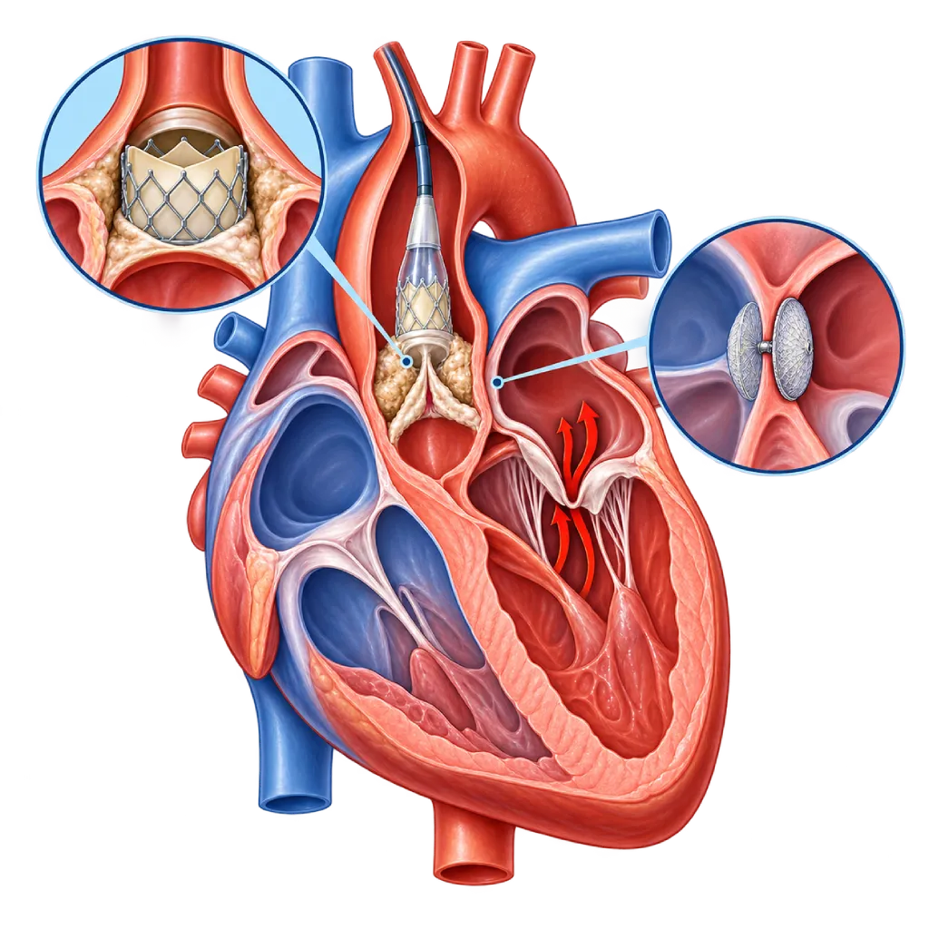
Adult structural heart disease means problems with the valves, the dividing walls (septum), or the walls of the heart in adults that can be repaired mechanically, now often through a catheter. Examples include a narrowed aortic valve, a leaking mitral valve, and a hole in the septum first found in adulthood.
SourcesSigns That Need Urgent Help
- Severe shortness of breath at rest or coughing up frothy sputum
- Fainting, especially during or after activity
- Severe chest pain or chest pain that does not ease with rest
- A very fast and irregular heartbeat with weakness
- Weakness on one side of the body, a drooping face, or slurred speech
If these signs appear, go to the nearest emergency department right away or call Heart Attack Center 1500 258.
01 / Understanding the condition
About Adult Structural Heart Disease
Structural heart disease is the term doctors use for problems in the physical parts of the heart, such as the valves, the septum (dividing wall), the walls of the ventricles, or the atria, that can be repaired mechanically. In adults, this field has grown rapidly because many problems can now be treated through a catheter inserted from a blood vessel in the groin, without opening the chest. This page covers the adult perspective and the treatment options. A general overview for all ages is covered on the Structural Heart Abnormalities page.
Common Problems in Adults
In older age, common problems are narrowing of the aortic valve due to calcification, covered on the Aortic Stenosis page, as well as leaking of the mitral and tricuspid valves. At younger ages, some people only discover a congenital problem, such as a hole in the wall between the atria or an aortic valve with two leaflets (bicuspid). People with congenital heart disease who had surgery as children also need lifelong monitoring because residual problems or new issues can appear. Other groups include leaks around an artificial valve, the small pouch of the left atrium (left atrial appendage) where clots often form in atrial fibrillation, and thickening of the septum that blocks blood flow in hypertrophic cardiomyopathy.
Decisions Are Made by a Heart Team
The 2025 ESC/EACTS and 2020 ACC/AHA guidelines recommend that treatment decisions be made by a heart team made up of interventional cardiologists, cardiac surgeons, cardiac imaging specialists, and when needed, anesthesiologists and geriatricians. The team weighs age, fitness, the anatomy seen on imaging, other health conditions, and the patient's wishes. The result may be regular monitoring, a catheter procedure, surgery, or medicines to ease symptoms.
Treatment Timing Determines the Outcome
Structural problems often worsen slowly, and the heart adapts until the load becomes too heavy. If treatment is delayed too long, the heart muscle can be permanently damaged even if the problem is repaired later. That is why people with moderate to severe problems are monitored with regular echocardiography, and treatment is considered as soon as symptoms or early signs of strain on the heart appear. Even after treatment, follow-up is still needed to monitor the new valve or the closure device that was placed.
02 / Understanding the condition
Symptoms to Recognize.
Common Symptoms
Many people have no symptoms for years. Symptoms usually appear gradually as the heart begins to be strained.
- Shortness of breath during activity, and later also when lying down
- Tiring quickly and reduced ability to exercise
- Palpitations or an irregular heartbeat
- Chest pain or heaviness during activity
- Dizziness or nearly fainting during activity
- Swollen ankles or a belly that feels like it is getting bigger
Symptoms in Older Adults
Symptoms are often mistaken for signs of aging, when they may actually come from a narrowed or leaking valve.
- Walking more slowly or stopping to rest more often
- Shortness of breath when climbing stairs or walking uphill
- Dizziness or unsteadiness when standing up or during activity
- Often waking at night because of shortness of breath
Symptoms in Adult Women
Pregnancy adds to the heart's workload, so a valve or septal problem that was previously quiet can start to cause symptoms.
- Shortness of breath during pregnancy that becomes worse than usual
- Palpitations or an irregular rhythm that first appears during pregnancy
- Swelling that worsens quickly along with shortness of breath
- Being unable to lie flat because of shortness of breath
03 / Understanding the condition
What Causes This Condition?
- Calcification and aging of the valves, especially the aortic and mitral valves
- Congenital problems that only cause symptoms in adulthood, such as a bicuspid aortic valve or a hole in the wall between the atria
- Residual problems or new issues after congenital heart surgery in childhood
- Valve damage from rheumatic fever, covered on the Rheumatic Heart Disease page.
- Enlargement of the ventricles or atria due to heart failure, a heart attack, or atrial fibrillation, so the valves do not close tightly
- Infection of a natural or artificial valve
- Thickening of the ventricular septum in hypertrophic cardiomyopathy that blocks blood flow out of the heart
04 / Understanding the condition
Who Is at Higher Risk?
Age
Aortic valve calcification and leaking of the mitral and tricuspid valves become increasingly common after age 65.
Medical
High blood pressure, atrial fibrillation, heart failure, heart attack, chronic kidney disease, and a history of radiation therapy to the chest area.
Genetic
A bicuspid aortic valve, which can run in families, hypertrophic cardiomyopathy, and connective tissue diseases such as Marfan syndrome.
Other
A history of rheumatic fever, congenital heart surgery in childhood, previous valve replacement, and valve infection.
Groups at Higher Risk
- Older Adults: Aortic valve calcification and leaking of the mitral and tricuspid valves become increasingly common after age 65.
- People Who Had Surgery for Congenital Heart Disease: Surgery in childhood is often not the end of treatment; residual problems, worn valves, or rhythm disorders can appear in adulthood.
- People with a Bicuspid Aortic Valve: This congenital valve problem can narrow or leak earlier, often from age 40 into the 60s, and sometimes comes with enlargement of the aorta.
- Rheumatic Fever Survivors: Valve damage from rheumatic fever may only start causing symptoms in adulthood or during pregnancy.
- People with Atrial Fibrillation: Besides the risk of stroke, enlarged atria can cause the mitral and tricuspid valves to leak.
05 / Understanding the condition
How Is It Diagnosed?
Your doctor starts by asking about your symptoms and how much activity you can manage, then listens to your heart for a murmur. Echocardiography is the key test because it shows which part has a problem, how severe it is, and how it affects the size and pumping strength of the heart. An ECG and an NT-proBNP blood test help assess the heart's rhythm and workload.
If treatment is being considered, testing becomes more detailed. Echocardiography through the esophagus (transesophageal) clearly shows the mitral valve, the wall between the atria, and the left atrial pouch. A cardiac CT scan is used to measure the valve and blood vessels before a valve replacement through a catheter. The coronary arteries are also checked, with CT or angiography, because any narrowing can be treated in the same plan. In people without symptoms, a supervised exercise test is sometimes used to confirm whether the problem already limits the heart's capacity. The results of all tests are then discussed by the heart team.
Echocardiography
The main test to assess the valves, septum, chamber size, and pumping strength. Echocardiography through the esophagus gives more detailed images before a procedure.
Electrocardiogram (ECG)
Assesses the rhythm, especially atrial fibrillation, as well as signs of thickening or enlargement of the heart chambers.
NT-proBNP and BNP Test
Raised levels show that the heart is starting to be strained and help your doctor decide when to treat.
Treadmill Stress Test
In severe valve problems without symptoms, a supervised exercise test can reveal hidden symptoms or an abnormal blood pressure response.
Cardiac CT Scan
Measures the aortic valve, the large blood vessels, and the blood vessel access route to plan a valve replacement through a catheter.
Cardiac MRI
Measures chamber size, the severity of valve leaks, and scarring of the heart muscle in detail.
Cardiac Catheterization
Measures the pressures inside the heart and lungs when other test results do not agree, and checks the coronary arteries before a procedure.
06 / Understanding the condition
Treatment Options.
Treatment is decided by a doctor after an assessment. The categories below can help you follow the conversation with your doctor.
Regular Monitoring
Mild to moderate problems are monitored with echocardiography at intervals your doctor sets, so that treatment is done before the heart is permanently damaged.
Medicines to Control Symptoms and Coexisting Conditions
Diuretics, blood pressure medicines, rhythm-control medicines, and anticoagulants for atrial fibrillation ease symptoms and prevent complications, but they do not repair the valve or septum.
Transcatheter Aortic Valve Replacement
TAVI places a new valve inside the old valve through a blood vessel. The heart team chooses it based on age, surgical risk, and anatomy, especially in older people.
Mitral and Tricuspid Valve Repair
Leaking can be reduced with valve repair surgery, or with a clip on the valve leaflets placed through a catheter in selected patients.
Catheter-Based Closure
A hole in the wall between the atria, a patent foramen ovale in certain situations, leaks around an artificial valve, and the left atrial pouch can be closed with a special device through a catheter.
Heart Surgery
Open or minimally invasive valve surgery is chosen when the anatomy is not suited to a catheter, there are several problems at once, or age and overall condition favor the long-term results of surgery.
07 / Understanding the condition
Complications to Watch For.
Heart Failure
A narrowed or leaking valve and a large septal hole make the heart work harder until its pumping weakens and fluid builds up.
Atrial Fibrillation
Enlarged atria are prone to an irregular rhythm, covered on the Atrial Fibrillation page.
Stroke
Clots can form in the left atrium in atrial fibrillation or pass through a septal hole, then travel to the brain.
Pulmonary Hypertension
A long-standing mitral valve leak or septal hole can permanently raise the pressure in the blood vessels of the lungs.
Infective Endocarditis
Damaged valves or artificial valves are more easily infected by bacteria, covered on the Infective Endocarditis page.
Fainting and Sudden Cardiac Arrest
Severe narrowing of the aortic valve can cause fainting during activity and, less often, cardiac arrest.
08 / Understanding the condition
What Can You Do?
- Control your blood pressure and cholesterol to reduce the strain on the valves and walls of the heart.
- If you have a valve problem or congenital heart disease, have echocardiography on schedule even if you have no symptoms.
- Keep your teeth and mouth clean and have regular dental checkups to lower the risk of valve infection.
- If you had heart surgery as a child, continue follow-up with a cardiologist as an adult.
- Discuss pregnancy plans with a cardiologist if you have a valve or septal problem.
- Tell your doctor if your ability to be active declines, and do not assume it is only due to age.
09 / Understanding the condition
When Should You See a Doctor?
Structural problems in adults often worsen slowly, so small changes in your ability to be active are worth having checked. If you have severe shortness of breath at rest, fainting, severe chest pain, or signs of a stroke, go to the nearest emergency room right away or call an ambulance (119).
Make an appointment with a cardiologist if:
- A doctor hears a heart murmur or echocardiography shows a valve or septal problem.
- You start to feel short of breath, tire quickly, feel dizzy, or have chest pain during activities that used to feel light.
- You have had congenital heart surgery or a valve replacement and have not had a checkup in the past year.
- You have atrial fibrillation but cannot take blood thinners long term.
- You have a valve or septal problem and are planning a pregnancy.
Don't Wait in an Emergency.
If symptoms are severe or come on suddenly, go to the nearest emergency department right away or call Heart Attack Center 1500 258, rather than waiting for an appointment.
10 / Common questions
Questions You May Have.
Frequently asked questions about structural heart disease in adults.
What does structural heart disease mean?
This term covers problems in the physical parts of the heart, such as the valves, septum, ventricular walls, and atria, that can be repaired mechanically. It differs from coronary artery disease, which involves the heart's blood vessels, and from rhythm disorders, which involve the electrical system. All three often occur together in adults, so doctors usually assess all of them before putting together a treatment plan.
What is a heart team and why are decisions made together?
A heart team is made up of interventional cardiologists, cardiac surgeons, cardiac imaging specialists, and other doctors as needed. They review test results together because each option, such as a catheter procedure or surgery, has advantages and limitations. The 2025 ESC/EACTS and 2020 ACC/AHA guidelines recommend this approach. Your views and priorities are also part of the decision.
Is a catheter procedure always better than surgery?
Not always. A catheter procedure generally offers a shorter recovery because the chest is not opened, and it is very helpful for older people or those at high risk from surgery. However, surgery remains more suitable for some people, for example those who are younger, those whose anatomy is not suited to a catheter, or those with several problems that need to be repaired at once.
Does older age rule out valve treatment?
Age alone is not a barrier. Many people over 80 have an aortic valve replacement through a catheter and return to their activities. What matters more is fitness, the ability to care for yourself, kidney and lung function, and the patient's expectations. The heart team assesses whether treatment is likely to improve quality of life, then discusses it openly with you and your family.
I had heart surgery as a child. Do I still need checkups?
Yes. The ESC 2020 and AHA/ACC 2018 guidelines recommend lifelong monitoring for people with congenital heart disease, including those who have had surgery. Residual leaks, new narrowing, worn valves, or rhythm disorders can appear years later without symptoms. How often you need checkups depends on the type of problem and surgery. Bring your old surgery records if you still have them.
Does a patent foramen ovale need to be closed?
Not always. This opening between the two atria is found in about one in four adults and usually causes no problems. Closure through a catheter is considered mainly in adults who are not yet elderly and have had a stroke with no other cause, after assessment together with a neurologist. A full explanation is covered on the Patent Foramen Ovale page.
Sources and Medical Review
Translated from the Indonesian edition medically reviewed by dr. Sri Diniharini, Sp.JP(K).
Sources as cited in the Indonesian edition.
- Praz F, Borger MA, Lanz J, Marin-Cuartas M, Abreu A, Adamo M, et al. 2025 ESC/EACTS Guidelines for the management of valvular heart disease. Eur Heart J. 2025;46(44):4635-736.
- Otto CM, Nishimura RA, Bonow RO, Carabello BA, Erwin JP, Gentile F, et al. 2020 ACC/AHA Guideline for the Management of Patients With Valvular Heart Disease: A Report of the American College of Cardiology/American Heart Association Joint Committee on Clinical Practice Guidelines. Circulation. 2021;143(5):e72-227.
- Stout KK, Daniels CJ, Aboulhosn JA, Bozkurt B, Broberg CS, Colman JM, et al. 2018 AHA/ACC Guideline for the Management of Adults With Congenital Heart Disease: A Report of the American College of Cardiology/American Heart Association Task Force on Clinical Practice Guidelines. Circulation. 2019;139(14):e698-800.
- Brida M, Chessa M, Celermajer D, Li W, Geva T, Khairy P, et al. Atrial septal defect in adulthood: a new paradigm for congenital heart disease. Eur Heart J. 2022;43(28):2660-71.
- Pristipino C, Sievert H, D'Ascenzo F, Mas JL, Meier B, Scacciatella P, et al. European position paper on the management of patients with patent foramen ovale. General approach and left circulation thromboembolism. EuroIntervention. 2019;14(13):1389-402.
- Van Gelder IC, Rienstra M, Bunting KV, Casado-Arroyo R, Caso V, Crijns HJGM, et al. 2024 ESC Guidelines for the management of atrial fibrillation developed in collaboration with the European Association for Cardio-Thoracic Surgery (EACTS). Eur Heart J. 2024;45(36):3314-414.
- Coffey S, Roberts-Thomson R, Brown A, Carapetis J, Chen M, Enriquez-Sarano M, et al. Global epidemiology of valvular heart disease. Nat Rev Cardiol. 2021;18(12):853-64.
Doctors Who Treat This Condition

Dr. dr. Faris Basalamah, Sp.JP(K)
Interventional Cardiology
Coronary Intervention Procedures +1 center
Clinical focusComplex Coronary Intervention · Cardiac Electrophysiology
View Profile
dr. Sri Diniharini, Sp.JP(K)
Echocardiography & Cardiovascular Imaging
Cardiovascular Diagnostic Center +1 center
Clinical focusAdvanced Echocardiography · Transesophageal Echocardiography
View Profile
Dr. dr. Dudy A. Hanafy, Sp.BTKV, Subsp. JD(K), MARS
Adult Cardiac Surgery
Advanced Cardiovascular Surgical Care +1 center
Clinical focusMinimally Invasive Cardiac Surgery · Coronary Artery Bypass Surgery
View Profile