
Quick answer
What Is Myocardial Perfusion Imaging?
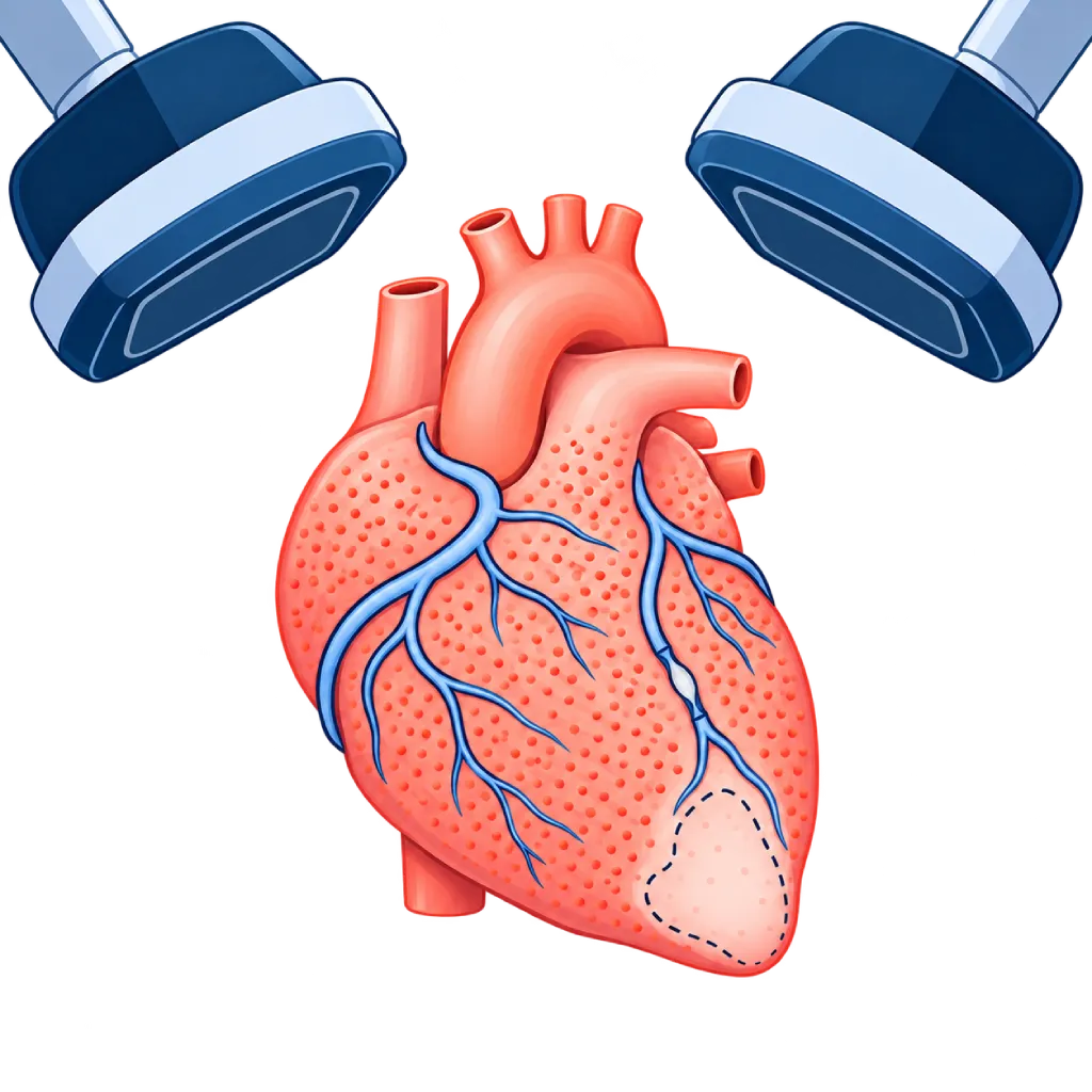
Myocardial perfusion imaging is a nuclear medicine test that shows blood flow to the heart muscle. A tracer is injected at rest and while the heart is stressed with a treadmill or medicine, then a special camera takes images of the heart. Areas with reduced blood flow only under stress indicate narrowed coronary arteries.
Sources01 / Understanding the procedure
About Myocardial Perfusion Imaging
What Is Myocardial Perfusion Imaging
Myocardial perfusion imaging, often called MPI or cardiac SPECT, is a nuclear medicine test to assess blood flow to the heart muscle. It helps your doctor find out whether any part of the heart muscle is not getting enough blood because the coronary arteries are narrowed, and how large that area is.
How It Works in Brief
A low-dose radioactive tracer is injected into a blood vessel and taken up by the heart muscle in proportion to the amount of blood flow that reaches it. A gamma camera captures the signal and builds images of the heart muscle layer by layer. Images are taken twice, at rest and after the heart is stressed. In a narrowed vessel, blood flow is still sufficient at rest but cannot increase when the heart is working hard, so that part of the muscle looks fainter only on the stress images.
What the Results Mean
- Normal: the tracer is spread evenly on both sets of images. The risk of a heart attack in the near future is usually low.
- Reduced only under stress: indicates ischemia, meaning the heart muscle does not get enough blood when working hard because of narrowed coronary arteries.
- Reduced at rest and under stress: usually indicates scar tissue from a previous heart attack.
When This Test Is Chosen
Guidelines recommend functional tests like this one for suspected coronary artery disease, especially when the ECG recording during a treadmill test is hard to read or you cannot exercise. This test also helps assess whether a known narrowing is severe enough to be treated with stent placement or bypass surgery. The size of the area that lacks blood also helps determine the next treatment steps.
02 / Understanding the procedure
When Is This Procedure Considered?
A doctor may consider this procedure in the situations below. The decision is always made after a thorough assessment.
- Chest Pain with Suspected Coronary Artery DiseaseTo look for areas of the heart muscle that do not get enough blood when working hard.
- Treadmill ECG That Is Hard to ReadFor example because of a heart conduction disorder or a pacemaker.
- Unable to ExerciseThe heart is stressed with medicine instead of a treadmill.
- Assessing a Known NarrowingHelps decide whether a stent or bypass surgery is needed.
- New Symptoms After a Stent or BypassTo check whether any part of the heart muscle is again not getting enough blood.
- Assessing Risk Before Major SurgeryIn certain patients with high heart risk, based on the doctor's judgment.
Situations to Consider First
- Recent Heart Attack or Unstable AnginaThe stress test is postponed until your condition is stable.
- Severe Asthma or Breathlessness from Narrowed AirwaysCertain stress medicines can trigger breathlessness; your doctor chooses another medicine.
- Severe Aortic Valve Narrowing with SymptomsA stress test is risky and is usually not done.
- Uncontrolled Rhythm Disturbances or Low Blood PressureThese need to be treated before the heart is stressed.
- Pregnancy and BreastfeedingUsually avoided during pregnancy; if you are breastfeeding, follow the staff's instructions.
03 / Before the procedure
How Should You Prepare?
The right preparation makes the test results reliable.
Avoid caffeine. Do not drink coffee, tea, chocolate, soft drinks, or energy drinks, including decaffeinated ones, for at least 12 to 24 hours before the test, because caffeine blocks the action of the stress medicine.
Fasting. You usually fast from food for about 4 hours beforehand; plain water is still allowed.
Medicines. Ask your doctor which medicines need to be stopped temporarily, for example beta blockers or nitrates. Do not stop any medicine on your own.
Clothing and history. Wear clothes and shoes that are comfortable for walking. Tell the staff if you have asthma, are pregnant, might be pregnant, or are breastfeeding.
Unsure about the preparation instructions? Ask our team before the procedure.
04 / During the procedure
How Is the Procedure Done?
With a one-day protocol, the whole test usually takes 3 to 4 hours, including waiting time. Some hospitals split it over two days. A small IV line is placed in your arm and ECG electrodes are attached to your chest.
Rest Images
The tracer is injected while you are at rest. After waiting about 30 to 60 minutes, you lie down with both arms above your head while the camera moves around your chest for 15 to 20 minutes.
Cardiac Stress Test
You walk on a treadmill that gets faster and steeper while your ECG and blood pressure are monitored. If you cannot exercise, medicine is given through the IV line to mimic the effect of exercise on the heart's blood vessels. The second tracer dose is injected when the stress reaches its peak.
Images After Stress
After a short break, the camera takes images of your heart again in the same way as the rest images.
Reading the Results
Your doctor compares the two sets of images, assesses the pumping function of the heart, and matches the findings with your symptoms and the ECG recording during stress.
05 / Benefits and risks
Benefits and Risks Should Be Discussed Together.
Every medical procedure has benefits and risks. Your doctor will explain both for your situation before the procedure.
Benefits You Can Expect
- Assessing Blood Flow, Not Just Vessel ShapeShows the effect of a narrowing on the heart muscle.
- Showing How Large the Affected Area IsHelps your doctor decide whether catheterization, a stent, or bypass surgery is needed.
- Medicine Option If You Cannot ExerciseSuitable for older adults or patients with limited mobility.
- Also Assesses Pumping FunctionECG-synchronized images show the movement of the heart walls.
- Reassuring Normal ResultsThe risk of heart events in the near future is usually low.
The benefits of a procedure are not a promise of a cure. Ask how the results will affect your care plan.
Risks You Should Know
- Radiation ExposureThe dose is usually about 5 to 10 mSv, depending on the protocol, and is kept as low as possible.
- Effects of the Stress MedicineA brief warm feeling in the face, headache, shortness of breath, or chest heaviness.
- Rhythm Disturbances or Chest Pain During StressRare, and the team monitors you throughout the stress test.
- False-Positive ResultsShadows from breast tissue or the diaphragm sometimes look like an abnormality.
Risks can differ from person to person. A full explanation and consent are given after an individual assessment.
06 / After the procedure
What Is Recovery Like?
You can go home right away and resume your usual activities, unless your doctor advises otherwise. The tracer decays within hours, and the majority of it leaves the body through urine and stool.
After the test. Drink more fluids and urinate regularly. A warm feeling in the face, mild headache, or chest heaviness caused by the stress medicine usually goes away within a few minutes. If you are breastfeeding or caring for a baby, follow the staff's instructions.
Warning signs. Go to the emergency room right away or contact the medical team if you have:
- Chest pain that does not go away with rest
- Shortness of breath that keeps getting worse
- A very fast or irregular heartbeat
- Severe dizziness or fainting
Know the Signs That Need Prompt Care.
Follow the team's guidance on warning signs. If severe or sudden symptoms appear after the procedure, go to the nearest emergency department right away or call Heart Attack Center 1500 258.
07 / Team and support
Doctors and Procedure Support
The doctors below are listed for this procedure. Listed by name, not by ranking.

Dr. med. dr. Denio A. Ridjab, Sp.JP(K)
Interventional Cardiology
Coronary Intervention Procedures +2 centers
Clinical focusInterventional Cardiology · Coronary Artery Disease
View Profile
Dr. dr. Jajang Sinardja, Sp.JP(K)
Interventional Cardiology
Coronary Intervention Procedures
Clinical focusAcute Coronary Syndrome · Primary Coronary Intervention
View Profile
dr. Puti S. Saus, Sp.JP(K)
Interventional Cardiology
Coronary Intervention Procedures
Clinical focusAcute Coronary Syndrome · Primary Coronary Intervention
View Profile08 / Common questions
Before You Plan the Procedure.
Frequently asked questions about myocardial perfusion imaging. What the results mean for your condition is explained by your doctor.
Is the radioactive substance used in this test safe?
The radiation dose is usually about 5 to 10 mSv, depending on the protocol used, and the team aims to use as low a dose as possible. The tracer decays within hours and leaves the body naturally. Your doctor only orders this test when the benefit of the information outweighs the small radiation risk.
How is it different from a regular treadmill test?
A regular treadmill test only assesses the ECG recording while the heart is working hard. Myocardial perfusion imaging adds images of blood flow to the heart muscle, so it is more accurate and can show which part lacks blood and how large that area is. This test is very useful if your ECG is hard to read from the start.
What if I cannot walk on the treadmill?
Your doctor can use a medicine given through an IV line to mimic the effect of exercise on the heart's blood vessels. For a few minutes, you may feel warmth in your face, a headache, shortness of breath, or chest heaviness. These symptoms usually go away quickly, and the team monitors your ECG and blood pressure the whole time.
Why can't I drink coffee before the test?
Caffeine blocks the action of the stress medicine often used in this test. As a result, the heart's blood vessels do not widen as they should, and a narrowing may go undetected. For this reason, avoid coffee, tea, chocolate, soft drinks, and energy drinks as instructed, including those labeled caffeine-free.
What happens if the results are abnormal?
Your doctor will explain whether the affected area indicates ischemia or scar tissue, and how large it is. If the ischemia is extensive or symptoms do not improve with medicines, your doctor may recommend cardiac catheterization to look at the coronary arteries directly. For mild ischemia, treatment with medicines and lifestyle changes is often enough.
How is it different from a cardiac PET scan or CT coronary angiography?
A cardiac PET scan also assesses blood flow to the heart muscle, with sharper images and a lower radiation dose, but the equipment is less widely available. CT coronary angiography shows the shape of the vessels and plaque, not the blood flow. Your doctor chooses the test that suits your symptoms and condition.
Sources and Medical Review
Translated from the Indonesian edition medically reviewed by Dr. dr. Jajang Sinardja, Sp.JP(K).
Sources as cited in the Indonesian edition.
- Vrints C, Andreotti F, Koskinas KC, et al. 2024 ESC Guidelines for the management of chronic coronary syndromes. Eur Heart J. 2024;45(36):3415-3537.
- Gulati M, Levy PD, Mukherjee D, et al. 2021 AHA/ACC/ASE/CHEST/SAEM/SCCT/SCMR Guideline for the Evaluation and Diagnosis of Chest Pain. Circulation. 2021;144(22):e368-e454.
- Henzlova MJ, Duvall WL, Einstein AJ, et al. ASNC imaging guidelines for SPECT nuclear cardiology procedures: stress, protocols, and tracers. J Nucl Cardiol. 2016;23(3):606-639.
- Dorbala S, Ananthasubramaniam K, Armstrong IS, et al. Single photon emission computed tomography (SPECT) myocardial perfusion imaging guidelines: instrumentation, acquisition, processing, and interpretation. J Nucl Cardiol. 2018;25(5):1784-1846.
- Mettler FA Jr, Huda W, Yoshizumi TT, Mahesh M. Effective doses in radiology and diagnostic nuclear medicine: a catalog. Radiology. 2008;248(1):254-263.