
Quick answer
What Is Cardiomyopathy?
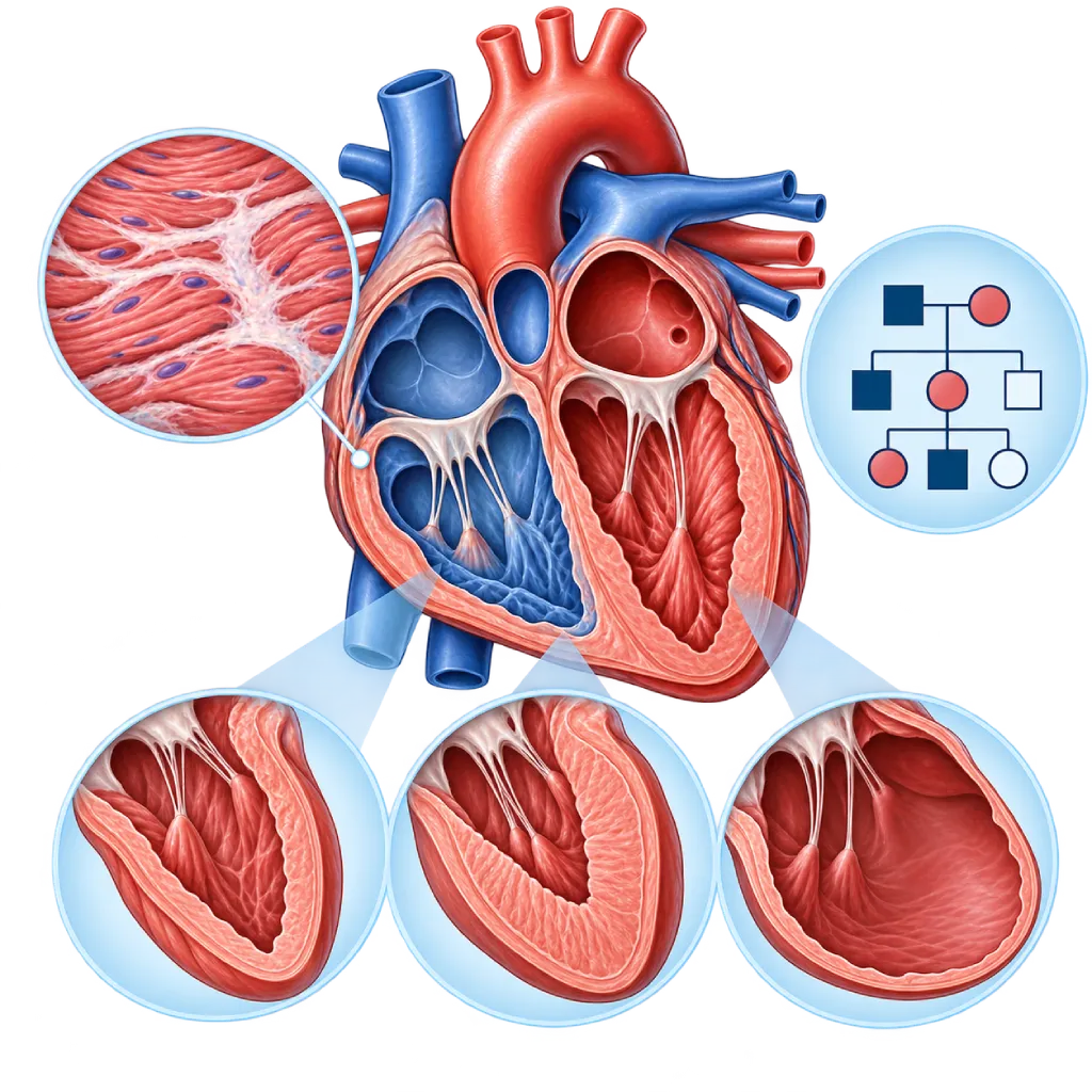
Cardiomyopathy is a group of heart muscle diseases that make the heart chambers enlarge, the walls thicken, or the muscle become stiff and scarred, which can impair the heart's ability to pump blood and keep a steady rhythm.
SourcesSigns That Need Urgent Help
- Severe shortness of breath at rest or being unable to lie down
- Fainting, especially during or after activity
- A very fast heartbeat with weakness, severe dizziness, or cold sweats
- Chest pain that lasts more than a few minutes
- Coughing up frothy or pink sputum
- Bluish lips or fingertips
- Sudden confusion or reduced consciousness
If these signs appear, go to the nearest emergency department right away or call Heart Attack Center 1500 258.
01 / Understanding the condition
About Cardiomyopathy
Cardiomyopathy is the name for a group of diseases that affect the heart muscle (myocardium). In cardiomyopathy, the shape or function of the heart muscle changes: the heart chambers can enlarge and the pumping can weaken, the walls can thicken, or the muscle can become stiff so it has difficulty expanding to fill with blood. Some people have no symptoms for years, while others experience shortness of breath, tiring easily, swelling, palpitations, or fainting.
Types of Cardiomyopathy
The 2023 European Society of Cardiology (ESC) guidelines group cardiomyopathies according to how the heart looks on imaging tests. Hypertrophic cardiomyopathy is marked by a thickened left ventricular wall. Dilated cardiomyopathy is marked by an enlarged left ventricle with weakened pumping. Restrictive cardiomyopathy makes the heart muscle stiff, so filling with blood is impaired. Arrhythmogenic cardiomyopathy causes part of the heart muscle to be replaced by scar tissue and fat, which easily triggers rhythm problems, covered on the Arrhythmogenic Right Ventricular Cardiomyopathy page. There is also non-dilated left ventricular cardiomyopathy, which is scarring or weakened pumping without enlargement of the chamber.
Other pages cover types recognized by their cause or circumstances: infiltrative cardiomyopathy due to a buildup of substances such as amyloid, peripartum cardiomyopathy around the time of giving birth, takotsubo cardiomyopathy triggered by severe stress, toxic cardiomyopathy due to alcohol or certain medicines, and left ventricular noncompaction cardiomyopathy. By the ESC definition, heart muscle abnormalities that are fully explained by coronary artery disease, hypertension, valve disease, or congenital heart disease are not classified as cardiomyopathy. Even so, doctors still commonly use the term ischemic cardiomyopathy for weakened pumping due to coronary artery disease, and nonischemic cardiomyopathy for weakened pumping that is not due to coronary disease.
Why Cardiomyopathy Happens
Some cardiomyopathies are genetic, meaning they are caused by changes in the genes that control heart muscle proteins and can be passed down in families. Others are acquired, for example after inflammation of the heart muscle (myocarditis), or due to heavy alcohol use over a long time, certain chemotherapy medicines, hormone disorders, pregnancy, or a buildup of abnormal substances in the heart muscle. In some people, genetic factors and outside triggers act together.
What It Means for You and Your Family
Because many types of cardiomyopathy are inherited, the ESC guidelines recommend checking close relatives, namely parents, siblings, and children, with an ECG and echocardiography, plus genetic testing when appropriate. Cardiomyopathy is generally a long-term condition. Treatment aims to relieve symptoms, prevent heart failure and dangerous rhythm problems, and maintain quality of life. Because treatment differs by type, knowing which type of cardiomyopathy you have is an important first step.
02 / Understanding the condition
Types and Related Conditions
The following conditions belong to the Cardiomyopathy group. Open each one for a full explanation.
03 / Understanding the condition
Symptoms to Recognize.
Common Symptoms
- Shortness of breath during activity, and later also when lying flat
- Tiring easily and a reduced ability to be active
- Swelling in the ankles, legs, or abdomen
- Palpitations or a heartbeat that feels irregular
- Dizziness or feeling close to fainting
- Chest pain or pressure during activity
- Waking at night because of shortness of breath or coughing
Symptoms in Adult Women
Peripartum cardiomyopathy can appear from late pregnancy up to several months after giving birth, and its symptoms are easily mistaken for ordinary tiredness after delivery.
- Shortness of breath that gets worse in late pregnancy or after giving birth
- Difficulty lying flat because breathing feels short
- Leg swelling that increases quickly after delivery
- Palpitations and severe fatigue that do not improve with rest
Symptoms in Children
In babies and children, symptoms are often noticed during feeding, eating, or play.
- Fast breathing or heavy sweating during breastfeeding or eating
- Difficulty gaining weight as expected for age
- Tiring quickly and falling behind peers during play
- Fainting during exercise, play, or hard crying
- A swollen-looking abdomen due to an enlarged liver
04 / Understanding the condition
What Causes This Condition?
- Changes in the genes that control heart muscle proteins, which are often passed down in families
- Inflammation of the heart muscle (myocarditis), for example after a viral infection or due to autoimmune disease
- Heavy alcohol use over a long period or the use of substances such as cocaine and amphetamines
- Certain chemotherapy medicines or radiation therapy to the chest area
- Buildup of abnormal substances in the heart muscle, such as amyloid or iron
- Changes in workload and hormones around pregnancy and after giving birth
- A heart rhythm that stays too fast for a long time
- Coronary artery disease that damages the heart muscle, called ischemic cardiomyopathy
05 / Understanding the condition
Who Is at Higher Risk?
Genetic
Having a parent, sibling, or child with cardiomyopathy, heart failure at a young age, a pacemaker or ICD implanted at a young age, or sudden cardiac arrest without a clear cause.
Lifestyle
Heavy drinking, using substances such as cocaine or amphetamines, and smoking can damage the heart muscle or increase its workload.
Medical
Hypertension, diabetes, coronary artery disease, thyroid disorders, autoimmune diseases, obesity, and a history of chemotherapy or chest radiation.
Age
Cardiomyopathy can appear at any age. Genetic types are often detected in children, teenagers, or young adults, while types caused by coronary artery disease and amyloidosis are more common in older age.
Other
Pregnancy and the period after giving birth, as well as systemic diseases such as amyloidosis, sarcoidosis, or iron overload due to repeated transfusions.
Groups at Higher Risk
- Family Members of People with Cardiomyopathy: Parents, siblings, and children of people with genetic cardiomyopathy may carry the same gene change even without symptoms.
- Cancer Survivors: Certain chemotherapy medicines, targeted therapies, and chest radiation can weaken the heart muscle, sometimes years after treatment ends.
- Pregnant Women and New Mothers: Peripartum cardiomyopathy can appear from late pregnancy up to several months after delivery.
- People with Coronary Artery Disease: A heart attack or widespread coronary narrowing can damage the heart muscle and reduce its pumping strength.
- Heavy Drinkers: Heavy alcohol use over many years can cause cardiomyopathy, which often improves when alcohol is stopped.
06 / Understanding the condition
How Is It Diagnosed?
The doctor starts by asking about your symptoms, medicines and habits, and your family history going back three generations, including relatives who had heart failure, used a pacemaker, or died suddenly at a young age. After a physical examination, an ECG and echocardiography are usually the first steps to look at the shape and function of the heart. The 2023 ESC guidelines recommend cardiac MRI with contrast in the initial evaluation because it can show scar tissue and patterns of damage that help distinguish the types of cardiomyopathy. If the heart's pumping is weakened, the doctor also checks whether coronary artery disease plays a role, through coronary CT or coronary angiography. Blood tests are used to assess strain on the heart and look for causes that can be treated. Genetic testing and family screening are recommended when the results may affect the care of you or your family.
Echocardiography
The main test to assess the size of the heart chambers, wall thickness, pumping strength, valve function, and any obstruction to blood flow out of the left ventricle.
Electrocardiogram (ECG)
Looks for rhythm problems, electrical conduction problems, and electrical patterns that point to certain types of cardiomyopathy.
Cardiac MRI
Assesses the heart muscle tissue in detail, including scar tissue, inflammation, or a buildup of substances, helping to determine the type and cause.
Laboratory Tests
Blood tests such as NT-proBNP, troponin, kidney, liver, and thyroid function, and iron to assess strain on the heart and look for treatable causes.
Holter Monitoring
Rhythm recording for 24 to 48 hours to find rhythm problems that come and go and to assess their risk.
Coronary Angiography
Assesses the coronary arteries to confirm whether coronary artery disease is the cause of weakened pumping.
Genetic testing from a blood sample in patients who meet the criteria for cardiomyopathy, to look for a genetic cause and guide family screening.
07 / Understanding the condition
Treatment Options.
Treatment is decided by a doctor after an assessment. The categories below can help you follow the conversation with your doctor.
Heart Failure Medicines
Medicine classes such as ARNIs or ACE inhibitors, beta blockers, mineralocorticoid antagonists, and SGLT2 inhibitors are chosen by your doctor according to the type of cardiomyopathy and the strength of the heart's pumping.
Type-Specific Treatment
Some types have specific medicines, for example cardiac myosin inhibitors for obstructive hypertrophic cardiomyopathy or transthyretin stabilizers for ATTR cardiac amyloidosis.
Pacemakers, ICDs, and CRT
An ICD is implanted when the risk of sudden cardiac arrest is high, a pacemaker when the heartbeat is too slow, and resynchronization therapy when the two ventricles do not contract in sync.
Antiarrhythmic Medicines and Ablation
Used to control rhythm problems that accompany cardiomyopathy, for example recurrent atrial fibrillation or ventricular tachycardia.
Treating Triggers and Coexisting Conditions
Stopping alcohol, controlling blood pressure and an overly fast rhythm, treating thyroid disorders, or adjusting chemotherapy together with an oncologist can improve heart muscle function.
Cardiac Rehabilitation
Supervised exercise and self-care education help improve fitness and confidence in being active.
Advanced Heart Failure Therapy
In advanced heart failure, your doctor may consider a left ventricular assist device (LVAD) or a heart transplant.
08 / Understanding the condition
Complications to Watch For.
Heart Failure
A weak or stiff heart muscle cannot pump or hold enough blood, so fluid builds up in the lungs and legs. Covered on the Heart Failure page.
Heart Rhythm Disorders
Changes and scar tissue in the heart muscle can trigger atrial fibrillation, ventricular rhythms that are too fast, or electrical conduction problems that make the heartbeat too slow.
Sudden Cardiac Arrest
A very fast ventricular rhythm can make the heart suddenly stop pumping. This risk is assessed in every type of cardiomyopathy to decide whether an ICD is needed.
Stroke and Blood Clots
Atrial fibrillation or slow blood flow in an enlarged heart chamber can form clots that travel to the brain or other organs.
Mitral Valve Leakage
An enlarged heart chamber can pull on the mitral valve leaflets so the valve does not close tightly and shortness of breath worsens.
09 / Understanding the condition
What Can You Do?
- If a family member has cardiomyopathy, have heart checkups as your doctor advises even if you have no symptoms
- Limit or stop alcohol, and avoid substances such as cocaine and amphetamines
- Control your blood pressure, blood sugar, and cholesterol
- Take your medicines as prescribed and do not stop them without talking to your doctor
- Weigh yourself every morning and note any rapid weight gain
- Ask about heart function monitoring before and during certain chemotherapy
- Discuss the type and intensity of exercise that is safe for you
10 / Understanding the condition
When Should You See a Doctor?
Make an appointment with a cardiologist if the following symptoms keep coming back, persist, or get worse. A checkup is also recommended even if you have no symptoms, if a close relative has been diagnosed with cardiomyopathy. If you faint, have prolonged chest pain, or have severe shortness of breath at rest, go to the emergency room right away.
- Shortness of breath or tiring easily during activities that used to feel easy
- Swelling in the legs or rapid weight gain over a few days
- Palpitations, dizziness, or nearly fainting
- A close relative with cardiomyopathy, heart failure at a young age, or sudden cardiac arrest
- Shortness of breath or swelling in late pregnancy or after giving birth
- About to start or currently having chemotherapy that can affect the heart
Don't Wait in an Emergency.
If symptoms are severe or come on suddenly, go to the nearest emergency department right away or call Heart Attack Center 1500 258, rather than waiting for an appointment.
11 / Common questions
Questions You May Have.
Frequently asked questions about cardiomyopathy.
Is cardiomyopathy the same as heart failure?
No. Cardiomyopathy is a disease of the heart muscle, while heart failure is a condition in which the heart cannot pump or hold blood as the body needs. Cardiomyopathy is one cause of heart failure, but not everyone with cardiomyopathy develops heart failure. Some people remain free of symptoms for years, especially when the condition is found early and monitored regularly.
Is cardiomyopathy inherited?
Some types, especially hypertrophic, dilated, and arrhythmogenic cardiomyopathy, are often caused by gene changes that can be inherited. In the common inheritance pattern, autosomal dominant, each child has about a 50 percent chance of inheriting the gene change, although not all gene carriers will develop symptoms. That is why your doctor may recommend counseling and genetic testing, followed by regular ECGs and echocardiography for close relatives.
Can cardiomyopathy be cured?
It depends on the type and cause. Cardiomyopathy triggered by alcohol, a heart rhythm that is too fast, pregnancy, or inflammation can improve when the trigger is treated. Genetic types usually last a lifetime but can be controlled with medicines, devices, and regular follow-up. Even after the heart's pumping has improved, medicines are usually continued because stopping them can make the condition worse again.
Can people with cardiomyopathy exercise?
Generally yes, and light to moderate exercise is recommended for most people with cardiomyopathy. The type and intensity of training need to be tailored to the type of cardiomyopathy, the risk of rhythm problems, and test results. Competitive or high-intensity sports need to be assessed by your doctor first. A cardiac rehabilitation program can help you train safely and under supervision.
Can women with cardiomyopathy get pregnant?
Many women with cardiomyopathy can have a safe pregnancy, but pregnancy adds to the heart's workload. A pregnancy should be planned and discussed early with a cardiologist and an obstetrician, because some heart medicines need to be adjusted by a doctor before pregnancy. In certain conditions, for example when the heart's pumping is very weak, pregnancy is high risk and requires special consideration.
What tests should my family have?
Close relatives, namely parents, siblings, and children, are usually advised to have a clinical examination, an ECG, and echocardiography. If the gene change causing the disease has been found in the patient, family members can have targeted genetic testing. Relatives who do not carry that gene change generally do not need regular checkups, while carriers are monitored regularly according to their age and the type of cardiomyopathy.
Sources and Medical Review
Translated from the Indonesian edition medically reviewed by dr. BRM Ario S. Kuncoro, Sp.JP(K).
Sources as cited in the Indonesian edition.
- Arbelo E, Protonotarios A, Gimeno JR, Arbustini E, Barriales-Villa R, Basso C, et al. 2023 ESC Guidelines for the management of cardiomyopathies. Eur Heart J. 2023;44(37):3503-626.
- McKenna WJ, Maron BJ, Thiene G. Classification, epidemiology, and global burden of cardiomyopathies. Circ Res. 2017;121(7):722-30.
- McDonagh TA, Metra M, Adamo M, Gardner RS, Baumbach A, Böhm M, et al. 2021 ESC Guidelines for the diagnosis and treatment of acute and chronic heart failure. Eur Heart J. 2021;42(36):3599-726.
- Zeppenfeld K, Tfelt-Hansen J, de Riva M, Winkel BG, Behr ER, Blom NA, et al. 2022 ESC Guidelines for the management of patients with ventricular arrhythmias and the prevention of sudden cardiac death. Eur Heart J. 2022;43(40):3997-4126.
- Wilde AAM, Semsarian C, Márquez MF, Sepehri Shamloo A, Ackerman MJ, Ashley EA, et al. European Heart Rhythm Association (EHRA)/Heart Rhythm Society (HRS)/Asia Pacific Heart Rhythm Society (APHRS)/Latin American Heart Rhythm Society (LAHRS) Expert Consensus Statement on the state of genetic testing for cardiac diseases. Europace. 2022;24(8):1307-67.
- Pelliccia A, Sharma S, Gati S, Bäck M, Börjesson M, Caselli S, et al. 2020 ESC Guidelines on sports cardiology and exercise in patients with cardiovascular disease. Eur Heart J. 2021;42(1):17-96.
- Regitz-Zagrosek V, Roos-Hesselink JW, Bauersachs J, Blomström-Lundqvist C, Cífková R, De Bonis M, et al. 2018 ESC Guidelines for the management of cardiovascular diseases during pregnancy. Eur Heart J. 2018;39(34):3165-241.
Doctors Who Treat This Condition

dr. Siska S. Danny, Sp.JP(K)
Interventional Cardiology
Coronary Intervention Procedures
Clinical focusPCI for Acute Coronary Syndrome · Primary Coronary Intervention
View Profile
dr. BRM Ario S. Kuncoro, Sp.JP(K)
Echocardiography & Cardiovascular Imaging
Cardiovascular Diagnostic Center +2 centers
Clinical focusAdvanced and 3D Echocardiography · Transesophageal Echocardiography
View Profile
Dr. dr. Dicky A. Hanafy, Sp.JP(K)
Arrhythmia and Electrophysiology
Arrhythmia and Device Center +1 center
Clinical focusElectrophysiology and Arrhythmia Ablation · Pacemaker and ICD Implantation
View Profile