
Quick answer
What Is Left Ventricular Noncompaction Cardiomyopathy?
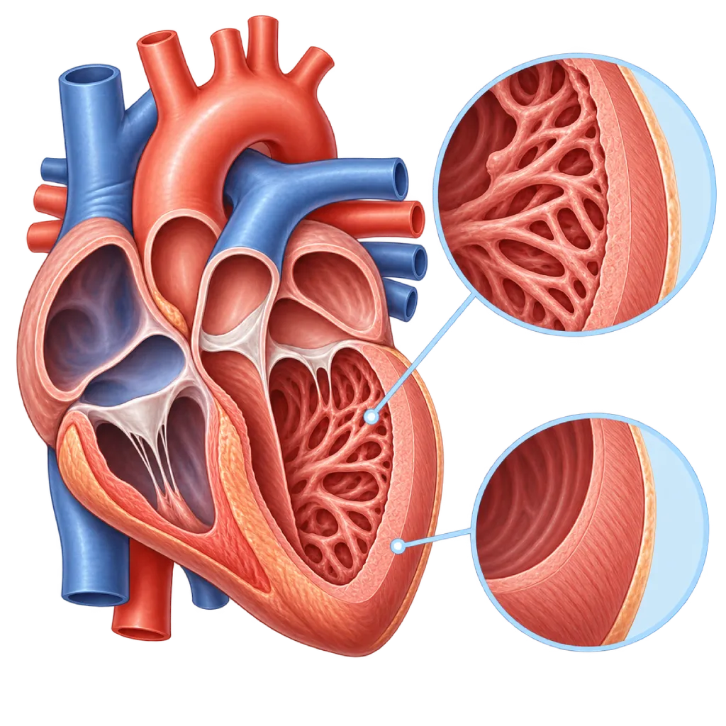
Left ventricular noncompaction cardiomyopathy is a condition in which the inner layer of the wall of the heart's left ventricle has many muscle ridges and deep spaces, making it look like a sponge. Some people stay healthy, while others are at risk of heart failure, rhythm disorders, or blood clots.
SourcesSigns That Need Urgent Help
- Severe shortness of breath at rest or being unable to lie down
- Sudden fainting, especially during exercise
- A very fast heartbeat with severe dizziness or weakness
- Chest pain that lasts more than a few minutes
- A frothy cough or noisy breathing when short of breath
- Sudden facial drooping, arm weakness, or slurred speech
If these signs appear, go to the nearest emergency department right away or call Heart Attack Center 1500 258.
01 / Understanding the condition
About Left Ventricular Noncompaction Cardiomyopathy
The wall of the heart's left lower chamber (left ventricle) is normally made of a compact layer of muscle with a relatively smooth inner surface. In left ventricular noncompaction cardiomyopathy, the inner layer is filled with muscle ridges (trabeculae) with deep spaces between them, while the compact outer layer looks thinner. This appearance is also called excessive trabeculation. In medical literature, the condition is known as left ventricular non-compaction (LVNC).
A Feature or a Disease?
Understanding of this condition has changed in recent years. The 2023 European Society of Cardiology (ESC) cardiomyopathy guidelines no longer classify it as a separate type of cardiomyopathy. Excessive trabeculation is now seen as a feature of the heart muscle's structure that can be found on its own or alongside other cardiomyopathies, such as dilated cardiomyopathy and hypertrophic cardiomyopathy.
A similar appearance can also occur in healthy people, for example athletes who train intensively, pregnant women, and people with sickle cell anemia or chronic kidney disease. In pregnancy, the trabeculation can decrease again after delivery. For this reason, a finding of trabeculation on a test result does not automatically mean you have heart disease. Heart changes in athletes are covered on the Athlete's Heart page.
When to Be Concerned
What determines the risk is the strength of the heart's pumping, whether there are rhythm disorders, blood clots, or scar tissue in the heart muscle, and family history. If the size and function of the left ventricle are normal and there are no symptoms, the course is usually uneventful. If the pump is weakened, the risks resemble those of dilated cardiomyopathy: heart failure, rhythm disorders, and blood clots that can travel to the brain.
Some cases are related to changes in genes for heart muscle proteins, so they can be found in several members of the same family. This condition can also accompany congenital heart defects, such as Ebstein anomaly, as well as certain nerve and muscle diseases. It can be found at any age, from infancy to old age.
Treatment Approach
Treatment is not aimed at the trabeculae themselves but at their consequences. People without symptoms and with normal heart function usually only need regular monitoring. If there is heart failure, a rhythm disorder, or a blood clot, treatment follows the guidelines for each of these conditions. An overview of this family of diseases is covered on the Cardiomyopathy page.
02 / Understanding the condition
Symptoms to Recognize.
Common Symptoms
Many people with excessive trabeculation have no symptoms. The following symptoms appear when the heart's pumping or rhythm is affected.
- Shortness of breath during activity or when lying down
- Tiring easily and a reduced ability to be active
- Swelling in the ankles and legs
- Palpitations or a feeling of an irregular heartbeat
- Dizziness or nearly fainting
- Fainting, especially during or after physical activity
Symptoms in Children
In babies and children, heart failure often shows up as feeding and growth problems rather than shortness of breath.
- Difficulty nursing or tiring quickly while feeding
- Sweating a lot while nursing or eating
- Fast breathing even at rest
- Poor weight gain for their age
- Tiring more quickly than other children of the same age during play
- Fainting while crying hard or exercising
03 / Understanding the condition
What Causes This Condition?
- Changes (variants) in genes for heart muscle proteins, which can be inherited in families
- Part of another cardiomyopathy, such as dilated or hypertrophic cardiomyopathy
- Certain congenital heart defects, for example Ebstein anomaly
- Nerve and muscle diseases or mitochondrial diseases that also affect the heart muscle
- Barth syndrome in boys, a rare inherited disorder that also weakens the body's muscles
- Adaptive trabeculation that can decrease again, for example in athletes, pregnancy, or sickle cell anemia
- In some people, no cause is found even after a complete evaluation
04 / Understanding the condition
Who Is at Higher Risk?
Genetic
Family members with cardiomyopathy, heart failure at a young age, or sudden death without a clear cause increase the likelihood of a genetic cause.
Medical
Congenital heart defects, nerve and muscle diseases, mitochondrial diseases, sickle cell anemia, and chronic kidney disease are associated with excessive trabeculation, sometimes as a disease and sometimes as an adaptation of the heart.
Age
This condition can be found in babies, children, and adults. The form that appears in babies is more often accompanied by congenital heart defects or inherited metabolic diseases.
Other
Years of intense physical training and pregnancy can cause trabeculation that resembles this condition. This appearance is also seen more often in people of African descent.
Groups at Higher Risk
- Family Members of People with Cardiomyopathy: Parents, siblings, and children of people with genetic cardiomyopathy are advised to have a heart examination even if they have no symptoms yet.
- Athletes in Intense Training: Trabeculation can appear as the heart's adaptation to training, so it needs to be carefully distinguished from cardiomyopathy to avoid a wrong label.
- Children with Congenital Heart Defects: Excessive trabeculation is more often found together with certain congenital defects and metabolic diseases in babies and children.
- People with Nerve and Muscle Diseases: Several muscle and mitochondrial diseases also affect the heart muscle, so regular heart checkups are recommended.
05 / Understanding the condition
How Is It Diagnosed?
This condition is often found by chance when echocardiography is done for another reason, or when family members are examined. The doctor will ask about your symptoms, any history of fainting, your exercise habits, pregnancy, and any family history of heart disease or sudden death, then perform a physical examination.
Echocardiography and cardiac MRI show the ratio of the spongy layer to the compact layer, the size of the chambers, and the pumping strength. However, no single number is enough to make the diagnosis. The 2023 ESC guidelines and an expert imaging panel emphasize that the results must be interpreted together with symptoms, heart function, scar tissue on MRI, and family history. An ECG and a rhythm recording over several days help look for rhythm disorders. If a genetic cause is suspected, the doctor may recommend counseling and genetic testing, followed by examinations for family members.
Echocardiography
Assesses the pattern of the trabeculae and the compact layer, the size of the left ventricle, the pumping strength, and the valves, and looks for blood clots inside the heart.
Cardiac MRI
Shows the muscle layers in more detail, measures ventricular function accurately, and looks for scar tissue associated with the risk of rhythm disorders.
Electrocardiogram (ECG)
Looks for electrical conduction problems, signs of enlarged heart chambers, and rhythm disorders that may accompany this condition.
Holter Monitoring
Records the heart rhythm for 24 to 48 hours or longer to look for rhythm disorders in people who have palpitations or have fainted.
NT-proBNP and BNP Test
A blood test that helps assess the strain on the heart and monitor heart failure if pumping function is reduced.
Genetic testing is considered when trabeculation comes with cardiomyopathy or a family history, preceded by counseling and followed by heart examinations for close family members.
06 / Understanding the condition
Treatment Options.
Treatment is decided by a doctor after an assessment. The categories below can help you follow the conversation with your doctor.
Regular Monitoring
People without symptoms and with normal pumping function usually only need routine checkups with echocardiography and ECG on the schedule set by the doctor.
Heart Failure Medications
If the pump is weakened, the doctor prescribes heart failure medication classes according to guidelines, for example beta blockers, renin-angiotensin system inhibitors, mineralocorticoid antagonists, and SGLT2 inhibitors.
Blood Thinners
Given if there is atrial fibrillation, a blood clot in the heart, or a history of embolism. When the pump is very weak, the doctor weighs the benefits and risks.
Pacing Devices and ICDs
An implantable defibrillator (ICD) is considered when the risk of cardiac arrest is high, and a resynchronization pacemaker (CRT) for heart failure with certain conduction problems.
Genetic Testing and Family Screening
Helps identify family members at risk before symptoms appear, so that monitoring can start earlier.
Heart Transplant
Considered for advanced heart failure that no longer improves with medication and devices.
07 / Understanding the condition
Complications to Watch For.
Heart Failure
If the muscle of the left ventricle weakens, the heart cannot pump enough blood, causing shortness of breath, swelling, and tiring easily. Covered on the Heart Failure page.
Heart Rhythm Disorders
Atrial fibrillation and fast rhythms from the lower chambers can develop, especially if the pump is weakened or there is scar tissue.
Blood Clots and Stroke
Slow blood flow in the spaces between the trabeculae, or in upper chambers that quiver irregularly, can form clots that travel to the brain or other organs.
Sudden Cardiac Arrest
Severe rhythm disorders from the lower chambers can suddenly stop the heart from pumping. The risk is assessed mainly from pumping function, scar tissue, and any history of fainting. Covered on the Sudden Cardiac Death page.
08 / Understanding the condition
What Can You Do?
- Have a heart examination if a family member has cardiomyopathy or died suddenly at a young age
- Follow your echocardiography checkup schedule even if you have no symptoms
- Discuss safe types and intensity of exercise with your cardiologist
- Note the time and circumstances when your heart races or you nearly faint
- Take your medications as prescribed and do not stop them on your own
- If you are planning a pregnancy, consult your cardiologist first
- Avoid smoking and limit alcohol to reduce the strain on your heart
09 / Understanding the condition
When Should You See a Doctor?
A finding of excessive trabeculation on a test result should be discussed with a cardiologist, even if you feel healthy. Go to the emergency room right away if you faint, have severe shortness of breath at rest, have chest pain that does not go away, or show signs of stroke. Make an appointment if you experience the following:
- An echocardiography or MRI report mentions noncompaction or excessive trabeculation
- Shortness of breath, tiring easily, or leg swelling that happens more and more often
- Palpitations, dizziness, or nearly fainting
- A family member has cardiomyopathy, heart failure at a young age, or sudden death
- You are pregnant or planning a pregnancy and have been diagnosed with this condition
- Your child has difficulty feeding, poor weight gain, or fast breathing
Don't Wait in an Emergency.
If symptoms are severe or come on suddenly, go to the nearest emergency department right away or call Heart Attack Center 1500 258, rather than waiting for an appointment.
10 / Common questions
Questions You May Have.
Frequently asked questions about left ventricular noncompaction cardiomyopathy.
Does excessive trabeculation mean I definitely have heart disease?
Not always. Excessive trabeculation can be found in healthy people, for example athletes, pregnant women, and people with sickle cell anemia. The 2023 ESC guidelines view it as a feature of the heart muscle's structure, not a diagnosis on its own. The doctor will assess your symptoms, the size and pumping strength of your heart, whether there is scar tissue on MRI, and your family history. If everything is fine, regular monitoring is usually enough.
Is this condition inherited?
Some cases are related to changes in genes for heart muscle proteins and can be inherited. If excessive trabeculation comes with cardiomyopathy, or there is a family history of cardiomyopathy and sudden death, the doctor may recommend counseling and genetic testing. The results help determine which family members need to be examined. Family screening usually involves an ECG and echocardiography, repeated regularly because abnormalities can appear later.
Can I still exercise?
In general, yes, but the type and intensity should be discussed with your cardiologist. People with normal pumping function, no rhythm disorders, and no history of fainting can usually exercise as normal with regular monitoring. If the pump is weakened, there is a rhythm disorder, or you have fainted before, the doctor may limit strenuous or competitive sports. The decision is made together based on your test results.
Do I need to take blood thinners?
Not everyone needs them. Blood thinners are recommended if there is atrial fibrillation, a blood clot inside the heart, or a history of stroke or embolism. When pumping function is very weak, the doctor will weigh the benefit of preventing clots against the risk of bleeding. Do not start or stop this medication without your doctor's guidance, because both can be dangerous.
What if I want to get pregnant?
Pregnancy increases the heart's workload. If your pumping function is normal, pregnancy can generally proceed with monitoring by a cardiologist and an obstetrician. If the pump is weakened or there is a rhythm disorder, the risk is higher and some heart medications need to be changed because they are not safe for the baby. For this reason, discuss your plans before becoming pregnant so that tests and medication adjustments can be done early.
Can this condition be cured?
The trabeculae themselves usually remain, except those caused by pregnancy or training, which can decrease again. However, heart function matters more. Many people continue their normal activities because their heart pumps well. If the pump is weakened, heart failure medications can improve symptoms and, in some people, improve pumping strength. Regular checkups help detect changes early.
Sources and Medical Review
Translated from the Indonesian edition medically reviewed by dr. BRM Ario S. Kuncoro, Sp.JP(K).
Sources as cited in the Indonesian edition.
- Arbelo E, Protonotarios A, Gimeno JR, Arbustini E, Barriales-Villa R, Basso C, et al. 2023 ESC Guidelines for the management of cardiomyopathies. Eur Heart J. 2023;44(37):3503-3626.
- Petersen SE, Jensen B, Aung N, Friedrich MG, McMahon CJ, Mohiddin SA, et al. Excessive trabeculation of the left ventricle: JACC: Cardiovascular Imaging expert panel paper. JACC Cardiovasc Imaging. 2023;16(3):408-425.
- Towbin JA, Lorts A, Jefferies JL. Left ventricular non-compaction cardiomyopathy. Lancet. 2015;386(9995):813-825.
- Ross SB, Jones K, Blanch B, Puranik R, McGeechan K, Barratt A, et al. A systematic review and meta-analysis of the prevalence of left ventricular non-compaction in adults. Eur Heart J. 2020;41(14):1428-1436.
- Aung N, Doimo S, Ricci F, Sanghvi MM, Pedrosa C, Woodbridge SP, et al. Prognostic significance of left ventricular noncompaction: systematic review and meta-analysis of observational studies. Circ Cardiovasc Imaging. 2020;13(1):e009712.
- Gati S, Papadakis M, Papamichael ND, Zaidi A, Sheikh N, Reed M, et al. Reversible de novo left ventricular trabeculations in pregnant women: implications for the diagnosis of left ventricular noncompaction in low-risk populations. Circulation. 2014;130(6):475-483.
- Zeppenfeld K, Tfelt-Hansen J, de Riva M, Winkel BG, Behr ER, Blom NA, et al. 2022 ESC Guidelines for the management of patients with ventricular arrhythmias and the prevention of sudden cardiac death. Eur Heart J. 2022;43(40):3997-4126.
Doctors Who Treat This Condition

dr. BRM Ario S. Kuncoro, Sp.JP(K)
Echocardiography & Cardiovascular Imaging
Cardiovascular Diagnostic Center +2 centers
Clinical focusAdvanced and 3D Echocardiography · Transesophageal Echocardiography
View Profile
Dr. dr. Dicky A. Hanafy, Sp.JP(K)
Arrhythmia and Electrophysiology
Arrhythmia and Device Center +1 center
Clinical focusElectrophysiology and Arrhythmia Ablation · Pacemaker and ICD Implantation
View Profile
dr. Radityo Prakoso, Sp.JP(K)
Pediatrics & Congenital Heart Disease
Congenital and Structural Heart Center
Clinical focusPediatric Cardiology Consultation · Congenital Heart Disease Care
View Profile