
Quick answer
What Is Double Outlet Right Ventricle?
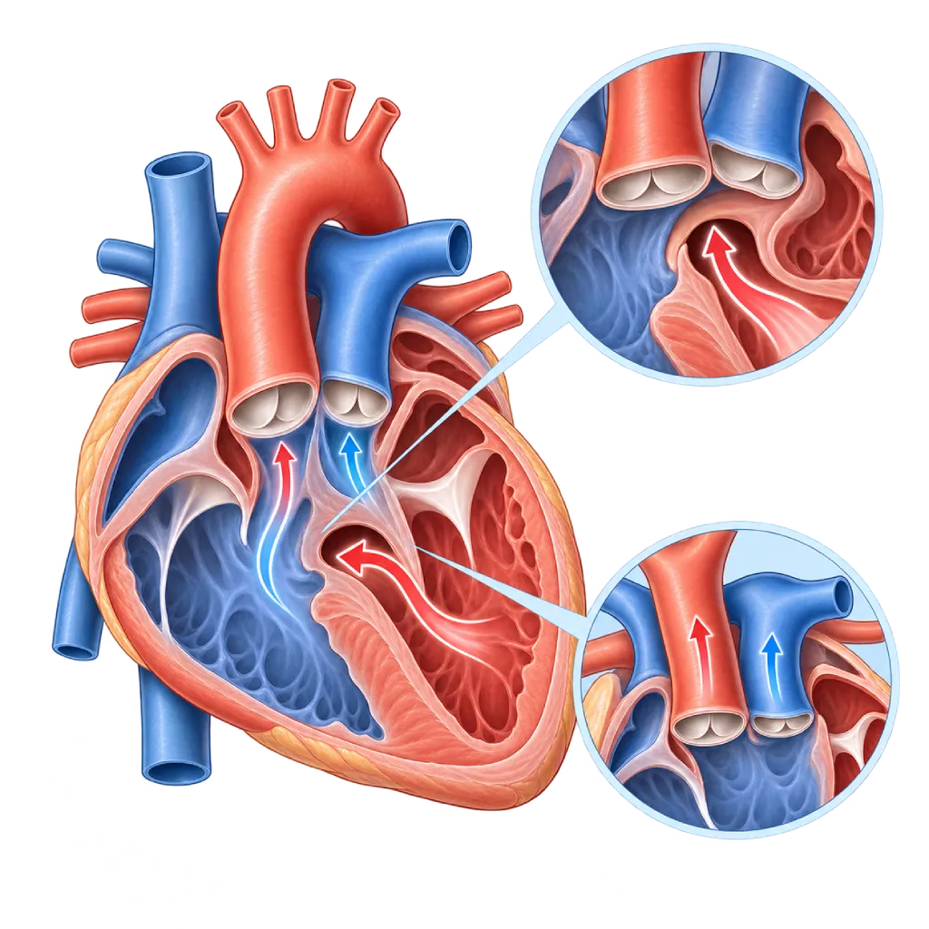
Double outlet right ventricle is a congenital heart defect in which the aorta and the pulmonary artery both arise from the right ventricle. Symptoms vary, from heart failure to a bluish color, depending on where the septal hole lies and whether the pathway to the lungs is narrowed.
SourcesSigns That Need Urgent Help
- Severe bluish color that keeps getting worse
- A baby who is very limp, hard to wake, or refuses to feed
- Very fast breathing with the chest wall pulling in
- Pale, cold skin and a weak pulse
- Fainting or seizures
If these signs appear, go to the nearest emergency department right away or call Heart Attack Center 1500 258.
01 / Understanding the condition
About Double Outlet Right Ventricle
In a normal heart, the aorta arises from the left ventricle and the pulmonary artery arises from the right ventricle. In double outlet right ventricle (DORV), both great arteries arise entirely or largely from the right ventricle. Blood from the left ventricle can leave it only through a hole in the wall between the ventricles. This defect is rare, about 1 to 2 of every 100 congenital heart defects, and forms in the early weeks of pregnancy. An overview of other congenital defects is covered on the Congenital Heart Disease page.
One Name, Several Forms
Symptoms and treatment depend greatly on the location of the hole and whether the pathway to the lungs is narrowed. If the hole lies below the aorta without narrowing, the condition resembles a large hole in the ventricular septum with too much blood flow to the lungs, covered on the Ventricular Septal Defect page. If the pathway to the lungs is narrowed, it resembles tetralogy of Fallot and the baby looks blue. If the hole lies below the pulmonary artery (Taussig-Bing anomaly), it resembles transposition of the great arteries and often comes with narrowing of the aorta. There are also holes located far from both vessels, which make repair more complex.
Surgery Is Planned Individually
The goal of surgery is to channel blood from the left ventricle to the aorta and blood from the right ventricle to the pulmonary artery. This can be done with a tunnel inside the ventricle from the hole to the aorta, combined with an arterial switch operation, widening of the pathway to the lungs, or placement of a conduit. If the anatomy does not allow the two ventricles to work separately, the doctor may choose a single-ventricle pathway with the Glenn and then the Fontan operation. Some babies need a temporary procedure first, for example to reduce excessive blood flow to the lungs or to increase blood flow in a baby who is very blue.
Lifelong Monitoring
After surgery, the outflow pathway to the aorta or to the lungs can narrow again, the septum can leak, and rhythm disorders can develop. The 2020 ESC and 2018 AHA/ACC guidelines recommend regular lifelong checkups at a service with expertise in congenital heart disease, including when teenagers move on to adult doctors.
02 / Understanding the condition
Symptoms to Recognize.
Common Symptoms
- Fast breathing or tiring quickly during feeding
- Sweating during feeding
- Difficulty gaining weight
- Bluish lips and skin in certain forms
- A heart murmur heard when examined by a doctor
- Low oxygen saturation on newborn screening
Symptoms in Newborns
Forms that resemble tetralogy of Fallot or transposition often look blue from the first days of life.
- Bluish color from birth
- Low oxygen saturation on screening
- A heart murmur at the first examination
- Difficulty feeding or falling asleep quickly while feeding
Symptoms in Infants
Forms with excessive blood flow to the lungs usually cause signs of heart failure at a few weeks of age.
- Fast breathing at rest or during sleep
- Feeding briefly and then stopping because of tiredness
- Heavy sweating on the head during feeding
- Not gaining weight
- Frequent coughs or respiratory infections
Symptoms in Children
After surgery, the following symptoms need to be checked because they may indicate renarrowing or an arrhythmia.
- Tiring more quickly than other children of the same age
- Shortness of breath while playing
- Palpitations, dizziness, or fainting
- Bluish color that comes back
03 / Understanding the condition
What Causes This Condition?
- Problems with the rotation and division of the outflow tracts of the fetal heart in the early weeks of pregnancy
- Chromosomal abnormalities such as trisomy 18, trisomy 13, or 22q11.2 deletion in some babies
- Changes in the genes that control heart development in some families
- In many babies, the exact cause is unknown
04 / Understanding the condition
Who Is at Higher Risk?
Genetic
Chromosomal abnormalities such as trisomy 18, trisomy 13, and 22q11.2 deletion, as well as a family history of congenital heart disease, increase the likelihood of this defect.
Medical
Uncontrolled maternal diabetes before and during pregnancy is associated with a higher risk of defects of the outflow tracts of the heart.
Lifestyle
Smoking and drinking alcohol during pregnancy increase the risk of congenital heart defects in general.
Groups at Higher Risk
- Babies with Chromosomal Abnormalities: Trisomy 18, trisomy 13, and 22q11.2 deletion are more often accompanied by this defect, so echocardiography is recommended for babies with these diagnoses.
- Babies of Mothers with Diabetes: Diabetes that is uncontrolled before pregnancy is associated with defects of the outflow tracts of the heart, including this defect.
- Children and Adults After Surgery: The outflow to the aorta and to the lungs can narrow again, the septum can leak, and arrhythmias can appear years later.
05 / Understanding the condition
How Is It Diagnosed?
This defect can be seen on a pregnancy ultrasound and confirmed with fetal echocardiography. After birth, doctors suspect it from a heart murmur, a bluish color, low oxygen saturation on screening, or signs of heart failure in the first few weeks.
Echocardiography is the main test. It assesses, among other things, the position of the septal hole relative to the aorta and pulmonary artery, the distance between them, whether there is narrowing of the pathway to the lungs or to the aorta, the position of the valves, and the shape of the aortic arch. Because these relationships determine the type of surgery, a cardiac CT scan or MRI is often used to create a three-dimensional image. Cardiac catheterization is done if the lung pressure needs to be measured or the blood vessels need to be mapped. Genetic testing may be recommended if there are other abnormalities outside the heart.
Fetal echocardiography and newborn oxygen saturation screening help find the defect early.
Echocardiography
The main test to assess the position of the septal hole relative to the aorta and pulmonary artery, narrowing of the outflow pathways, the valves, and the aortic arch.
Cardiac CT Scan
Creates a three-dimensional image of how the septal hole relates to the great arteries, to plan the tunnel inside the ventricle.
Cardiac MRI
Assesses the anatomy and function of the ventricles without X-rays, especially in older children and adults after surgery.
Cardiac Catheterization
Measures lung pressure and maps the blood vessels when needed before surgery or a repeat procedure.
Electrocardiogram (ECG)
Assesses the rhythm and the strain on the ventricles, including heart block that can appear after surgery on the septum.
06 / Understanding the condition
Treatment Options.
Treatment is decided by a doctor after an assessment. The categories below can help you follow the conversation with your doctor.
Heart Failure Medicines and Nutritional Support
In babies with excessive blood flow to the lungs, the doctor may give diuretics and other medicines, plus high-calorie milk if needed, while waiting for surgery.
Temporary Procedures
Banding of the pulmonary artery to reduce excessive blood flow, or a surgical shunt or stent to increase blood flow to the lungs in babies who are very blue.
Two-Ventricle Repair
A tunnel is created inside the ventricle to direct blood from the left ventricle to the aorta, combined with widening of the pathway to the lungs, an arterial switch, or a conduit. Covered on the Congenital Heart Surgery page.
Single-Ventricle Pathway
If two-ventricle repair is not possible, the Glenn and then the Fontan operations are performed so that venous blood flows directly to the lungs without being pumped.
Repeat Procedures by Catheter or Surgery
A narrowing or a conduit can be widened with a balloon, fitted with a valve through a catheter, or repaired again with surgery. Covered on the Transcatheter Pulmonary Valve Replacement page.
Lifelong Checkups
Regular echocardiography, ECG, MRI or CT, and exercise tests to detect renarrowing, septal leaks, and arrhythmias early.
07 / Understanding the condition
Complications to Watch For.
Heart Failure in Babies
In forms without narrowing of the pathway to the lungs, excessive blood flow to the lungs makes the baby breathless and causes difficulty feeding and growing.
Severe Cyanosis
In forms that resemble tetralogy of Fallot or transposition, oxygen levels can be low and require earlier treatment.
Pulmonary Hypertension
Long-standing excessive blood flow damages the blood vessels of the lungs and can make surgery riskier. Covered on the Pulmonary Hypertension page.
Narrowing of the Outflow to the Aorta
The tunnel from the septal hole to the aorta can narrow over time and require repeat surgery.
Narrowing of the Pathway to the Lungs or the Conduit
The pathway to the lungs or the conduit can narrow or leak as the child grows and need to be widened or replaced.
Rhythm Disorders and Heart Block
Arrhythmias or heart block can appear after surgery; some require a pacemaker. Covered on the Heart Rhythm Disorders / Arrhythmia page.
08 / Understanding the condition
What Can You Do?
- This defect cannot be prevented with certainty, but a healthy pregnancy helps lower the risk of congenital heart defects
- Control diabetes before and during pregnancy, take folic acid as recommended, and avoid smoking and alcohol
- Have your scheduled pregnancy ultrasounds, and fetal echocardiography if there is a family history or a suspected problem
- Make sure your baby has oxygen saturation screening before going home from the place of birth
- Keep the teeth and mouth clean to reduce the risk of heart infection
- Follow the lifelong checkup schedule after surgery even if your child looks healthy
09 / Understanding the condition
When Should You See a Doctor?
Take your baby to the emergency room right away if they look increasingly blue, are very limp, are hard to wake, are breathing very fast, or refuse to feed. Make an appointment with a pediatric cardiologist if:
- A pregnancy ultrasound shows an abnormality of the great arteries or the septum of the fetal heart.
- Your baby's oxygen saturation screening result is abnormal.
- Your baby breathes fast, sweats during feeding, or is not gaining weight.
- The doctor finds a heart murmur in your baby or child.
- Your child tires more quickly after surgery, becomes short of breath while playing, has palpitations, or has fainted.
- A teenager or adult who has had surgery has not had a checkup for a long time.
Don't Wait in an Emergency.
If symptoms are severe or come on suddenly, go to the nearest emergency department right away or call Heart Attack Center 1500 258, rather than waiting for an appointment.
10 / Common questions
Questions You May Have.
Frequently asked questions about double outlet right ventricle.
Why can symptoms differ between children with this defect?
This defect actually includes several forms. If the septal hole lies below the aorta without narrowing of the pathway to the lungs, the baby tends to develop heart failure. If the pathway to the lungs is narrowed, the baby looks blue, as in tetralogy of Fallot. If the hole lies below the pulmonary artery, it resembles transposition of the great arteries. That is why the doctor will explain which form your child has and the appropriate plan.
When is surgery usually done?
The timing of surgery depends on the form of the defect and the baby's condition. Forms that resemble transposition are usually operated on in the first few weeks. Forms with excessive blood flow to the lungs are generally repaired within the first few months, before the lung blood vessels are damaged. Forms that resemble tetralogy of Fallot are often repaired in the first year. If the baby is not yet ready, a temporary procedure may be done first. The pediatric heart team will decide the appropriate timing.
Can my child have two ventricles that work separately after surgery?
For many children, yes. Two-ventricle repair surgery makes the left ventricle pump to the aorta again and the right ventricle pump to the pulmonary artery. However, if the septal hole is located far from the aorta, the ventricles are unequal in size, or a valve is in a position that gets in the way, this repair may not be safe. In that situation, the doctor may choose a single-ventricle pathway with the Glenn and then the Fontan operation.
Will my child need surgery again later?
Some children need a repeat procedure. The tunnel to the aorta can narrow, the conduit to the pulmonary artery needs to be replaced as the child grows, or a remaining hole needs to be closed. Some repeat procedures can be done through a catheter, while others require surgery. Regular checkups help the doctor find problems before symptoms appear, so that procedures can be planned at the right time.
Is the next pregnancy at risk?
The chance of a heart defect in the next pregnancy is slightly higher than in the general population, especially if there is a chromosomal abnormality or a family history of congenital heart disease. Genetic counseling can help estimate this chance. In the next pregnancy, fetal echocardiography is recommended so that any defect can be found early and the delivery can be well planned.
Sources and Medical Review
Translated from the Indonesian edition medically reviewed by dr. Radityo Prakoso, Sp.JP(K).
Sources as cited in the Indonesian edition.
- Baumgartner H, De Backer J, Babu-Narayan SV, et al. 2020 ESC Guidelines for the management of adult congenital heart disease. Eur Heart J. 2021;42(6):563-645.
- Stout KK, Daniels CJ, Aboulhosn JA, et al. 2018 AHA/ACC Guideline for the Management of Adults With Congenital Heart Disease: A Report of the American College of Cardiology/American Heart Association Task Force on Clinical Practice Guidelines. Circulation. 2019;139(14):e698-e800.
- Walters HL, Mavroudis C, Tchervenkov CI, et al. Congenital Heart Surgery Nomenclature and Database Project: double outlet right ventricle. Ann Thorac Surg. 2000;69(4 Suppl):S249-S263.
- Ebadi A, Spicer DE, Backer CL, et al. Double-outlet right ventricle revisited. J Thorac Cardiovasc Surg. 2017;154(2):598-604.
- Obler D, Juraszek AL, Smoot LB, et al. Double outlet right ventricle: aetiologies and associations. J Med Genet. 2008;45(8):481-497.
- Martin GR, Ewer AK, Gaviglio A, et al. Updated Strategies for Pulse Oximetry Screening for Critical Congenital Heart Disease. Pediatrics. 2020;146(1):e20191650.
Doctors Who Treat This Condition

dr. Radityo Prakoso, Sp.JP(K)
Pediatrics & Congenital Heart Disease
Congenital and Structural Heart Center
Clinical focusPediatric Cardiology Consultation · Congenital Heart Disease Care
View Profile
dr. Salomo Purba, Sp.BTKV, Subsp.JPK(K)
Pediatric and Congenital Heart Surgery
Congenital and Structural Heart Center +2 centers
Clinical focusPediatric Heart Surgery · Congenital Heart Defect Surgery
View Profile
dr. BRM Ario S. Kuncoro, Sp.JP(K)
Echocardiography & Cardiovascular Imaging
Cardiovascular Diagnostic Center +2 centers
Clinical focusAdvanced and 3D Echocardiography · Transesophageal Echocardiography
View Profile