
Quick answer
What Is Tetralogy of Fallot?
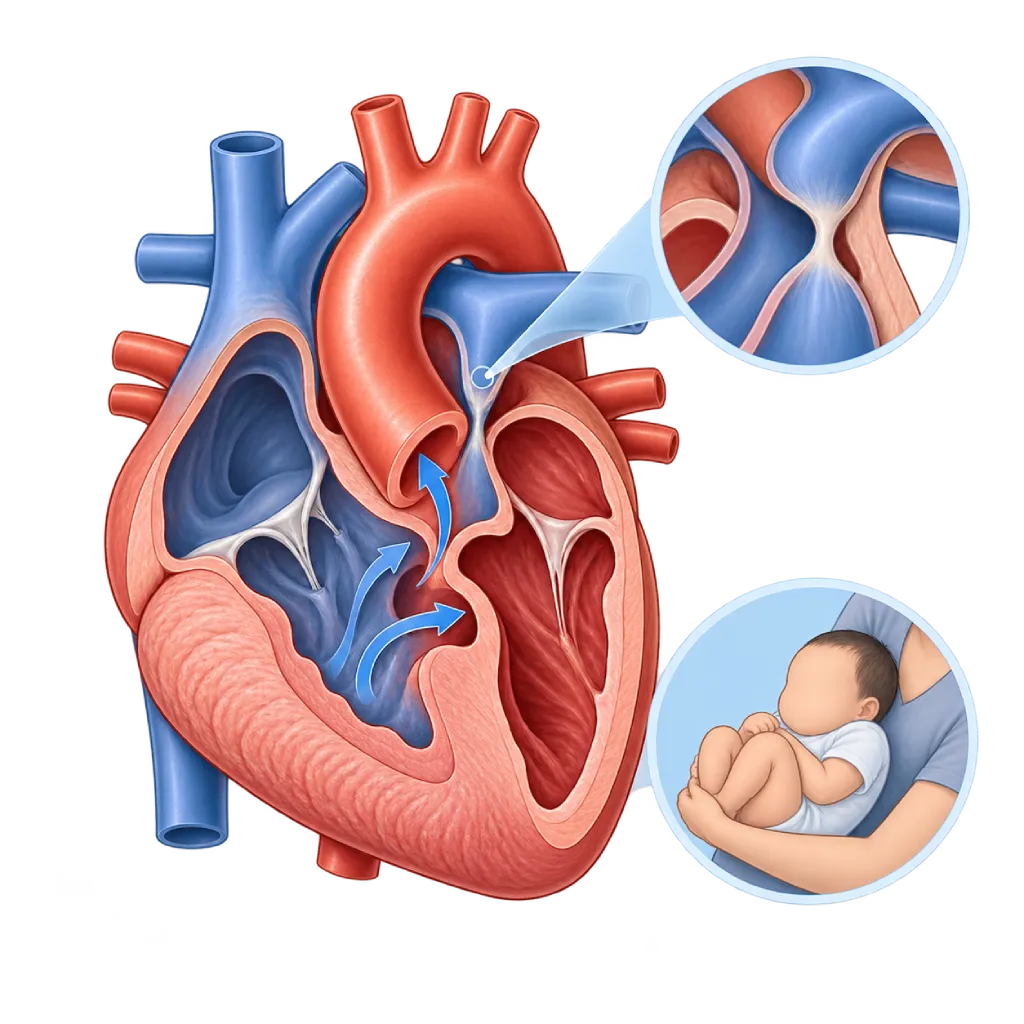
Tetralogy of Fallot is a congenital heart defect with four features: a hole in the wall between the ventricles, a narrowed outflow to the lungs, a displaced aorta, and a thickened right ventricular muscle. As a result, some oxygen-poor blood circulates to the body.
SourcesSigns That Need Urgent Help
- A blue spell that does not improve within a few minutes
- A child who is limp, unresponsive, or faints
- Seizures
- Very fast, deep breathing with severe blueness
- Fainting or a very fast heartbeat in adults who have had surgery
- Severe shortness of breath at rest
If these signs appear, go to the nearest emergency department right away or call Heart Attack Center 1500 258.
01 / Understanding the condition
About Tetralogy of Fallot
Tetralogy of Fallot is a common type of congenital heart disease that causes bluish lips and skin (cyanosis), found in about 3 to 5 of every 10,000 births. It is called a tetralogy because four abnormalities occur together: a large hole in the wall between the two ventricles (ventricular septal defect), a narrowing in the right ventricle's outflow tract or the pulmonary valve (pulmonary stenosis), an aorta that is shifted over the hole so it receives blood from both ventricles, and thickening of the right ventricular muscle because it must pump against the narrowing. The defect forms in the early weeks of pregnancy, when the outflow of the fetal heart is dividing. Some children also have a chromosome 22q11.2 deletion or Down syndrome.
Why the Child Looks Blue
Because the path to the lungs is narrowed, some oxygen-poor blood from the right ventricle is diverted through the hole and into the aorta. The more severe the narrowing, the more blood does not receive oxygen, so the child becomes bluer. Some babies are only slightly bluish, while others are very blue from birth. Babies can also have sudden blue spells, often called tet spells, usually when crying, feeding, or just after waking up.
Corrective Surgery
The main treatment is complete corrective surgery, usually in the first year of life and often at 3 to 6 months of age. The hole is closed with a patch and the outflow to the lungs is widened. This surgery uses a heart-lung machine and is usually followed by intensive care for several days. If the baby is too blue before being ready for correction, the doctor may perform a temporary procedure to increase blood flow to the lungs. The long-term results of this surgery are good; the majority of children grow into adulthood and lead active lives.
In Adulthood
Corrective surgery does not make the heart completely normal. The pulmonary valve often leaks after its outflow has been widened, which over time causes the right ventricle to enlarge. Heart rhythm disorders can also appear years later. The 2020 ESC and 2018 AHA/ACC guidelines recommend lifelong routine checkups, with periodic cardiac MRI to determine the timing of pulmonary valve replacement. In some people, this valve replacement can be done through a catheter without opening the chest. Other congenital defects are covered on the Congenital Heart Disease page.
02 / Understanding the condition
Symptoms to Recognize.
Common Symptoms
- Bluish lips, tongue, and nails, more noticeable when crying
- A heart murmur heard from infancy
- Fast breathing or tiring quickly when feeding or playing
- Difficulty gaining weight and height
- Rounded fingertips and toes (clubbing) if surgery is delayed for a long time
Symptoms in Newborns
Severity depends on the narrowing of the path to the lungs; some babies only look blue after a few weeks.
- Bluish color from birth when the narrowing is severe
- Low oxygen saturation on newborn screening
- A loud heart murmur at the first examination
- Difficulty feeding or tiring quickly while feeding
Symptoms in Infants
Blue spells often appear at 2 to 6 months of age, especially in the morning after waking up or when crying.
- Suddenly very fussy and crying nonstop
- Fast, deep breathing
- Blueness that gets worse within a few minutes
- Looking limp or floppy after crying
Symptoms in Children
Mainly in children who have not had surgery or who had surgery at an older age.
- Often squatting to rest after running or playing
- Tiring quickly and shortness of breath during activity
- Bluish lips and nails
- Fingers and toes that look rounded at the tips
Symptoms in Adults
In adults who had surgery as children, symptoms often appear gradually or are not felt at all.
- Tiring more quickly or getting out of breath during activity
- Palpitations or a heartbeat that feels irregular
- Dizziness or near fainting
- Swelling in the legs or abdomen
- Reduced exercise capacity compared with before
03 / Understanding the condition
What Causes This Condition?
- Abnormal development of the septum and outflow tract of the fetal heart in the early weeks of pregnancy
- A shift of the septum below the pulmonary valve that causes both the narrowing and the hole in the ventricular septum
- Chromosome 22q11.2 deletion (DiGeorge syndrome) in some children
- Down syndrome and other chromosomal abnormalities
- In many children, the exact cause is unknown
04 / Understanding the condition
Who Is at Higher Risk?
Genetic
22q11.2 deletion, Down syndrome, and a history of congenital heart disease in a parent or sibling increase the likelihood of tetralogy of Fallot.
Medical
Uncontrolled diabetes in the mother, uncontrolled phenylketonuria in the mother, and rubella infection in early pregnancy are linked to a higher risk of congenital heart disease.
Lifestyle
Drinking alcohol and smoking during pregnancy increase the risk of congenital heart defects in general.
Groups at Higher Risk
- Children with 22q11.2 Deletion: This chromosomal disorder is often accompanied by heart defects of the ventricular outflow, including tetralogy of Fallot, so a heart evaluation is recommended.
- Children with Down Syndrome: Down syndrome is often accompanied by congenital heart disease, so echocardiography is recommended for all babies with this syndrome.
- Adults Who Had Surgery for Tetralogy of Fallot: A leaking pulmonary valve, right ventricular enlargement, and arrhythmias can appear decades after surgery, often without early symptoms.
- Women Who Had Surgery and Are Planning Pregnancy: Pregnancy adds to the workload of the right ventricle, so assessment before pregnancy and genetic counseling help with planning.
05 / Understanding the condition
How Is It Diagnosed?
Tetralogy of Fallot is often already visible on a pregnancy ultrasound and is confirmed with fetal echocardiography. After birth, the clues are a heart murmur, bluish color, or low oxygen saturation on newborn screening.
Echocardiography is the main test. It shows all four abnormalities, the severity of the narrowing of the path to the lungs, the size of the pulmonary arteries, the position of the coronary arteries, and the side of the aortic arch. A chest X-ray may show a boot-shaped heart, and an ECG shows thickening of the right ventricle. A cardiac CT scan or cardiac catheterization is used when the lung or coronary vessels need to be mapped in more detail before surgery. Genetic testing for 22q11.2 deletion is often recommended.
After surgery, cardiac MRI becomes an important test to measure the size and function of the right ventricle and the leakage of the pulmonary valve. Holter monitoring and exercise testing help detect rhythm disorders and assess physical capacity.
Fetal echocardiography and newborn oxygen saturation screening help find the defect before the baby goes home from the place of birth.
Echocardiography
The main test, showing all four abnormalities, the severity of the narrowing, the size of the pulmonary arteries, the position of the coronary arteries, and the side of the aortic arch.
Electrocardiogram (ECG)
Shows thickening of the right ventricle before surgery, then monitors the width of the heart's electrical waves and arrhythmias after surgery.
Cardiac MRI
The reference test for measuring right ventricular volume and function and pulmonary valve leakage in older children and adults after surgery.
Cardiac CT Scan
Maps the pulmonary arteries, coronary arteries, and collateral vessels before surgery or a valve procedure.
Cardiac Catheterization
Measures pressures and maps the lung vessels when other imaging is not enough, or as part of a catheter-based valve procedure.
Holter Monitoring
Records the heart rhythm for 24 to 48 hours to look for arrhythmias in teenagers and adults after surgery.
06 / Understanding the condition
Treatment Options.
Treatment is decided by a doctor after an assessment. The categories below can help you follow the conversation with your doctor.
Treating Blue Spells
The baby is calmed and the knees are bent toward the chest to increase flow to the lungs. In the hospital, the doctor may give oxygen, fluids, and medication, then consider beta blocker medication while waiting for surgery.
Temporary Procedures for Babies
If the baby is very blue before being ready for correction, flow to the lungs can be increased with a surgical shunt, a stent in the ductus or the right ventricular outflow tract, or balloon widening of the pulmonary valve.
Complete Corrective Surgery
The hole is closed with a patch and the outflow to the lungs is widened, usually in the first year of life. Covered on the Congenital Heart Surgery page.
Pulmonary Valve Replacement
In teenagers and adults with a severely leaking or narrowed pulmonary valve, the valve is replaced through surgery or through a catheter. Covered on the Transcatheter Pulmonary Valve Replacement page.
Treatment of Rhythm Disorders
Antiarrhythmic medication, ablation, or an ICD is considered depending on the type of arrhythmia and the risk assessment. Covered on the Arrhythmia Management page.
Lifelong Follow-Up
Regular checkups with ECG, echocardiography, cardiac MRI, Holter monitoring, and exercise testing at a service experienced in adult congenital heart disease.
07 / Understanding the condition
Complications to Watch For.
Blue Spells (Tet Spells)
Oxygen levels drop suddenly when flow to the lungs becomes more obstructed. If a spell lasts a long time, the brain and other organs may not get enough oxygen.
Pulmonary Valve Leakage
After the outflow has been widened, the pulmonary valve often leaks. Over time, the right ventricle enlarges and weakens. Covered on the Pulmonary Regurgitation page.
Heart Rhythm Disorders
Atrial and ventricular arrhythmias can appear years after surgery and, in a small number of people, increase the risk of sudden cardiac death.
Residual Hole or Narrowing
A small leak at the septal patch or remaining narrowing in the right ventricular outflow tract sometimes requires a repeat procedure.
Aortic Root Enlargement
The aortic root can enlarge in some adults, so its size is monitored during regular checkups.
Infective Endocarditis
Infection of the inner lining of the heart or an artificial valve occurs more easily. Good dental hygiene and antibiotics before certain procedures, as advised by your doctor, help prevent it.
08 / Understanding the condition
What Can You Do?
- Tetralogy of Fallot cannot be reliably prevented, but good prenatal care helps lower the risk of congenital heart defects
- Control diabetes, complete rubella vaccination before pregnancy, take folic acid as recommended, and avoid smoking and alcohol
- Have scheduled pregnancy ultrasounds, and fetal echocardiography if there is a family history or a concern
- Learn how to calm your baby and use the knee-to-chest position during a blue spell, then seek help right away
- Make sure your baby drinks enough, especially during fever, vomiting, or diarrhea, because dehydration can trigger blue spells
- Keep teeth and mouth clean and have regular dental checkups to reduce the risk of endocarditis
- Continue lifelong checkups after surgery, including when moving from a pediatric to an adult doctor during the teenage years
09 / Understanding the condition
When Should You See a Doctor?
Take your child to the emergency room right away if a blue spell does not improve within a few minutes, or if your child is limp, unresponsive, has a seizure, or faints. In adults who have had surgery, fainting or a very fast heartbeat also needs urgent help. Make an appointment with a cardiologist if:
- Your baby looks bluish, has difficulty feeding, or has trouble gaining weight.
- A doctor finds a heart murmur or low oxygen saturation on screening.
- Your child often squats to rest or tires quickly while playing.
- You have had surgery for tetralogy of Fallot and your exercise capacity has declined or you often have palpitations.
- You have not had a checkup for more than a year after surgery, or you are planning a pregnancy.
Don't Wait in an Emergency.
If symptoms are severe or come on suddenly, go to the nearest emergency department right away or call Heart Attack Center 1500 258, rather than waiting for an appointment.
10 / Common questions
Questions You May Have.
Frequently asked questions about tetralogy of Fallot.
What should I do when my baby has a blue spell?
Stay calm and soothe your baby. Hold your baby with the knees bent toward the chest, or lay your baby on their side with the knees bent; this position helps increase blood flow to the lungs. If the spell does not improve within a few minutes, your baby becomes limp, or the blueness gets worse, go to the emergency room right away. Report every spell to your doctor, because blue spells are usually a reason to schedule surgery sooner.
When is the right time for corrective surgery?
Many heart centers perform complete corrective surgery at 3 to 6 months of age, or within the first year of life. The exact timing depends on how blue the baby is, whether there are blue spells, body weight, and the shape of the pulmonary and coronary arteries. Babies who are very blue early may need a temporary procedure first. The pediatric heart team will explain the options and the right timing for your child.
Can a child live a normal life after surgery?
The majority of children who have had surgery grow up, go to school, exercise, and work like their peers. Long-term studies show that many patients live into adulthood with a good quality of life. However, their heart is still different from a normal heart. The pulmonary valve, right ventricle size, and heart rhythm need lifelong monitoring, so regular checkups are still needed even when the child feels well.
Why do adults who had surgery as children still need cardiac MRI?
A leaking pulmonary valve makes the right ventricle enlarge slowly, often without symptoms. Cardiac MRI can accurately measure the size and strength of the right ventricle and the amount of blood leaking back, without X-rays. These regular measurements help your doctor decide when to replace the pulmonary valve before the right ventricle becomes permanently weakened. Your doctor will set the MRI schedule based on your condition.
Can women with tetralogy of Fallot get pregnant?
Many women who have had surgery can have a safe pregnancy after being assessed by a doctor. The assessment before pregnancy covers pulmonary valve leakage, right ventricle size and function, heart rhythm, and current medications. If there is a 22q11.2 deletion, the chance of passing it on to a child is about 50 percent, so genetic counseling is recommended. During pregnancy, checkups are more frequent and fetal echocardiography is recommended.
Sources and Medical Review
Translated from the Indonesian edition medically reviewed by dr. Radityo Prakoso, Sp.JP(K).
Sources as cited in the Indonesian edition.
- Baumgartner H, De Backer J, Babu-Narayan SV, et al. 2020 ESC Guidelines for the management of adult congenital heart disease. Eur Heart J. 2021;42(6):563-645.
- Stout KK, Daniels CJ, Aboulhosn JA, et al. 2018 AHA/ACC Guideline for the Management of Adults With Congenital Heart Disease: A Report of the American College of Cardiology/American Heart Association Task Force on Clinical Practice Guidelines. Circulation. 2019;139(14):e698-e800.
- Apitz C, Webb GD, Redington AN. Tetralogy of Fallot. Lancet. 2009;374(9699):1462-1471.
- Gatzoulis MA, Balaji S, Webber SA, et al. Risk factors for arrhythmia and sudden cardiac death late after repair of tetralogy of Fallot: a multicentre study. Lancet. 2000;356(9234):975-981.
- Smith CA, McCracken C, Thomas AS, et al. Long-term Outcomes of Tetralogy of Fallot: A Study From the Pediatric Cardiac Care Consortium. JAMA Cardiol. 2019;4(1):34-41.
- Geva T. Repaired tetralogy of Fallot: the roles of cardiovascular magnetic resonance in evaluating pathophysiology and for pulmonary valve replacement decision support. J Cardiovasc Magn Reson. 2011;13(1):9.
Doctors Who Treat This Condition

dr. Radityo Prakoso, Sp.JP(K)
Pediatrics & Congenital Heart Disease
Congenital and Structural Heart Center
Clinical focusPediatric Cardiology Consultation · Congenital Heart Disease Care
View Profile
dr. Salomo Purba, Sp.BTKV, Subsp.JPK(K)
Pediatric and Congenital Heart Surgery
Congenital and Structural Heart Center +2 centers
Clinical focusPediatric Heart Surgery · Congenital Heart Defect Surgery
View Profile
dr. Suko Adiarto, Sp.JP(K), Ph.D.
Interventional Cardiology
Vascular Diagnostic and Therapeutic Center +1 center
Clinical focusCoronary Intervention · Peripheral Vascular Intervention
View Profile