
Quick answer
What Is Patent Ductus Arteriosus?
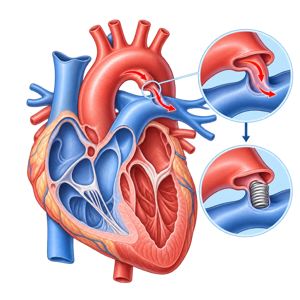
Patent ductus arteriosus is a channel between the aorta and the pulmonary artery that stays open after birth. A small channel often causes no symptoms, while a large one strains the heart and lungs and needs to be closed.
SourcesSigns That Need Urgent Help
- Very fast breathing, gasping, or the chest pulling inward with each breath
- A baby who refuses to feed or is very limp
- Pale grayish skin, with cold hands and feet
- Bluish color, especially in the legs and toes
- Severe shortness of breath or fainting in children or adults
If these signs appear, go to the nearest emergency department right away or call Heart Attack Center 1500 258.
01 / Understanding the condition
About Patent Ductus Arteriosus
In the womb, a baby does not yet breathe with the lungs. Blood from the right side of the heart is routed past the lungs through a short vessel called the ductus arteriosus, which connects the pulmonary artery to the aorta. After the baby is born and starts breathing, this channel normally narrows within one to three days and closes permanently within a few weeks. If it stays open, the condition is called patent ductus arteriosus, or PDA.
After birth, the pressure in the aorta is higher than in the pulmonary artery. As a result, some oxygen-rich blood from the aorta flows back to the lungs through this channel. The lungs receive too much blood, while the left atrium and left ventricle have to hold and pump the extra volume.
In Premature and Full-Term Babies
PDA is common in premature babies because the channel is not yet mature enough to close. In very premature babies, PDA can worsen breathing problems and is usually managed by the neonatal intensive care team. Many of these close on their own in the first weeks. In full-term babies, a PDA that is still open after a few months rarely closes on its own and is usually planned for closure.
Channel Size Determines Its Effects
A very small channel often cannot be heard and is found only on echocardiography. A small to moderate channel causes a characteristic heart murmur that is heard continuously, like the sound of a machine. A large channel can cause heart failure in babies and, if left for years, can damage the blood vessels of the lungs. In adults, PDA is sometimes found only because of a heart murmur, an enlarged left heart, or a rhythm disturbance.
How PDA Is Treated
Treatment depends on age, the size of the channel, and its effects on the heart and lungs. In premature babies, the medical team may monitor first or give medicine that helps the channel close. In full-term babies, children, and adults, a channel that strains the heart is generally closed with a coil or a plug device through a catheter, without opening the chest. Ligation surgery is chosen when the catheter approach is not suitable. Once the channel is closed and there are no other problems, most children can be as active as their peers.
02 / Understanding the condition
Symptoms to Recognize.
Common Symptoms
- A continuous, machine-like heart murmur heard during an examination
- Often no symptoms when the channel is small
- Tiring easily or shortness of breath during activity with a moderate to large channel
- A strong, bounding pulse
- Palpitations or an irregular heartbeat in adults
Symptoms in Newborns
Mainly in premature babies cared for in the neonatal intensive care unit.
- Fast breathing or an increasing need for breathing support
- A strong, bounding pulse in the hands and feet
- Low diastolic (bottom number) blood pressure
- A heart murmur that is first heard after a few days
- A bloated belly or difficulty tolerating milk feeds
Symptoms in Infants
- Fast breathing, especially during feeding
- Sweating and tiring quickly during feeding
- Poor weight gain
- Frequent coughs and colds or pneumonia
03 / Understanding the condition
What Causes This Condition?
- The ductus arteriosus does not close after birth as it should
- Premature birth, because the channel is not yet mature enough to close
- Low oxygen levels after birth, for example in babies born at high altitude
- Rubella infection in the mother during early pregnancy
- Certain genetic or chromosomal disorders, such as Down syndrome and Char syndrome
- In full-term babies, it often occurs without a clear cause
04 / Understanding the condition
Who Is at Higher Risk?
Other
Babies born very prematurely or with a very low birth weight have PDA more often. Being born at high altitude is also associated with PDA. In full-term babies, PDA is more common in girls.
Medical
Rubella infection in the mother in early pregnancy and severe breathing problems in newborns are associated with a ductus that stays open.
Genetic
A family history of PDA and genetic syndromes such as Down syndrome increase the likelihood of PDA.
Groups at Higher Risk
- Premature Babies: The more premature the baby, the greater the chance that the ductus stays open and affects breathing.
- Babies with Congenital Rubella Syndrome: Rubella in early pregnancy can cause PDA together with eye and hearing problems.
- Children with Down Syndrome: Heart defects, including PDA, often occur with Down syndrome, so echocardiography is recommended after birth.
- Adults with an Unclosed PDA: At risk of left heart enlargement, atrial fibrillation, endarteritis, and high lung pressure, so regular checkups are needed.
05 / Understanding the condition
How Is It Diagnosed?
In premature babies, PDA is often suspected because breathing gets worse, the pulse feels strong, or a heart murmur appears a few days after birth. In older babies and children, PDA is usually found because the doctor hears the characteristic continuous murmur during a routine checkup.
Echocardiography confirms the diagnosis. This test shows the size of the channel, the direction of flow, the size of the left atrium and left ventricle, and an estimate of lung pressure. The results help the doctor judge whether the PDA is significant for the circulation. An ECG and a chest X-ray complete the assessment. Cardiac catheterization is needed when lung pressure is suspected to be high, and it is often used at the same time to close the channel. In adults, a CT scan can help assess the shape of the channel and any calcification.
Physical Examination
The doctor hears a continuous murmur below the left collarbone and feels a strong, bounding pulse.
Echocardiography
Confirms that the channel is open and measures its diameter, the direction of flow, the size of the left heart, and the estimated lung pressure.
Electrocardiogram (ECG)
Assesses the heart rhythm and signs of strain on the left heart.
Chest X-Ray
Shows heart enlargement and excess blood flow to the lungs.
Cardiac Catheterization
Measures lung pressure when it is suspected to be high and provides the route for closing the channel with a device.
Cardiac CT Scan
In adults, helps assess the shape of the channel and any calcification before a procedure.
06 / Understanding the condition
Treatment Options.
Treatment is decided by a doctor after an assessment. The categories below can help you follow the conversation with your doctor.
Regular Monitoring
A very small PDA without a murmur and without enlargement of the left heart generally only needs monitoring.
Ductus-Closing Medicine for Premature Babies
In certain premature babies, doctors may give a nonsteroidal anti-inflammatory drug (NSAID) or acetaminophen (paracetamol) to help the channel close. The decision and the dose are determined by the neonatal intensive care team.
Catheter Closure
The channel is closed with a coil or a plug device through a catheter inserted from a blood vessel in the groin. This is the main option for children and adults, and it is also done in some premature babies.
Ductus Ligation Surgery
The channel is tied off or clipped through a small incision on the side of the chest, mainly in very small babies or when the shape of the channel is not suitable for a catheter.
Symptom Management
Diuretic medicines and fluid management can ease shortness of breath while waiting for closure. The type and dose are determined by the doctor.
07 / Understanding the condition
Complications to Watch For.
Heart Failure
A large channel overloads the left heart with blood volume, so the baby becomes short of breath, sweats, and has trouble gaining weight.
Lung Problems in Premature Babies
Excess blood flow to the lungs can prolong the need for breathing support in very premature babies.
Pulmonary Hypertension
If a large channel is left untreated, the blood vessels of the lungs can be damaged and lung pressure rises permanently. In advanced stages, the flow reverses and the legs look bluish. Covered on the Pulmonary Hypertension page.
Infective Endarteritis
An infection of the channel wall, similar to endocarditis. The risk is low, but it should be recognized when a person with PDA has a prolonged fever.
Atrial Fibrillation in Adults
A left atrium that has been enlarged for years can trigger an irregular rhythm in adults.
08 / Understanding the condition
What Can You Do?
- Get regular prenatal checkups to reduce the risk of premature birth
- Make sure you are immune to rubella before you become pregnant
- Avoid smoking and alcohol during pregnancy
- Follow the schedule for follow-up echocardiography if your baby was born prematurely with PDA
- Keep the teeth and mouth clean if you or your child has a PDA that has not been closed
09 / Understanding the condition
When Should You See a Doctor?
If your baby was born prematurely, the doctor will monitor the PDA during the hospital stay. After going home, or for full-term babies, see a doctor if the following signs appear. Go to the emergency room right away if your baby is very short of breath, turns bluish, or refuses to feed.
- Your baby breathes fast or sweats during feeding
- Your baby has trouble gaining weight
- A doctor hears a heart murmur during a routine checkup
- Your child often has pneumonia or tires easily
- A person with PDA has a prolonged fever with no clear cause
- An adult with PDA has palpitations, shortness of breath, or tires easily
Don't Wait in an Emergency.
If symptoms are severe or come on suddenly, go to the nearest emergency department right away or call Heart Attack Center 1500 258, rather than waiting for an appointment.
10 / Common questions
Questions You May Have.
Questions parents often ask about patent ductus arteriosus.
Can a PDA close on its own?
In premature babies, many PDAs close on their own within the first weeks, so doctors often monitor first. In full-term babies, a channel that is still open after about three months of age rarely closes on its own. For this reason, in children, a PDA that causes a murmur or strains the heart is usually planned for closure through a catheter or surgery.
Is catheter closure of a PDA safe for children?
Catheter closure has been performed widely in children and adults, with good results in the majority of patients and a short recovery. The device is inserted through a blood vessel in the groin, so there is no incision in the chest. As with any procedure, there are still risks, such as the device shifting or problems with the blood vessel in the groin, although these are rare. The doctor will explain the benefits and risks based on your child's size and the shape of the channel.
Do premature babies with PDA always need medicine or a procedure?
Not always. Research in recent years shows that many PDAs in premature babies close on their own, and early treatment does not always bring benefit. The neonatal intensive care team usually assesses the size of the channel and its effects on breathing, blood pressure, and digestion. Medicine or a procedure is given when the PDA is judged to be significant for the baby's circulation.
I am an adult and just found out I have a PDA. Does it have to be closed?
It depends on its size. A very small PDA without a murmur and without left heart enlargement generally only needs monitoring. If the left heart is enlarged because of the shunt flow and lung pressure is not yet too high, the 2020 ESC guidelines recommend closure, usually through a catheter. If lung pressure is already high, a complete evaluation is needed first to make sure closure is still safe.
Can my child be active as usual after the PDA is closed?
Generally, yes. After catheter closure, children can usually return to light activity within a few days and to full activity once the doctor allows it. A follow-up echocardiogram is done to make sure the channel is fully sealed and the heart size returns to normal. If there is no remaining flow or other problem, most children do not need long-term activity restrictions.
Sources and Medical Review
Translated from the Indonesian edition medically reviewed by dr. Radityo Prakoso, Sp.JP(K).
Sources as cited in the Indonesian edition.
- Baumgartner H, De Backer J, Babu-Narayan SV, et al. 2020 ESC Guidelines for the management of adult congenital heart disease. Eur Heart J. 2021;42(6):563-645.
- Stout KK, Daniels CJ, Aboulhosn JA, et al. 2018 AHA/ACC Guideline for the Management of Adults With Congenital Heart Disease: A Report of the American College of Cardiology/American Heart Association Task Force on Clinical Practice Guidelines. Circulation. 2019;139(14):e698-e800.
- Schneider DJ, Moore JW. Patent ductus arteriosus. Circulation. 2006;114(17):1873-82.
- Backes CH, Hill KD, Shelton EL, et al. Patent ductus arteriosus: a contemporary perspective for the pediatric and adult cardiac care provider. J Am Heart Assoc. 2022;11(17):e025784.
- Hamrick SEG, Sallmon H, Rose AT, et al. Patent ductus arteriosus of the preterm infant. Pediatrics. 2020;146(5):e20201209.
- Mitra S, Florez ID, Tamayo ME, et al. Association of placebo, indomethacin, ibuprofen, and acetaminophen with closure of hemodynamically significant patent ductus arteriosus in preterm infants: a systematic review and meta-analysis. JAMA. 2018;319(12):1221-38.
Doctors Who Treat This Condition

dr. Radityo Prakoso, Sp.JP(K)
Pediatrics & Congenital Heart Disease
Congenital and Structural Heart Center
Clinical focusPediatric Cardiology Consultation · Congenital Heart Disease Care
View Profile
dr. Salomo Purba, Sp.BTKV, Subsp.JPK(K)
Pediatric and Congenital Heart Surgery
Congenital and Structural Heart Center +2 centers
Clinical focusPediatric Heart Surgery · Congenital Heart Defect Surgery
View Profile
dr. BRM Ario S. Kuncoro, Sp.JP(K)
Echocardiography & Cardiovascular Imaging
Cardiovascular Diagnostic Center +2 centers
Clinical focusAdvanced and 3D Echocardiography · Transesophageal Echocardiography
View Profile