
Quick answer
What Is Valve Regurgitation / Insufficiency?
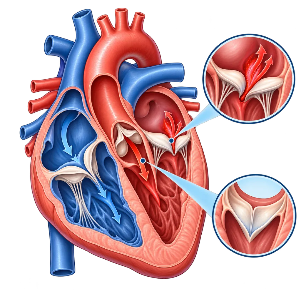
Valve regurgitation, or valve insufficiency, is a condition in which a heart valve does not close tightly, so some blood flows backward. Mild leaking is often harmless, but severe leaking puts a strain on the heart.
SourcesSigns That Need Urgent Help
- Sudden severe shortness of breath, or shortness of breath at rest
- Coughing up pink, frothy sputum
- Severe chest pain, especially pain that spreads to the back
- Pale, cold, sweaty skin with weakness or confusion
- Fainting or nearly fainting
- Fever with rapidly worsening shortness of breath in people with an artificial valve
If these signs appear, go to the nearest emergency department right away or call Heart Attack Center 1500 258.
01 / Understanding the condition
About Valve Regurgitation / Insufficiency
Heart valves should close tightly after blood passes through, like a one-way door. In regurgitation, also called insufficiency or a leaky valve, the valve does not close tightly. Some blood flows back into the previous chamber, so the heart has to pump the same blood again.
Very mild leaking is fairly often seen on echocardiography in healthy people, especially in the tricuspid and pulmonary valves, and usually does not need treatment. What your doctor assesses is which valve is affected, the degree of leaking (mild, moderate, or severe), its cause, and its effect on the size and strength of the ventricles.
Types of Valve Regurgitation
Any valve can leak, and each one has its own page. Mitral regurgitation makes blood flow back from the left ventricle into the left atrium. Aortic regurgitation makes blood flow back from the aorta into the left ventricle. Tricuspid regurgitation and pulmonary regurgitation occur on the right side of the heart and are often linked to high lung pressure or congenital heart disease.
Primary and Secondary
The 2021 ESC/EACTS and 2020 ACC/AHA guidelines distinguish two mechanisms. In primary regurgitation, the valve leaflets or their supporting structures are damaged, for example by prolapse, rheumatic heart disease, infection, or a congenital problem. In secondary or functional regurgitation, the leaflets are actually normal, but an enlarged ventricle or atrium pulls on the valve ring so the valve cannot close. This distinction matters because the treatment is different.
Chronic and Acute
In the majority of cases, regurgitation develops slowly. The heart adapts by enlarging, so symptoms are only felt after years. That is why monitoring the size and function of the ventricles with regular echocardiography is the basis for deciding when to treat, often before symptoms appear.
In contrast, severe leaking that appears suddenly gives the heart no time to adapt. Causes include a tear in the muscle that supports the mitral valve after a heart attack, an infection that damages the valve leaflets, or a tear in the wall of the aorta. This causes severe shortness of breath and is an emergency.
Treatment depends on the affected valve, the mechanism, and the degree of leaking. Mild to moderate leaking usually only needs monitoring. Severe leaking is treated with valve repair or replacement, either through surgery or through a catheter. In secondary regurgitation, the first step is to treat the underlying heart muscle disease or rhythm disorder.
02 / Understanding the condition
Types and Related Conditions
The following conditions belong to the Valve Regurgitation / Insufficiency group. Open each one for a full explanation.
03 / Understanding the condition
Symptoms to Recognize.
Common Symptoms
- Shortness of breath during activity
- Tiring easily and reduced stamina
- Palpitations or a strong, pounding heartbeat, especially when lying down
- Shortness of breath when lying flat, so you need to sleep propped up on high pillows
- Swelling in the ankles or legs
- A bloated or swollen belly due to fluid buildup
- A heart murmur that the doctor hears during an examination
04 / Understanding the condition
What Causes This Condition?
- Mitral valve prolapse or rupture of the cords that support the valve (chordae)
- Rheumatic heart disease, which makes the valve leaflets thicken and shrink
- Valve infection (infective endocarditis) that damages or makes holes in the valve leaflets
- Enlargement of the ventricles in cardiomyopathy or after a heart attack
- Enlargement of the atria in long-standing atrial fibrillation
- Enlargement of the aortic root or a bicuspid (two-leaflet) aortic valve
- High pressure in the lung blood vessels that strains the right side of the heart
- Previous congenital heart surgery, for example Tetralogy of Fallot repair
05 / Understanding the condition
Who Is at Higher Risk?
Age
Changes in valve tissue and enlargement of the atria become more common with age, so mitral and tricuspid regurgitation are found more often in older adults.
Medical
A history of heart attack, cardiomyopathy, heart failure, atrial fibrillation, high blood pressure, pulmonary hypertension, rheumatic fever, or endocarditis increases the risk of valve leaking.
Genetic
Mitral valve prolapse, a bicuspid aortic valve, Marfan syndrome, and some connective tissue disorders can be found in more than one family member.
Other
Having an artificial valve, a history of congenital heart surgery, chest radiation therapy, and injection drug use, which increases the risk of valve infection.
Groups at Higher Risk
- Heart Attack Survivors: Damage to the heart muscle can shift the supports of the mitral valve, causing the valve to leak.
- People with Heart Failure or Atrial Fibrillation: Enlarged ventricles and atria pull on the rings of the mitral and tricuspid valves so the valves do not close tightly.
- Rheumatic Fever Survivors: Rheumatic inflammation can slowly damage the valve leaflets over many years.
- People Who Had Surgery for Congenital Heart Disease: For example, after Tetralogy of Fallot repair, the pulmonary valve often leaks and needs lifelong monitoring.
- Older Adults: Changes in valve tissue and enlargement of the atria make valve leaking more common in older age.
06 / Understanding the condition
How Is It Diagnosed?
Doctors usually first suspect a leaking valve from a heart murmur heard through a stethoscope. Echocardiography then becomes the main test. With a heart ultrasound and Doppler, your doctor sees which valve is leaking, its cause, the degree of leaking, and the size and strength of the ventricles. Guidelines recommend assessing the degree with several measurements together, not just one number.
Transesophageal echocardiography, an ultrasound through the esophagus, gives more detailed images of the valve, for example when planning mitral valve repair or looking for a valve infection. Cardiac MRI helps measure the volume of leaking blood and the size of the ventricles when echocardiography results are unclear. An exercise test assesses your ability to be active. When leaking appears suddenly, tests are done quickly in the emergency room while the patient's condition is stabilized.
Physical Examination
Listening for a heart murmur, checking the pulse and blood pressure, and looking for signs of fluid buildup such as leg swelling or crackles in the lungs.
Echocardiography
The main test to identify the leaking valve, its mechanism (primary or secondary), the degree of leaking, and the size and function of the ventricles.
Transesophageal echocardiography (through the esophagus) shows the valve leaflets in more detail to plan valve repair or look for a valve infection.
Cardiac MRI
Measures the leak volume and ventricle size in detail, especially useful in aortic and pulmonary regurgitation.
Electrocardiogram (ECG)
Looks for atrial fibrillation, other rhythm disorders, and signs of enlarged heart chambers.
NT-proBNP and BNP Test
A blood test for markers of heart strain that helps assess disease progression and the timing of treatment.
Cardiac Catheterization
Done when test results do not match the symptoms, or to check the coronary arteries before valve surgery.
07 / Understanding the condition
Treatment Options.
Treatment is decided by a doctor after an assessment. The categories below can help you follow the conversation with your doctor.
Regular Monitoring
Mild to moderate leaking without symptoms is monitored with regular checkups and echocardiography; visits become more frequent as the leak becomes more severe.
Medicines for Symptoms and the Underlying Disease
Diuretics relieve fluid buildup. In secondary regurgitation, guideline-based heart failure medicines are the first step. Your doctor decides the type and dose.
Valve Repair
The natural valve is repaired so it closes tightly again, especially the mitral and tricuspid valves, through surgery or a clip placed through a catheter.
Valve Replacement
A valve that cannot be repaired is replaced with a mechanical or biological artificial valve, through surgery or, for certain valves, through a catheter.
Treatment of Sudden Leaking
Severe leaking that appears suddenly is treated in the intensive care unit to stabilize the circulation and often requires urgent surgery.
08 / Understanding the condition
Complications to Watch For.
Heart Failure
The extra volume of blood makes the ventricles enlarge and then weaken, causing shortness of breath, swelling, and tiring quickly. Covered on the Heart Failure page.
Atrial Fibrillation
Atria enlarged by mitral or tricuspid leaking are prone to an irregular rhythm, which increases the risk of stroke. Covered on the Atrial Fibrillation page.
Pulmonary Hypertension
Blood flowing back into the left atrium raises the pressure in the lung blood vessels and strains the right side of the heart. Covered on the Pulmonary Hypertension page.
Infective Endocarditis
A leaking valve is more easily infected when germs are in the bloodstream, and infection can make the leak worse. Covered on the Infective Endocarditis page.
Ventricular Rhythm Disorders
Enlarged and weakened ventricles are more prone to rhythm disorders, so the heart rhythm is also monitored during checkups.
09 / Understanding the condition
What Can You Do?
- Treat sore throats in children completely to prevent rheumatic fever
- Keep your teeth and mouth clean and have regular dental checkups
- Control your blood pressure so the load on the aortic and mitral valves does not increase
- Manage coronary artery disease, heart failure, and atrial fibrillation as your doctor advises
- Avoid injection drugs, which can carry germs to the heart valves
- Follow your regular echocardiography schedule even if you have no symptoms yet
10 / Understanding the condition
When Should You See a Doctor?
See a cardiologist if a general practitioner finds a heart murmur, or if you start tiring quickly and getting short of breath during normal activities. People with a known valve leak should have a checkup earlier than scheduled if their symptoms increase. Go to the emergency room right away if severe shortness of breath comes on suddenly, you cough up pink frothy sputum, or you faint.
- Shortness of breath during activity or when lying flat
- Palpitations, especially if the rhythm feels irregular
- Leg swelling or rapid weight gain over a few days
- Fever with no clear cause in people with an artificial valve or who have had endocarditis
- Echocardiography results that show moderate or severe leaking
Don't Wait in an Emergency.
If symptoms are severe or come on suddenly, go to the nearest emergency department right away or call Heart Attack Center 1500 258, rather than waiting for an appointment.
11 / Common questions
Questions You May Have.
Frequently asked questions about valve regurgitation or insufficiency.
Is mild valve leaking dangerous?
Very mild leaking is often found on echocardiography in healthy people, especially in the tricuspid and pulmonary valves, and is usually harmless. Mild leaking in the mitral or aortic valve usually only needs monitoring. What matters is the cause and whether the leak increases over time. Your doctor will suggest a checkup schedule based on your findings.
What is the difference between valve regurgitation and valve insufficiency?
Both refer to the same thing: a valve that does not close tightly, so blood flows backward. The term regurgitation emphasizes the backward flow, while insufficiency emphasizes that the valve does not close enough. In everyday language, patients often call it a leaky valve. This is different from a hole in the wall between the heart's chambers, which is often called a hole in the heart.
When does a leaking valve need a procedure?
A procedure is generally considered when the leak is already severe and causing symptoms, or when there are no symptoms yet but echocardiography shows the ventricle starting to enlarge or weaken beyond a certain threshold. Newly developed atrial fibrillation or rising lung pressure can also be reasons. The decision is made by the heart team, weighing the benefits and risks of the procedure for you.
Can a leaking valve be repaired without open-chest surgery?
For some patients, yes. Mitral valve leaking can be reduced with a clip placed through a catheter from a blood vessel in the groin, especially when the surgical risk is high. The pulmonary valve in people with congenital heart disease can be replaced through a catheter. Surgery through a small incision between the ribs is also an option for certain anatomy. Suitability is determined from test results.
Can I get pregnant if I have a leaking valve?
Pregnancy with mild to moderate valve regurgitation can generally go well, because valve leaking tends to be better tolerated during pregnancy than a narrowed valve. Severe leaking with a weakened ventricle needs to be assessed first. It is a good idea to plan pregnancy together with a cardiologist and an obstetrician, including reviewing the medicines you are taking.
Do I need lifelong checkups?
In general, yes. Valve leaking can slowly increase without symptoms, so regular checkups help detect changes before the ventricles are damaged. Even after valve repair or replacement, checkups are still needed to monitor valve function, heart rhythm, and medicines. Your doctor sets how often you need checkups based on the degree of leaking and the type of valve.
Sources and Medical Review
Translated from the Indonesian edition medically reviewed by Dr. dr. Dafsah A. Juzar, Sp.JP(K).
Sources as cited in the Indonesian edition.
- Vahanian A, Beyersdorf F, Praz F, Milojevic M, Baldus S, Bauersachs J, et al. 2021 ESC/EACTS Guidelines for the management of valvular heart disease. Eur Heart J. 2022;43(7):561-632.
- Otto CM, Nishimura RA, Bonow RO, Carabello BA, Erwin JP 3rd, Gentile F, et al. 2020 ACC/AHA Guideline for the Management of Patients With Valvular Heart Disease: A Report of the American College of Cardiology/American Heart Association Joint Committee on Clinical Practice Guidelines. Circulation. 2021;143(5):e72-e227.
- Zoghbi WA, Adams D, Bonow RO, Enriquez-Sarano M, Foster E, Grayburn PA, et al. Recommendations for Noninvasive Evaluation of Native Valvular Regurgitation: A Report from the American Society of Echocardiography Developed in Collaboration with the Society for Cardiovascular Magnetic Resonance. J Am Soc Echocardiogr. 2017;30(4):303-371.
- Lancellotti P, Pibarot P, Chambers J, La Canna G, Pepi M, Dulgheru R, et al. Multi-modality imaging assessment of native valvular regurgitation: an EACVI and ESC council of valvular heart disease position paper. Eur Heart J Cardiovasc Imaging. 2022;23(5):e171-e232.
- McDonagh TA, Metra M, Adamo M, Gardner RS, Baumbach A, Bohm M, et al. 2021 ESC Guidelines for the diagnosis and treatment of acute and chronic heart failure. Eur Heart J. 2021;42(36):3599-3726.
- Delgado V, Ajmone Marsan N, de Waha S, Bonaros N, Brida M, Burri H, et al. 2023 ESC Guidelines for the management of endocarditis. Eur Heart J. 2023;44(39):3948-4042.
Doctors Who Treat This Condition

dr. Widya T. Suwatri, Sp.BTKV, Subsp. JD(K)
Adult Cardiac Surgery
Advanced Cardiovascular Surgical Care +1 center
Clinical focusMinimally Invasive Cardiac Surgery · Coronary Artery Bypass Surgery
View Profile
Dr. med. dr. Denio A. Ridjab, Sp.JP(K)
Interventional Cardiology
Coronary Intervention Procedures +2 centers
Clinical focusInterventional Cardiology · Coronary Artery Disease
View Profile
Dr. dr. Dafsah A. Juzar, Sp.JP(K)
Interventional Cardiology
Coronary Intervention Procedures +1 center
Clinical focusComplex Coronary Intervention · Cardiac Catheterization
View Profile