
Quick answer
What Is Atrial Septal Defect?
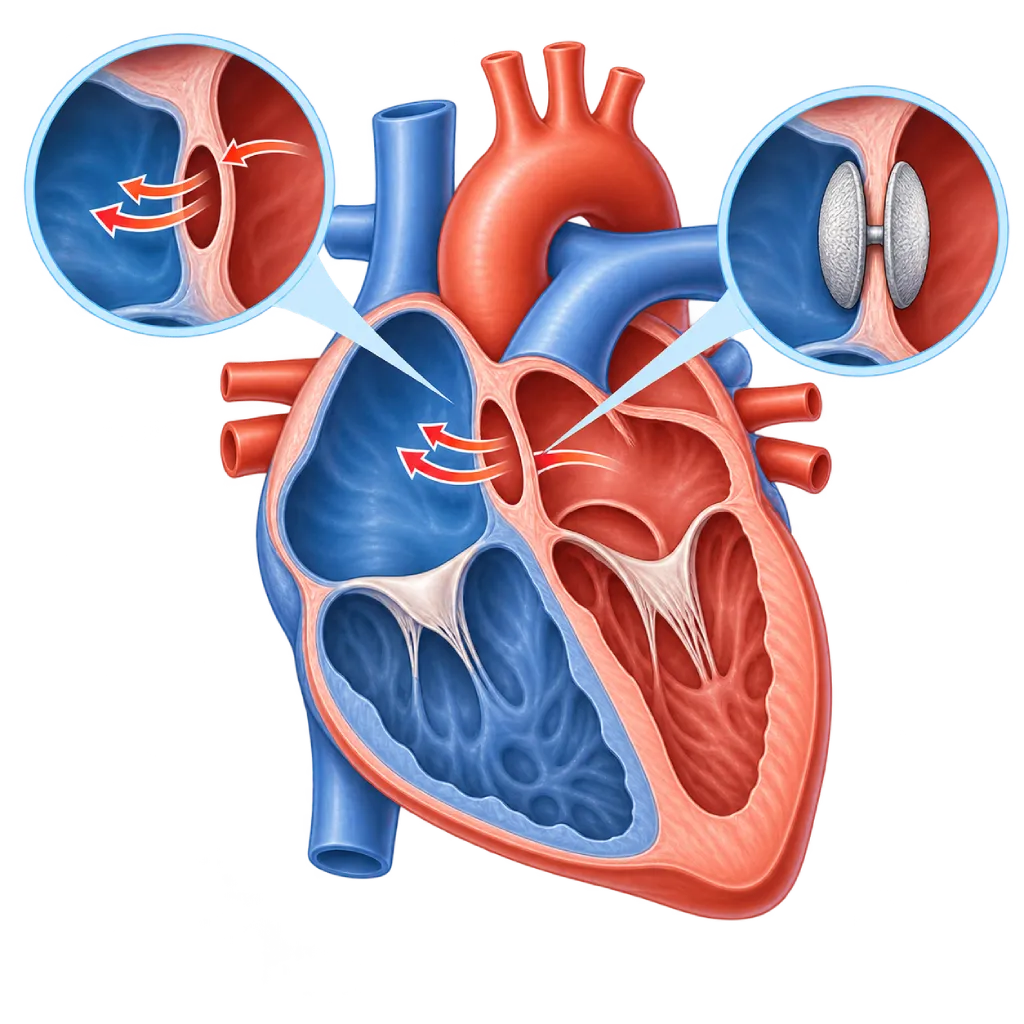
An atrial septal defect is a hole present at birth in the wall between the heart's two upper chambers. Small holes often close on their own, while large holes can strain the right side of the heart and the lungs, so they need to be closed.
SourcesSigns That Need Urgent Help
- A very fast or irregular heartbeat that does not settle
- Fainting or nearly fainting
- Severe shortness of breath, including at rest or when lying down
- Bluish lips and nails
- Sudden facial drooping, slurred speech, or weakness on one side of the body
- Swelling of the legs and abdomen that increases quickly
If these signs appear, go to the nearest emergency department right away or call Heart Attack Center 1500 258.
01 / Understanding the condition
About Atrial Septal Defect
The heart has two upper chambers (atria) separated by a thin wall called the atrial septum. In an atrial septal defect (ASD), this wall has a hole that has been there since birth. Because the pressure in the left atrium is slightly higher, oxygen-rich blood flows from the left atrium to the right atrium through the hole. This blood is then pumped back to the lungs, so the right side of the heart and the lung blood vessels receive more blood than they should. Over time, this extra load causes the right side of the heart to enlarge.
Types of Atrial Septal Defect
The common type is secundum ASD, a hole in the middle of the septum. This type accounts for about 70 to 80 percent of cases and can often be closed with a catheter. Other types are primum ASD in the lower part of the septum, which often comes with a mitral valve abnormality; sinus venosus ASD near the openings of the large veins, which frequently comes with lung veins that drain to an abnormal place; and the rare coronary sinus ASD. Types other than secundum usually require surgery. An ASD is different from a patent foramen ovale, a small gap left over from the circulation before birth, which is covered on the Patent Foramen Ovale page.
Why It Is Often Found Only in Adulthood
Children with an ASD usually look healthy and grow normally. Their heart murmur is soft, so it is easily missed. As a result, ASD is one of the congenital heart defects often found only in adulthood. Symptoms such as tiring quickly, shortness of breath during activity, or palpitations often appear only after age 30 to 40, when the right side of the heart has already enlarged. Rhythm problems such as atrial fibrillation and atrial flutter also become more common with age.
When the Hole Needs to Be Closed
Small holes, under about 5 millimeters, often close on their own in the first years of life. According to the 2020 ESC and 2018 AHA/ACC guidelines, closure is recommended when the right side of the heart is enlarged because of the shunt, as long as the pressure in the lung blood vessels is not too high, with or without symptoms. In children, closure is usually planned at preschool age. If the lung pressure is already very high, closure can actually be harmful, so the decision is made after a full evaluation.
02 / Understanding the condition
Symptoms to Recognize.
Common Symptoms
- Often no symptoms for many years
- Tiring quickly or shortness of breath during activity
- Palpitations or a heartbeat that feels irregular
- A gradual decline in the ability to exercise
- A soft heart murmur heard during a doctor's examination
Symptoms in Children
Most children with an ASD look healthy. The following symptoms are more likely with a large hole.
- Tiring more quickly than peers during play
- Frequent coughs and colds or repeated pneumonia
- Some difficulty gaining weight
- A heart murmur found during a school checkup or vaccination visit
03 / Understanding the condition
What Causes This Condition?
- The wall between the two upper chambers does not form completely in the early weeks of pregnancy
- In most cases, the exact trigger is not known
- Changes in certain genes, for example NKX2-5, GATA4, or TBX5 (Holt-Oram syndrome)
- Chromosome abnormalities such as Down syndrome, especially for primum ASD
- Pregnancy factors such as rubella infection, uncontrolled diabetes, and exposure to alcohol and smoking
04 / Understanding the condition
Who Is at Higher Risk?
Genetic
A history of ASD or other congenital heart disease in a parent or sibling, as well as genetic syndromes such as Down syndrome and Holt-Oram syndrome, increases the likelihood of ASD.
Medical
Uncontrolled diabetes, rubella infection, and the use of certain medicines in early pregnancy are linked to the risk of heart defects in the baby.
Lifestyle
Smoking and drinking alcohol during pregnancy are linked to the risk of birth defects, including defects of the walls inside the heart.
Age
If an ASD is not closed, the risk of rhythm problems, enlargement of the right side of the heart, and high lung pressure increases with age, especially after 40.
Other
ASD is found more often in women than in men, at a ratio of about 2 to 1.
Groups at Higher Risk
- Children with Down Syndrome: About 40 to 50 percent of children with Down syndrome have a heart defect, including primum ASD, so echocardiography is recommended after birth.
- Women of Childbearing Age: ASD is more common in women, and some cases are found only during pregnancy because pregnancy adds to the heart's workload.
- Adults with an Unclosed ASD: The risk of atrial fibrillation, atrial flutter, and high lung pressure increases if the hole has not been closed, especially after age 40.
- Family Members of People with ASD: Siblings and children of people with an ASD, especially when it is linked to a gene change, may be considered for a heart checkup.
05 / Understanding the condition
How Is It Diagnosed?
An ASD is often first suspected when a doctor hears a soft heart murmur and a second heart sound that stays split, or when an ECG and chest X-ray show signs of strain on the right side of the heart. In adults, an ASD can also be found while looking for the cause of palpitations, shortness of breath, or a stroke.
Echocardiography through the chest wall confirms the diagnosis, determines the type and size of the hole, and assesses enlargement of the right side of the heart and the estimated lung pressure. Before catheter closure, the doctor usually performs a transesophageal echocardiogram to measure the hole and its rims. If the type of hole is unclear or a lung vein is suspected to drain to an abnormal place, cardiac MRI or CT helps. Right heart catheterization is needed if the lung pressure is suspected to be high, because the results determine whether the hole can be closed.
Physical Examination
The doctor hears a soft murmur and a second heart sound that stays split, a typical clue of ASD.
Echocardiography
Echocardiography through the chest wall shows the hole, the direction of flow, and enlargement of the right side of the heart. Transesophageal echocardiography (through the esophagus) assesses the rims of the hole before catheter closure.
Electrocardiogram (ECG)
Assesses the rhythm and signs of strain on the right side of the heart, including atrial fibrillation or flutter in adults.
Chest X-Ray
May show an enlarged right side of the heart and lung blood vessels that look fuller.
Cardiac MRI
Measures the size and function of the right ventricle and looks for lung veins that drain to an abnormal place, especially in sinus venosus ASD.
Right Heart Catheterization
Measures the pressure and resistance of the lung blood vessels if high lung pressure is suspected, to make sure the hole is safe to close.
06 / Understanding the condition
Treatment Options.
Treatment is decided by a doctor after an assessment. The categories below can help you follow the conversation with your doctor.
Regular Monitoring
Small holes without enlargement of the right side of the heart only need monitoring with echocardiography. Many small holes in babies close on their own.
Catheter Closure
A secundum ASD with adequate rims is usually closed with a double-disc device inserted through a blood vessel in the groin, without opening the chest. Covered on the Atrial Septal Defect (ASD) Closure page.
Surgical Closure
Primum, sinus venosus, coronary sinus, or overly large secundum ASDs are closed with stitches or a patch during surgery, in certain patients through a small incision.
Medicines to Relieve Symptoms
Diuretics, rhythm-control medicines, or blood thinners may be given for shortness of breath, rhythm problems, or clot prevention. Your doctor determines the type and dose.
Treatment of Rhythm Problems
Atrial fibrillation or flutter can be treated with medicines or ablation, sometimes done before or at the same time as closing the hole.
Follow-Up After Closure
Regular echocardiography checks the device position, the size of the right side of the heart, and the rhythm. Antiplatelet medicine is usually given for several months as your doctor advises.
07 / Understanding the condition
Complications to Watch For.
Right Heart Enlargement
The extra blood flow causes the right atrium and right ventricle to enlarge. If the hole is closed in time, the right side of the heart can usually shrink back.
Atrial Fibrillation and Flutter
Enlarged atria are prone to irregular rhythms, especially after age 40, and these can persist even after the hole is closed. Covered on the Atrial Fibrillation page.
Pulmonary Hypertension
In a small number of patients, years of excess flow damage the lung blood vessels. Its advanced form, Eisenmenger syndrome, reverses the flow and makes the body look bluish.
Right Heart Failure
A right ventricle that is constantly overloaded can weaken and cause leg swelling, a swollen abdomen, and shortness of breath.
Stroke from Paradoxical Embolism
A clot from a vein can cross through the hole to the left side of the heart and then travel to the brain. This is rare, but it is one reason an ASD needs careful evaluation.
08 / Understanding the condition
What Can You Do?
- Have regular prenatal checkups and ask about a fetal heart ultrasound if there is a family history of congenital heart disease
- Make sure you are immune to rubella before pregnancy
- Control your blood sugar before and during pregnancy
- Avoid smoking and alcohol during pregnancy
- If you have an ASD, keep your follow-up appointments even without symptoms
- Talk with a cardiologist before planning a pregnancy
09 / Understanding the condition
When Should You See a Doctor?
Because an ASD often causes no symptoms, small findings during routine checkups should be followed up. Make an appointment with a cardiologist if you or your child has any of the following. Go to the emergency room right away if signs of stroke, fainting, or a very fast heartbeat that does not settle appear.
- A doctor hears a heart murmur, or an ECG shows strain on the right side of the heart
- Tiring quickly or shortness of breath during activities that used to feel easy
- Palpitations or a heartbeat that feels irregular
- Your child often gets pneumonia or tires more quickly than peers
- You have an ASD and are planning a pregnancy
Don't Wait in an Emergency.
If symptoms are severe or come on suddenly, go to the nearest emergency department right away or call Heart Attack Center 1500 258, rather than waiting for an appointment.
10 / Common questions
Questions You May Have.
Frequently asked questions about atrial septal defect.
Can an ASD close on its own?
Yes, especially small holes found in babies. Holes smaller than about 5 millimeters often close or shrink in the first years of life. Larger holes, or those still open after a few years of age, rarely close on their own. Your doctor will monitor with regular echocardiography and recommend closure if the right side of the heart starts to enlarge.
Does ASD closure always require surgery?
Not always. The majority of secundum ASDs can be closed with a catheter inserted through a blood vessel in the groin, without opening the chest, and recovery is short. Surgery is needed for the primum, sinus venosus, and coronary sinus types, or for secundum holes that are too large or have insufficient rims. The method is chosen after a transesophageal echocardiogram assesses the size and shape of the hole.
I only found out I have an ASD as an adult. Does it still need to be closed?
Closure can still be beneficial. The 2020 ESC guidelines recommend closure when the right side of the heart is enlarged and the pressure in the lung blood vessels is not too high, regardless of age. Closure in adults can reduce symptoms and improve the ability to be active. However, the risk of rhythm problems does not always go away, so follow-up is still needed. If the lung pressure is already very high, your doctor will first assess whether closure is safe.
Can women with an ASD get pregnant?
The majority of women with a small ASD or one that has already been closed can become pregnant with low risk. Pregnancy adds to the load on the right side of the heart, so a large hole that has not been closed can cause rhythm problems or shortness of breath. If there is severe pulmonary hypertension, pregnancy is high risk and needs to be discussed with your doctor. A consultation before pregnancy helps decide whether the hole should be closed first.
What should I watch for after catheter closure of an ASD?
You can usually return to light activities within a few days. Your doctor usually prescribes antiplatelet medicine for several months until the device is covered by body tissue. A follow-up echocardiogram is done to check the device position and the size of the right side of the heart. Contact your doctor right away if you develop chest pain, palpitations, shortness of breath, or fainting, because device problems, although rare, need to be recognized quickly.
Sources and Medical Review
Translated from the Indonesian edition medically reviewed by dr. Radityo Prakoso, Sp.JP(K).
Sources as cited in the Indonesian edition.
- Baumgartner H, De Backer J, Babu-Narayan SV, et al. 2020 ESC Guidelines for the management of adult congenital heart disease. Eur Heart J. 2021;42(6):563-645.
- Stout KK, Daniels CJ, Aboulhosn JA, et al. 2018 AHA/ACC Guideline for the Management of Adults With Congenital Heart Disease: A Report of the American College of Cardiology/American Heart Association Task Force on Clinical Practice Guidelines. Circulation. 2019;139(14):e698-e800.
- Brida M, Chessa M, Celermajer D, et al. Atrial septal defect in adulthood: a new paradigm for congenital heart disease. Eur Heart J. 2022;43(28):2660-71.
- Geva T, Martins JD, Wald RM. Atrial septal defects. Lancet. 2014;383(9932):1921-32.
- Silvestry FE, Cohen MS, Armsby LB, et al. Guidelines for the Echocardiographic Assessment of Atrial Septal Defect and Patent Foramen Ovale: From the American Society of Echocardiography and Society for Cardiac Angiography and Interventions. J Am Soc Echocardiogr. 2015;28(8):910-58.
- Du ZD, Hijazi ZM, Kleinman CS, et al. Comparison between transcatheter and surgical closure of secundum atrial septal defect in children and adults: results of a multicenter nonrandomized trial. J Am Coll Cardiol. 2002;39(11):1836-44.
Doctors Who Treat This Condition

dr. Radityo Prakoso, Sp.JP(K)
Pediatrics & Congenital Heart Disease
Congenital and Structural Heart Center
Clinical focusPediatric Cardiology Consultation · Congenital Heart Disease Care
View Profile
dr. BRM Ario S. Kuncoro, Sp.JP(K)
Echocardiography & Cardiovascular Imaging
Cardiovascular Diagnostic Center +2 centers
Clinical focusAdvanced and 3D Echocardiography · Transesophageal Echocardiography
View Profile
Dr. dr. Dudy A. Hanafy, Sp.BTKV, Subsp. JD(K), MARS
Adult Cardiac Surgery
Advanced Cardiovascular Surgical Care +1 center
Clinical focusMinimally Invasive Cardiac Surgery · Coronary Artery Bypass Surgery
View Profile