
Quick answer
What Is Mitral Regurgitation?
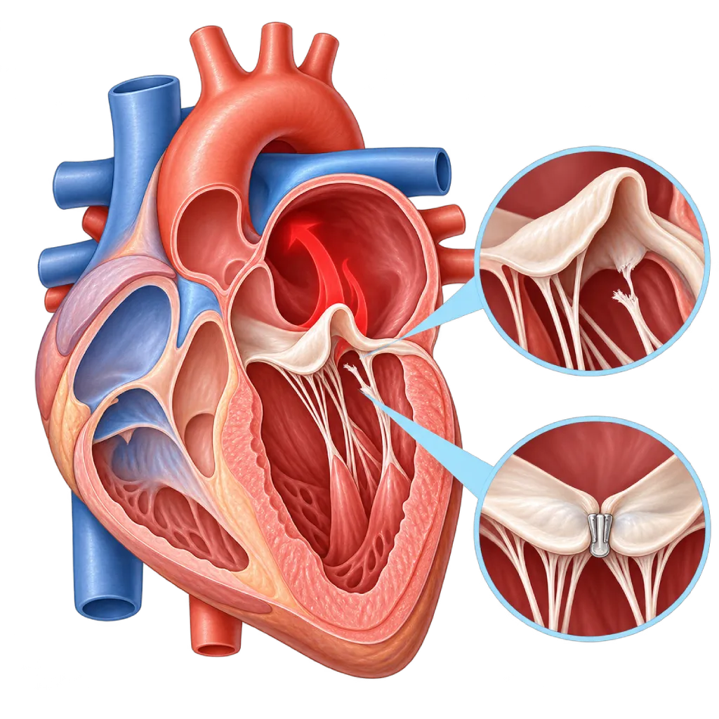
Mitral regurgitation is a leak in the mitral valve that lets some blood flow back from the left ventricle into the left atrium with each heartbeat. A severe leak can cause shortness of breath, palpitations, and heart failure.
SourcesSigns That Need Urgent Help
- Sudden severe shortness of breath, especially within a few days after a heart attack
- Coughing up pink, frothy sputum
- Being unable to lie down and needing to sit up to breathe
- Pale, cold, sweaty skin with weakness or confusion
- A very fast heartbeat with dizziness or shortness of breath
- Sudden weakness on one side of the body, facial drooping, or slurred speech
If these signs appear, go to the nearest emergency department right away or call Heart Attack Center 1500 258.
01 / Understanding the condition
About Mitral Regurgitation
The mitral valve sits between the left atrium and the left ventricle. It has two leaflets held in place by supporting cords (chordae) and small muscles in the ventricle wall. When the left ventricle pumps, both leaflets close tightly so that blood flows only into the aorta. In mitral regurgitation, the valve does not close tightly, so some blood flows back into the left atrium. This condition belongs to the group of leaky valves covered on the Valve Regurgitation / Insufficiency page.
Primary and Secondary
The 2021 ESC/EACTS and 2020 ACC/AHA guidelines divide mitral regurgitation into two types. In primary regurgitation, the damage is in the valve itself: the leaflets are too floppy and bulge into the atrium (prolapse), a supporting cord breaks, or there is rheumatic heart disease, a valve infection, or a congenital abnormality. In secondary regurgitation, the leaflets are actually normal, but a left ventricle enlarged by a heart attack or cardiomyopathy, or a left atrium enlarged by atrial fibrillation, pulls on the valve so that it cannot close.
Why This Distinction Matters
In severe primary regurgitation, repairing the valve can fix the main problem. Valve repair is preferred over replacement when the result is expected to last. In secondary regurgitation, the main problem lies in the heart muscle, so the first step is guideline-directed heart failure medicines and, when appropriate, a resynchronization pacemaker. A valve procedure is considered if symptoms persist.
Course of the Disease
Chronic mitral regurgitation develops slowly. The left atrium and ventricle enlarge to hold the extra blood, so symptoms often appear only after years. Monitoring the size and function of the left ventricle, heart rhythm, and lung pressure helps determine the right time for a procedure before the heart muscle weakens. New atrial fibrillation or rising lung pressure can be a sign that the leak is starting to strain the heart. A mild leak on its own is fairly common and usually not dangerous.
In contrast, a severe leak that appears suddenly, for example because a muscle supporting the valve tears after a heart attack, a supporting cord breaks, or the valve becomes infected, causes severe shortness of breath because fluid quickly builds up in the lungs. This requires emergency treatment and often urgent surgery.
02 / Understanding the condition
Symptoms to Recognize.
Common Symptoms
- Shortness of breath during activity
- Tiring easily and a reduced ability to be active
- Palpitations, often with an irregular rhythm
- Shortness of breath when lying flat or waking up at night short of breath
- Swelling in the ankles or legs
- A heart murmur heard by the doctor during a routine checkup
03 / Understanding the condition
What Causes This Condition?
- Mitral valve prolapse: the leaflets are too floppy and bulge into the left atrium
- A ruptured supporting cord of the valve (chorda), often due to age-related changes in valve tissue
- Rheumatic heart disease, which makes the leaflets thick and shrunken
- Valve infection (infective endocarditis) that damages the leaflets or makes holes in them
- An enlarged left ventricle after a heart attack or in cardiomyopathy
- An enlarged left atrium in long-standing atrial fibrillation
- A tear in a muscle that supports the valve as a complication of a heart attack
- Calcium buildup in the mitral valve ring in older adults or people with chronic kidney disease
04 / Understanding the condition
Who Is at Higher Risk?
Age
Changes in valve tissue, prolapse, and atrial enlargement make mitral regurgitation more common with age.
Medical
A history of heart attack, cardiomyopathy, heart failure, atrial fibrillation, high blood pressure, rheumatic fever, or endocarditis increases the risk.
Genetic
Mitral valve prolapse can be found in several members of the same family. Marfan syndrome and other connective tissue disorders also make the leaflets floppier.
Other
Chest radiation therapy for cancer and injecting drugs, which increases the risk of valve infection, can damage the mitral valve.
Groups at Higher Risk
- People with Mitral Valve Prolapse: A small share of prolapse cases progress to a severe leak, so regular echocardiography is needed. Covered on the Mitral Valve Prolapse page.
- Heart Attack Survivors: Damage to the left ventricle muscle can shift the valve's supports and cause a leak.
- People with Heart Failure or Cardiomyopathy: An enlarged left ventricle pulls on the mitral valve so that it does not close tightly.
- People with Long-Standing Atrial Fibrillation: An enlarged left atrium can stretch the mitral valve ring, causing the valve to leak.
- Rheumatic Fever Survivors: The mitral valve is the valve usually affected first in rheumatic heart disease.
05 / Understanding the condition
How Is It Diagnosed?
Your doctor usually hears a murmur as the ventricle pumps, heard clearly over the apex of the heart. Echocardiography is the main test to see which part of the valve has a problem, distinguish primary from secondary regurgitation, grade the leak using several measurements at once, and assess the size of the atrium and ventricle, pumping strength, and lung pressure.
Transesophageal echocardiography shows the leaflets and their supporting cords in more detail and is important when planning valve repair by surgery or catheter clip. Cardiac MRI helps when the severity of the leak is unclear. Echocardiography during exercise can assess changes in the leak and lung pressure. An NT-proBNP blood test and rhythm recordings help assess strain on the heart and atrial fibrillation. Before a procedure, the coronary arteries are checked with angiography or CT.
Echocardiography
The main test to determine the cause and severity of the leak, the size of the left atrium and ventricle, pumping strength, and estimated lung pressure.
Transesophageal echocardiography (through the esophagus) shows the valve leaflets and supporting cords in detail to plan valve repair.
Stress Echocardiography
Assesses changes in the leak and lung pressure during exercise when symptoms do not match the results at rest.
Cardiac MRI
Measures the leak volume and left ventricle size when echocardiography results are not clear enough.
Electrocardiogram (ECG)
Looks for atrial fibrillation and signs of an enlarged left atrium or ventricle.
NT-proBNP and BNP Test
A blood test for a marker of heart strain that helps assess how the disease is progressing.
Coronary Angiography
Checks the coronary arteries before valve surgery, or looks for coronary artery disease as a cause of secondary regurgitation.
06 / Understanding the condition
Treatment Options.
Treatment is decided by a doctor after an assessment. The categories below can help you follow the conversation with your doctor.
Regular Monitoring
A mild to moderate leak without symptoms is monitored with regular echocardiography; a severe leak without symptoms is monitored more closely.
Guideline-Directed Heart Failure Medicines
In secondary regurgitation, guideline-directed heart failure medicines are the foundation of treatment. Diuretics relieve fluid buildup. Your doctor determines the type and dose.
Atrial Fibrillation Management
Controlling heart rhythm or heart rate, along with blood thinners to prevent stroke, based on your doctor's assessment.
Surgical Valve Repair
The preferred option for severe primary regurgitation when the valve can be repaired with a lasting result; in selected patients it can be done through a small incision between the ribs.
Transcatheter Valve Clip
A clip is inserted through a blood vessel in the groin to bring the leaflets together, for patients at high surgical risk or with certain types of secondary regurgitation that remain symptomatic despite medicines.
Mitral Valve Replacement
The valve is replaced with a mechanical or biological artificial valve when repair is not possible or its result is not expected to last.
Resynchronization Pacemaker
In heart failure with certain electrical conduction disorders, this device can make the ventricles contract in sync and reduce secondary regurgitation.
07 / Understanding the condition
Complications to Watch For.
Atrial Fibrillation
An enlarged left atrium is prone to an irregular rhythm, which raises the risk of stroke. Covered on the Atrial Fibrillation page.
Heart Failure
Years of extra blood volume make the left ventricle enlarge and then weaken. Covered on the Heart Failure page.
Pulmonary Hypertension
Rising pressure in the left atrium is passed on to the lung blood vessels and strains the right ventricle. Covered on the Pulmonary Hypertension page.
Infective Endocarditis
A leaky valve is more easily infected when germs are in the bloodstream. Covered on the Infective Endocarditis page.
Pulmonary Edema
With a sudden leak, fluid quickly builds up in the lungs, causing severe shortness of breath. Covered on the Cardiogenic Pulmonary Edema page.
08 / Understanding the condition
What Can You Do?
- Control your blood pressure, cholesterol, and blood sugar, and quit smoking to protect your heart muscle
- Treat heart failure and atrial fibrillation as your doctor advises
- Treat throat infections in children completely to prevent rheumatic fever
- Keep your teeth and mouth clean and have regular dental checkups
- If you have mitral valve prolapse, follow your regular echocardiography schedule
- Get checked promptly if new symptoms appear, especially after a heart attack
09 / Understanding the condition
When Should You See a Doctor?
See a cardiologist if a heart murmur or mitral valve prolapse has been found, or if you start to tire quickly and feel short of breath. If you already know you have mitral regurgitation, come in for a checkup earlier than scheduled if you develop an irregular heartbeat or your symptoms get worse. Go to the emergency room right away if severe shortness of breath comes on suddenly, especially after a heart attack.
- Shortness of breath during activity or when lying flat
- Palpitations with an irregular rhythm
- Leg swelling or rapid weight gain over a few days
- A reduced ability to be active compared with before
- Fever without a clear cause in a person with a leaky valve or an artificial valve
Don't Wait in an Emergency.
If symptoms are severe or come on suddenly, go to the nearest emergency department right away or call Heart Attack Center 1500 258, rather than waiting for an appointment.
10 / Common questions
Questions You May Have.
Frequently asked questions about mitral regurgitation.
Is mitral valve prolapse the same as mitral regurgitation?
No. Prolapse means the valve leaflets bulge into the left atrium as they close. Many people with prolapse have only a mild leak or no leak at all and do not need a procedure. A small share progress to severe mitral regurgitation, so regular monitoring is recommended. Prolapse is covered on the Mitral Valve Prolapse page.
Why is valve repair preferred over replacement?
In primary mitral regurgitation, valve repair keeps your own valve and its supports, so left ventricle function is better preserved and many patients do not need long-term blood thinners. Long-term studies show better results than replacement when repair can be done well. Whether repair is possible is assessed with transesophageal echocardiography.
What is a transcatheter mitral valve clip?
A small clip is carried through a catheter from a vein in the groin to the heart, then clamped onto both valve leaflets to reduce the leak. The procedure does not require opening the chest and is guided by transesophageal echocardiography. This option is considered for patients at high surgical risk, or for certain types of secondary regurgitation that remain symptomatic despite guideline-directed medicines.
Do I need a procedure if I have no symptoms yet?
In severe primary mitral regurgitation, a procedure may be recommended even before symptoms appear, for example if the left ventricle starts to enlarge or weaken, atrial fibrillation develops, or lung pressure rises. If the valve is very likely to be repaired well, earlier surgery may also be considered. The goal is to prevent heart muscle damage that is hard to reverse. The heart team will explain their reasoning.
Can mitral regurgitation improve with medicines?
In primary regurgitation, medicines do not repair the damaged valve, but they can relieve symptoms. In secondary regurgitation, guideline-directed heart failure medicines and, when appropriate, a resynchronization pacemaker can make the left ventricle smaller, which reduces the leak. That is why a reassessment is done after treatment has been underway, before deciding on a valve procedure.
Can I get pregnant if I have mitral regurgitation?
Women with mild to moderate mitral regurgitation can generally go through pregnancy well. A severe leak, especially with a weakened left ventricle or high lung pressure, needs to be assessed before pregnancy, and sometimes the valve is repaired first. Plan your pregnancy together with your cardiologist and obstetrician, including a review of the medicines you are taking.
Sources and Medical Review
Translated from the Indonesian edition medically reviewed by dr. BRM Ario S. Kuncoro, Sp.JP(K).
Sources as cited in the Indonesian edition.
- Vahanian A, Beyersdorf F, Praz F, Milojevic M, Baldus S, Bauersachs J, et al. 2021 ESC/EACTS Guidelines for the management of valvular heart disease. Eur Heart J. 2022;43(7):561-632.
- Otto CM, Nishimura RA, Bonow RO, Carabello BA, Erwin JP 3rd, Gentile F, et al. 2020 ACC/AHA Guideline for the Management of Patients With Valvular Heart Disease: A Report of the American College of Cardiology/American Heart Association Joint Committee on Clinical Practice Guidelines. Circulation. 2021;143(5):e72-e227.
- Zoghbi WA, Adams D, Bonow RO, Enriquez-Sarano M, Foster E, Grayburn PA, et al. Recommendations for Noninvasive Evaluation of Native Valvular Regurgitation: A Report from the American Society of Echocardiography Developed in Collaboration with the Society for Cardiovascular Magnetic Resonance. J Am Soc Echocardiogr. 2017;30(4):303-371.
- Lazam S, Vanoverschelde JL, Tribouilloy C, Grigioni F, Suri RM, Avierinos JF, et al. Twenty-Year Outcome After Mitral Repair Versus Replacement for Severe Degenerative Mitral Regurgitation: Analysis of a Large, Prospective, Multicenter, International Registry. Circulation. 2017;135(5):410-422.
- Stone GW, Lindenfeld J, Abraham WT, Kar S, Lim DS, Mishell JM, et al. Transcatheter Mitral-Valve Repair in Patients with Heart Failure. N Engl J Med. 2018;379(24):2307-2318.
- Obadia JF, Messika-Zeitoun D, Leurent G, Iung B, Bonnet G, Piriou N, et al. Percutaneous Repair or Medical Treatment for Secondary Mitral Regurgitation. N Engl J Med. 2018;379(24):2297-2306.
- McDonagh TA, Metra M, Adamo M, Gardner RS, Baumbach A, Bohm M, et al. 2021 ESC Guidelines for the diagnosis and treatment of acute and chronic heart failure. Eur Heart J. 2021;42(36):3599-3726.
Doctors Who Treat This Condition

dr. BRM Ario S. Kuncoro, Sp.JP(K)
Echocardiography & Cardiovascular Imaging
Cardiovascular Diagnostic Center +2 centers
Clinical focusAdvanced and 3D Echocardiography · Transesophageal Echocardiography
View Profile
Dr. dr. Dudy A. Hanafy, Sp.BTKV, Subsp. JD(K), MARS
Adult Cardiac Surgery
Advanced Cardiovascular Surgical Care +1 center
Clinical focusMinimally Invasive Cardiac Surgery · Coronary Artery Bypass Surgery
View Profile
Dr. dr. Faris Basalamah, Sp.JP(K)
Interventional Cardiology
Coronary Intervention Procedures +1 center
Clinical focusComplex Coronary Intervention · Cardiac Electrophysiology
View Profile